
Autologous platelet-rich plasma (PRP) gel has been suggested for wound repair. However, there is insufficient evidence to support its usage in animals with cutaneous wounds. Therefore, this study aimed to evaluate the effect of PRP gel on cutaneous wound healing in rescued horses. Twelve horses were divided into two groups: group A (PRP-treated) and group B (control). The group A (n=6) animals were treated with autologous PRP gel, while the group B (n=6) animals were treated with sterile saline. Full-thickness (3 x 3 cm2) skin wounds were selected in each horse on either left or right region of back. Neovascularization was assessed on days 14 and 28 by color Doppler ultrasonography and cytokeratin staining. Wound re-epithelialization and the number of fibroblasts were assessed by cytokeratin staining. All data were statistically analyzed. We observed that PRP-wounds had highly significantly (P<0.01) increased levels of neovascularization on days 14 and 28 than control wounds. PRP-wounds had highly significant (P<0.01) increases in re-epithelialization levels and fibroblast numbers at days 14 and 28 than control wounds. In conclusion, PRP-treated wounds accelerated cutaneous wound healing by fostering re-epithelialization and neovascularization in rescued horses than control wounds. Therefore, this study suggests the use of autologous platelet-rich plasma gel to treat cutaneous wounds in rescued horses, which is a safe and effective method. The color Doppler ultrasonography can be used to assess neovascularization during wound healing.
Research Article · Volume 56, Issue 2, 16 January 2024 · Open Access
Autologous Platelets-Rich Plasma Gel Promotes Neo-Vascularization and Re-Epithelization in Cutaneous Wound Healing in Rescued Horses
· · · · · ·
Affiliations & Notes ⌄
- 1University of Veterinary and Animal Sciences, Faculty of Veterinary Sciences, Department of Veterinary Surgery and Pet Sciences, 54000 Lahore, Pakistan 2University of Veterinary and Animal Sciences, Faculty of Veterinary Sciences, Department of Pathology, 54000 Lahore, Pakistan 3University of Veterinary and Animal Sciences, Faculty of Veterinary Sciences, Department of Epidemiology and Public Health, 54000, Lahore, Pakistan 4Northeast Agricultural University, College of Veterinary Medicine, Depa
Article Info ⌄
- Journal:
- Pakistan Journal of Zoology
- Published:
- 16 January 2024

From the article
- We also found that PRP led to increased neovascularization around the wound and enhanced the effective secretion of VEGF in the wound tissue, confirming its beneficial effects on angiogenesis.
- In conclusion, PRP-treated wounds accelerated cutaneous wound healing by fostering re-epithelialization and neovascularization in rescued horses compared to control wounds.
- Therefore, this study suggested the use of autologous PRP gel to treat cutaneous wounds in rescued horses, which is a safe and effective method.
- The color Doppler ultrasonography can be used to assess neovascularization during wound healing.
Selected from the article's own text.
Abstract
INTRODUCTION
The skin is the largest organ in animals and plays a vital role in protecting them from pathogenic microorganisms and wounds . Different kinds of skin wounds are routinely encountered in veterinary clinics. Recovery of severe wounds can be influenced by a variety of factors, such as the duration of therapy and the treatment strategy, which affect the time and speed of healing . Despite the advances in wound closure techniques and devices, wound healing is still considered a challenge for surgeons . Horses, donkeys, and mules commonly develop cutaneous wounds. A horse’s performance is severely compromised by chronic non-healing wounds and abundant granulation tissue (proud flesh) and is a serious concern to its owners. The cost of wound restoration and delayed wound healing can pose a threat to proper wound healing. There are various growth factors, chemical mediators, cytokines, and types of cells involved in wound healing. Any alteration to this mechanism may result in chronic, non-healing wounds . The term chronic wounds refers to wounds that are not treated timely or do not heal in the appropriate way . As a result, there is a critical need for newer materials capable of enhancing the healing process in order to achieve optimal results.
Platelet-rich plasma (PRP) is a plasma component with 3-7 times the number of platelets than whole blood ; Bos-Mikic et al., 2018; ; . It also contains supraphysiological growth factors (GF) [insulin-like GF I, II, epidermal(GF, connective tissue GF, platelet derived GF, neural GF, vascular endothelium GF, hepatocyte GF, interleukin 8 (IL8), fibroblasts GF and transformed GF] and histamine, serotonin, calcium, zinc, superoxide dismutase (SOD) and adenosine triphosphate (ATP). Due to these important factors, it is successfully used in mammalian reproduction, dermatology and orthopaedics ; ; Irmak et al., 2020).
The platelet has a hemostatic function and high concentrations of growth factors and cytokines, which are essential for wound healing . Higher concentrations of growth factors promote epithelial and endothelial cell regeneration, as well as angiogenesis and healing processes . PRP gel speeds up the healing process of wounds that are difficult to treat. Growth factors (GFs) secreted by platelets contribute to the healing process. A natural platelet activator, thrombin, promotes wound healing and is used to activate platelets. As a result, it reduces wound size, increases the ratio of fibroblasts to macrophages, and promotes fibroblast proliferation . Numerous studies have confirmed the importance of PRP in wound healing and tissue regeneration. Some researchers report that PRP has an effective effect on the formation of new blood vessels. Pufe et al. (2005) point out that angiogenesis is controlled by naturally elevated levels of VEGF, a potent angiogenesis promoter. In addition to containing VEGF, PRP has been shown to increase VEGF synthesis , suggesting that it can promote neovascularization . Previous studies have shown that color Doppler ultrasonography (CDU) can be used to diagnose tendon injuries and assess their repair processes ; . However, according to a literature review, CDU has not been used to study the dynamic regulation of wound vasculature in equine full-thickness skin wound healing. Therefore, we used CDU to assess angiogenesis in an equine full-thickness skin wound healing model.
To the best of our knowledge, the efficacy of PRP gel for cutaneous wound healing has not been evaluated in rescued horses in Pakistan using ultrasonography or histopathology. Therefore, this study aimed to explore the repair of cutaneous wounds following topical application of autologous PRP in the form of a gel using color Doppler ultrasound and histological analysis.
MATERIALS AND METHODS
Animals
The research was carried out on rescued horses with chronic skin wounds in Pakistan’s Punjab province’s Lahore district in accordance with rules and regulations of the Local Ethical Review Committee (Ethical Approval No. DR/458; Dated: 07/10/2020) at the Department of Veterinary Surgery and Pet Sciences, University of Veterinary and Animal Sciences, Lahore, Pakistan. Throughout the experiment, all animals were housed in the Indoor University Stables as well as the Indoor Stables of the Society for the Prevention of Cruelty to Animals in Lahore. Horses were subjected to plenty of drinking water, grass, and concentrated feedstuff prior to the start of the experimental trials.
Preparation of platelet-rich plasma (PRP) gel
According to the method of , the horse PRP was prepared using two blood tubes containing 10 mL of whole blood collected on the same day as the surgical operation. Blood was collected from each horse as part of the wound-healing study. We placed blood into two Falcon tubes (15 mL each) with 10% sodium citrate anticoagulant. We centrifuged the tubes at 300 g/ 10 m (SCILOGEX swing out centrifuge) to isolate the plasma red cells. Platelets remained in the blood’s upper portion, and an intermediate zone containing large platelets and white cells, mostly leukocytes, ran between the upper and lower layers. As a result, 500 µL of plasma from each tube was removed and transferred to tube A. It was then used to generate autogenous thrombin. PRP was made by transferring the remaining plasma and the intermediate zone to a separate tube called B. It was then incubated at room temperature. Then, tube A was added with 300 µL of 10% calcium gluconate, stirred well, and incubated at 37 °C for 15 min. Then, both tubes (A and B) were centrifuged at room temperature at 640 g/ 10m. We used the entire volume of tube A, which contained thrombin-rich substrate. Thrombin was added in a 2:1 ratio to tube A (2 mL of PRP: 1 mL of thrombin) after tube B had been discharged halfway. In 40 min at room temperature, the PRP gel formed. Cell visualisation and differentiation were improved with Neubauer’s camara and trypan blue , which were used to count the platelets. The optimal platelet enrichment was determined by multiplying 4.0 times’ baseline values. PRP platelets were on average 15.8 x 10⁵, while whole blood platelets were on average 3.9 x 10⁵.
Experimental design and treatment
Twelve rescued horses weighing between 350 and 450 kg and aged 5 to 10 years were selected in this study. The animals were housed in the indoor stables of the Lahore Society for the Prevention of Cruelty to Animals. A sufficient supply of water and dry fodder was provided. All horses were given access to mineral salt throughout the trial period. 12 horses were divided into two groups: the group A (PRP-treated) and the group B (control). Each animal’s wound in group A (n=6) was treated with autologous PRP gel, whereas animals in group B (n=6) received sterile saline.
Horses were sedated intravenously with Xylazine HCl (Xylaz@Farvet Holland) at a dose rate of 1.1 mg/Kg . Each full-thickness skin wound (3 x 3 cm2) on either right or left region of back was dehaired and shaved from the edges before being separated from the subcutaneous tissue with scissors 25CM/10 (Noorani Surgical). After cleansing each site with sterile saline (NaCl 0.9 percent, Geofman), a thick coating of PRP gel was placed, which was then wrapped in sterile gauze and covered with a dressing. The bandage was removed after two days, and the wound was cleansed in sterile saline, coated with PRP gel, and wrapped. The treatment was given every four days until the 26th day. To limit granulation tissue overgrowth, semi-occlusive gauze containing PRP gel was applied to the skin wound with a soft bandage to allow fresh air. The PRP gel was reapplied to the wound every 8 days after the first 26 days. Similarly, following washing with Povidone-Iodine Solution (Pyodine Sol, Brookes Pharma), sterile saline was applied to the cutaneous wound in control Group B animals, and the bandage was placed using the same approach as in Group A animals. Over the wounds, sterile non-adherent semi-occlusive gauze was applied. Bandages were applied to the wounds (Surgitex, Rehman Rainbow (PVT.) LTD.). Prophylactic systemic antibiotics such as Biocon 5gm Inj. contain benzylpenicillin, procaine penicillin, and streptomycin sulphate (Vetcon Pharma) were administered intramuscularly twice a day for animal welfare, and the horses were kept in hygienic stables with limited exercise.
Color doppler ultrasonography (CDU)
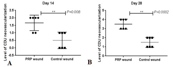
All skin wounds were scanned 14 and 28 days after surgery. Throughout the experiment, ultrasonography was performed by an experienced sonographer who was unaware of treatment options. The color Doppler ultrasound device MyLab Gamma® was used to perform ultrasound assessments. Ultrasound scans were performed using a high frequency 7.5-13 MHz linear array transducer. Wounds were examined in the transverse plane using transducers without stents and with a thin layer of acoustic coupling gel at the interface. The pressure applied by the sensor is kept to a minimum. Each CDU session was recorded and saved as a video clip for later assessment of vascularity using a semi-quantitative grading scale adapted from . On this scale, a scale of 0 means no detectable vessels, a scale of 1 means 1 to 2 small vessels, a scale of 2 means some small vessels or 1 to 2 larger vessels, and a scale of 3 means some larger vessels, while grade 4 indicates diffuse vascularization. Clips were blinded and analyzed independently by two observers. For statistical purposes, the scores of the two observers were averaged.
Histopathology
All horses were sedated prior to surgery and biopsy site were scrubbed with saline solution and gauze. Full-thickness specimens were collected using a 6 mm surgical biopsy punch. At both wound edges, we performed biopsies of a 3 to 4 mm diameter area of uninjured skin and a 4 to 5 mm depth of subcutaneous tissue. Prior to being transferred to 70% alcohol, the samples were fixed for 24 hours in 10% neutral buffered formalin. To assess neovascularization and re-epithelialization, tissues were fixed in paraffin, separated at 1.5 m thickness, and stained with cytokeratin. Using a semi-quantitative grading scale, two observers blindly evaluated all cytokeratin-stained slides. The degree of re-epithelialization, neovascularization, and fibroblasts scoring was based a semi-quantitative scoring scale: 0 (absent), 1 (minimal), 2 (mild), 3 (moderate), and 4 (marked) .
Statistical analysis
All data were statistically assessed by student t-test between the both groups using Graphpad prism (version 7.04, graphpad software Inc., San Diego, CA). All data were showed as mean ± standard error (Mean± SEM). The level of significance “*” showed (P<0.05) and “**” showed (P<0.01).
RESULTS
Color doppler ultrasonographical evaluation
At day 14, the PRP-treated wound showed mild development of blood vessels, whereas the control wound showed no development of neo-vascularization. Similarly, PRP-treated wounds showed increased development of neo-vascularization on day 28, whereas control wounds showed mild development of neo-vascularization on day 28 as shown in (Fig. 1). Statistically, PRP-treated wounds had a significantly (P<0.01) higher score of neo-vascularization at day 14 and day 28 when compared to control wounds (Fig. 2A, B).
Histopathology evaluation
Neo-vascularization
Histopathological results for neovascularization indicated that PRP treated wound obtained at day 14 showed mild development of blood vessels, while Control wound showed no development of neo-vascularization at day 14. Similarly, PRP treated wound showed increased development of neo-vascularization at day 28, while saline treated wound at day 28 showed mild development of neo-vascularization as indicated in (Fig. 3). Statistically,
compared with the Control wounds, PRP-treated wounds had highly significantly (P<0.01) increased level of neo-vascularization at day 14 and day 28 as indicated in (Fig. 4A, B). Similarly, number of fibroblasts scoring in the PRP-treated wounds were highly significantly (P<0.01) higher at day 14 and day 28 compared with the Control wounds as indicated in (Fig. 4C, D).
Re-epithelization
Histopathological results for re-epithelization indicated that PRP treated wounds obtained at 14 day indicated accelerated epithelial growth, increased proliferation of connective tissue, while saline treated wounds showed no development of epithelial growth, no basal lamina growing, and mild proliferation of connective tissue. At day 28, the PRP treated wound showed increased epidermal growth, intact basal layer, multiple layers of stratum spinosum visible, stratum corneum also visible, and increased fibrous connective tissue, while saline treated wounds indicated mild epithelial growth, basal lamina growing, and mild proliferation of connective tissue as indicated in (Fig. 5). Statistically, PRP-treated wounds had a highly significant (P<0.01) enhanced level of basal epithelial growth scoring at day 14 and day 28 compared to control wounds, as shown in (Fig. 6A, B).
DISCUSSION
Ultrasonography offers the advantages of being widely accessible, non-invasive, and ability to process real-time data (Anderson et al., 2001). Because of these advantages, sonography is excellent for time course monitoring. Thus, we aimed to assess neo-vascularization in a rescued equine full-thickness cutaneous wound healing model using Color Doppler ultrasonography and cytokeratin stain.
Recent research has shown that an absence of oxygen supply and an initial inflammatory response after tissue injury induce a distinct response in which a microvascular network arises in the wounds ; . As wounds heal, endothelial capillaries sprouts within the fibrin/fibronectin-rich wound clot and, within a few days, form a microvascular network within scar tissue . The density of blood vessels decreases as collagen builds up in granulation tissues and scar tissue forms . Blood vessel forms around the borders of the wound provide the necessary nutrition and oxygen for wound healing . The quality of wound healing is determined by the quantity and quality of neovascularization. Through binding to the vascular endothelium, VEGF acts as a growth factor that promotes the proliferation of endothelial cells and angiogenesis, mimicking wound angiogenesis ; . A recent study has shown that platelet-rich plasma can enhance angiogenesis after a cutaneous wound by increasing endothelial cell proliferation and VEGF expression. We also found that PRP led to increased neovascularization around the wound and enhanced the effective secretion of VEGF in the wound tissue, confirming its beneficial effects on angiogenesis. Unfortunately, the results of this study cannot be directly connected to VEGF since there is no suitable quantitative test method available for horse VEGF. In the present study, PRP-treated wounds had a significantly (P<0.01) higher level of neovascularization at day 14 and day 28 compared to control wounds, which was consistent with previous research described by ; ; .
PRP has been shown to enhance cutaneous wound repair, and it is commonly used to treat chronic wound. Skin wound repair is dependent on four factors: swelling, vasculature, scarring, and re-epithelialization ; ; . However, there has been no comprehensive examination of the impact of PRP on these parameters. In this study, we found that the beneficial effects of PRP gel on re-epithelialization and neo-vascularization worked together to improve skin wound healing in rescued horses.
Re-epithelialization is essential for successful cutaneous wound repair. The proliferation and differentiation of epidermal cells initiates the re-epithelialization process, in which multiple growth factors play a critical roles including VEGF , epidermal growth factor (EGF) (Choi et al., 2017), FGF (Choi et al., 2017; , TGF-β ; , PDGF and IGF-1 . PRP treatment has initially been shown to accelerate re-epithelialization and epidermal differentiation in equine wounds . PRP has the potential to significantly improve wound re-epithelialization by increasing the thickness and length of the neo-epidermal tissue . Our findings were agreement with the previous study described by .
Strukova et al. (2001) have indicated that activated platelets decrease wound size by raising the fibroblast to macrophage ratio as well as promoting proliferative fibroblasts. It might also be due to VEGF release, a trigger of angiogenesis that promotes both endothelial and fibroblast proliferation ; . In current study, the number of fibroblast were increased at 14 days and 28 days compared with the control animals that supporting that PRP gel promoted re-epithelization and angiogenesis in the cutaneous wound healing in rescued horses.
CONCLUSION
In conclusion, PRP-treated wounds accelerated cutaneous wound healing by fostering re-epithelialization and neovascularization in rescued horses compared to control wounds. Therefore, this study suggested the use of autologous PRP gel to treat cutaneous wounds in rescued horses, which is a safe and effective method. The color Doppler ultrasonography can be used to assess neovascularization during wound healing.
Acknowledgment
We acknowledged the Dr. Ghulam Mustafa and Shehla Gul Bokhari who helped us to evaluate the histopathological findings of cytokeratin stain and color Doppler ultrasonography.
Funding
This study has no financial support.
IRB approval
The study was IBR approved on 25/11/2020 (DAS: 1432).
Ethical approval
This study and all procedures were approved and carried out following the Ethical Review Committee’s rules and regulations (Ethical Approval No. DR/458; Dated: 07/10/2020) at the Department of Veterinary Surgery and Pet Sciences, University of Veterinary and Animal Sciences, Lahore, Pakistan.
Availability of data and materials
All data supporting the study were obtained from the corresponding authors on reasonable request.
Statement of conflicts of interest
The authors have declared no conflict of interest.
Figures
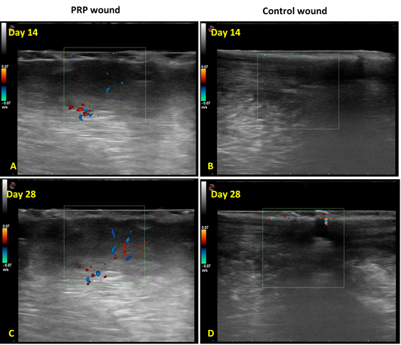
Ultrasonographical evaluation of neo-vascularization in the cutaneous wound of both groups. A; PRP treated wound obtained at day 14 showed mild development of blood vessels. B; Control wound showed no development of neo-vascularization at day 14. C; PRP treated wound at day 28 showed increased development of neo-vascularization at day 28. D; Control wound at day 28 showed mild development of neo-vascularization.
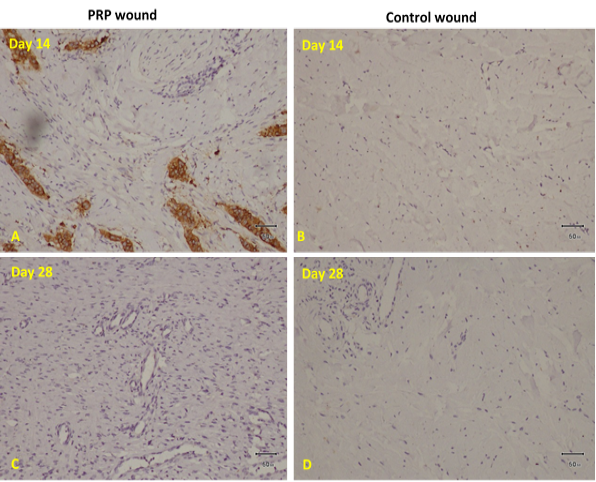
Histopathological evaluation of neo-vascularization in the cutaneous wound of both groups. Bar=50um A, PRP treated wound obtained at day 14 showed mild development of blood vessels. B, Control wound showed no development of neo-vascularization at day 14. C, PRP treated wound at day 28 showed increased development of neo-vascularization at day 28. D, Control wound at day 28 showed mild development of neo-vascularization.
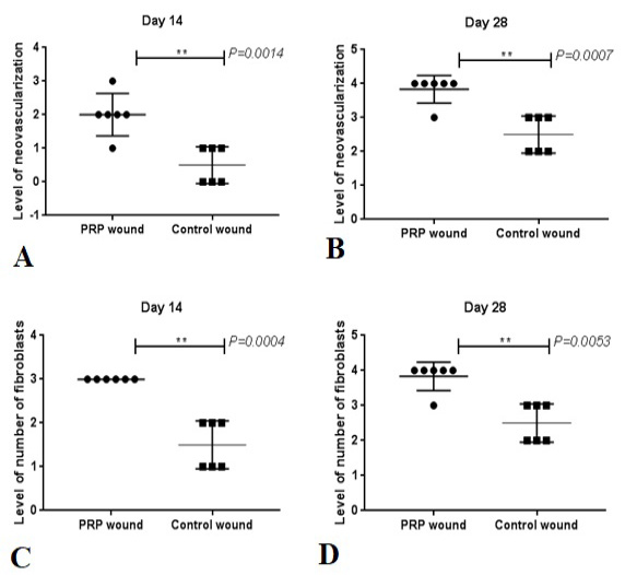
Histopathological scoring of neo-vascularization and fibroblast between the both groups. A, indicates level of neovascularization in PRP and Control wounds at day 14. B, indicates neovascularization in both groups at 28 days. C, indicates level of number of fibroblasts in PRP and Control wounds at day 14. D, indicates level of number of fibroblasts in both groups at 28 days. **, indicated the differences were significant in the PRP-treated group compared with the control group at day 28 (P<0.01).
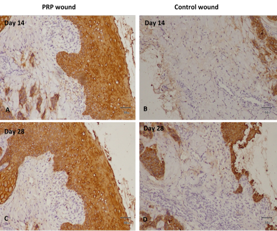
Cytokeratin staining in tissues obtained at Days 14 and 28 after wounding for Re-epithelization in both groups. A, PRP gel treated wound tissue displays accelerated epithelial growth, increased proliferation of connective tissue at day 14. B, Control wound tissue displays no development of epithelial growth, no basal lamina growing, and mild proliferation of connective tissue at day 14. C, PRP gel-treated wound tissue displays increased epidermal growth, intact basal layer, multiple layers of stratum spinosum visible, stratum corneum also visible, and increased fibrous connective tissue on day 28. D, Control wound tissue mild epithelial growth, basal lamina growing, and mild proliferation of connective tissue at 28 days.
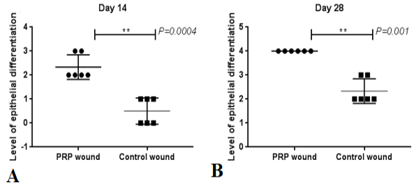
Histopathological scoring of re-epithelization between the both groups. A, indicates level of epithelial differentiation in PRP and Control wounds at day 14. B, indicates level of epithelial differentiation in both groups. ‘**’ indicates the differences were significant in the PRP-treated group compared with the control group at day 28 (P<0.01).
References
- Anderson, C.R., Hu, X., Tlaxca, J., Decleves, A.E., Houghtaling, R., Sharma, K., Lawrence, M., Ferrara, K., and Rychak, J.J., 2011. Ultrasound molecular imaging of tumor angiogenesis with an integrin targeted microbubble contrast agent. Invest. Radiol., 46: 215. https://doi.org/10.1097/RLI.0b013e3182034fed[CrossRef]
- Bao, P., Kodra, A., Tomic-Canic, M., Golinko, M.S., Ehrlich, H.P., and Brem, H., 2009. The role of vascular endothelial growth factor in wound healing. J. Surg. Res., 153: 347–358. https://doi.org/10.1016/j.jss.2008.04.023[CrossRef]
- Bosch, G., van-Schie, H.T., de-Groot, M.W., Cadby, J.A., van-de-Lest, C.H., Barneveld, A., and van-Weeren, P.R., 2010. Effects of plateletrich plasma on the quality of repair of mechanically induced core lesions in equine superficial digital flexor tendons: A placebo-controlled experimental study. J. Orthop. Res., 28: 211–217. https://doi.org/10.1002/jor.20980[CrossRef]
- Bos-Mikich, A., de-Oliveira, R., and Frantz, N., 2018. Platelet-rich plasma therapy and reproductive medicine. J. Assist. Reprod. Genet., 35: 753-756. https://doi.org/10.1007/s10815-018-1159-8[CrossRef]
- Brissett, A.E., and Hom, D., 2003. The effects of tissue sealants, platelet gels, and growth factors on wound healing. Current opinion in otolaryngol. Head. Neck. Surg., 11: 245–250. https://doi.org/10.1097/00020840-200308000-00005[CrossRef]
- Carmeliet, P., and Jain, R.K., 2011. Molecular mechanisms and clinical applications of angiogenesis. Nature, 473: 298-307. https://doi.org/10.1038/nature10144[CrossRef]
- Carter, C.A., Jolly, D.G., Worden-Sr, C.E., Hendren, D.G., and Kane, C.J., 2003. Platelet-rich plasma gel promotes differentiation and regeneration during equine wound healing. Exp. Mol. Path., 74: 244-255. https://doi.org/10.1016/S0014-4800(03)00017-0[CrossRef]
- Choi, S.M., Lee, K.M., Kim, H.J., Park, I.K., Kang, H.J., Shin, H.C., Baek, D., Choi, Y., Park, K.H., and Lee, J.W., 2018. Effects of structurally stabilized EGF and bFGF on wound healing in type I and type II diabetic mice. Acta Biomat., 66: 325-334. https://doi.org/10.1016/j.actbio.2017.11.045[CrossRef]
- Chong, D.L., Trinder, S., Labelle, M., Rodriguez-Justo, M., Hughes, S., Holmes, A.M., Scotton, C.J., and Porter, J.C., 2020. Platele-derived transforming growth factor-β1 promotes keratinocyte proliferation in cutaneous wound healing. J. Tissue Eng. Reg. Med., 14: 645-649. https://doi.org/10.1002/term.3022[CrossRef]
- DeRossi, R., Coelho, A.C., Mello, G.S., Frazílio, F.O., Leal, C.R., Facco, G.G., and Brum, K.B., 2009. Effects of platelet-rich plasma gel on skin healing in surgical wound in horses. Acta Cirúrg. Brasil., 24: 276-281. https://doi.org/10.1590/S0102-86502009000400006[CrossRef]
- Desai, C., Mahindra, U., Kini, Y., and Bakshi, M., 2013. Use of platelet-rich fibrin over skin wounds: Modified secondary intention healing. J. Cut. Aesthetic Surg., 6: 35–37. https://doi.org/10.4103/0974-2077.110096[CrossRef]
- DeVos, R.J., Weir, A., Cobben, L.P., and Tol, J.L., 2007. The value of power Doppler ultrasonography in Achilles tendinopathy: A prospective study. Am. J. Sports. Med., 35: 1696–1701. https://doi.org/10.1177/0363546507303116[CrossRef]
- Eppley, B.L., Woodell, J.E., and Higgins, J., 2004. Platelet quantification and growth factor analysis from platelet-rich plasma: Implications for wound healing. Plast. Reconstr. Surg., 114: 1502–1508. https://doi.org/10.1097/01.PRS.0000138251.07040.51[CrossRef]
- Gartner, M.H., Benson, J.D., and Caldwell, M.D., 1992. Insulin-like growth factors I and II expression in the healing wound. J. Surg. Res., 52: 389–394. https://doi.org/10.1016/0022-4804(92)90121-F[CrossRef]
- Irmak, G., Demirtas, T.T., and Gumusderelioglu, M., 2020. Sustained release of growth factors from photoactivated platelet rich plasma (PRP). Eur. J. Pharm. Biopharm., 148: 67-76. https://doi.org/10.1016/j.ejpb.2019.11.011[CrossRef]
- Jee, C.H., Eom, N.Y., Jang, H.M., Jun, H.W., Choi, E.S., Won, J.H., Hong, I.H., Kang, B.T., Jeong, D.W., and Jung, D.I., 2016. Effect of autologous platelet-rich plasma application on cutaneous wound healing in dogs. J. Vet. Sci., 17: 79-87. https://doi.org/10.4142/jvs.2016.17.1.79[CrossRef]
- Kliche, S., and Waltenberger, J., 2001. VEGF receptor signaling and endothelial function. IUBMB Life., 52: 61–66. https://doi.org/10.1080/15216540252774784[CrossRef]
- Kristoffersen, M., Ohberg, L., Johnston, C., and Alfredson, H., 2005. Neovascularization in chronic tendon injuries detected with colour Doppler ultrasound in horse and man. Imp. Res. Treat. Knee Surg. Sports Traumatol. Arthrosc., 13: 505–508. https://doi.org/10.1007/s00167-005-0648-3[CrossRef]
- Le, M., Naridze, R., Morrison, J., Biggs, L.C., Rhea, L., Schutte, B.C., Kaartinen, V., and Dunnwald, M., 2012. Transforming growth factor Beta 3 is required for excisional wound repair in vivo. PLoS One, 7: e48040. https://doi.org/10.1371/journal.pone.0048040[CrossRef]
- Marx, R.E., 2004. Platelet-rich plasma: Evidence to support its use. J. Oral. Maxillofac. Surg., 62: 489-496. https://doi.org/10.1016/j.joms.2003.12.003[CrossRef]
- Merchán, W., G´omez, L.A., Chasoy, M.E., Alfonso-Rodríguez, C.A., and Mu˜noz, A.L., 2019. Platelet-rich plasma, a powerful tool in dermatology. J. Tissue. Eng. Regen. Med., 13: 892-901. https://doi.org/10.1002/term.2832[CrossRef]
- Mickelson, M.A., Mans, C., and Colopy, S.A., 2016. Principles of wound management and wound healing in exotic pets. The veterinary clinics of north America. Exotic. Anim. Pract., 19: 33–53. https://doi.org/10.1016/j.cvex.2015.08.002[CrossRef]
- Ohberg, L., Lorentzon, R., and Alfredson, H., 2001. Neovascularisation in Achilles tendons with painful tendinosis but not in normal tendons: An ultrasonographic investigation. Knee. Surg. Sports Traumatol. Arthrosc., 9: 233–238. https://doi.org/10.1007/s001670000189[CrossRef]
- Potente, M., Gerhardt, H., and Carmeliet, P., 2011. Basic and therapeutic aspects of angiogenesis. Cell, 146: 873–887. https://doi.org/10.1016/j.cell.2011.08.039[CrossRef]
- Pufe, T., Petersen, W.J., Mentlein, R., and Tillmann, B.N., 2005. The role of vasculature and angiogenesis for the pathogenesis of degenerative tendons disease. Scand. J. med. Sci. Sports, 15: 211–222. https://doi.org/10.1111/j.1600-0838.2005.00465.x[CrossRef]
- Qi, D.D., Ding, M.Y., Wang, T., Hayat, M.A., Liu, T., and Zhang, J.T., 2021. The therapeutic effects of oral intake of hydrogen rich water on cutaneous wound healing in dogs. Vet. Sci., 8: 264. https://doi.org/10.3390/vetsci8110264[CrossRef]
- Sabol, F., Dancakova, L., Gal, P., Vasilenko, T., Novotny, M., Smetana, K., and Lenhardt, L., 2012. Immunohistological changes in skin wounds during the early periods of healing in a rat model. Vet. Med., 57: 77-82. https://doi.org/10.17221/5253-VETMED[CrossRef]
- Sadek, Y., Elballal, S., Sharshar, A., and Elsunsafty, M., 2020. Platelets rich plasma accelerates wound healing: Histopathological study. J. Cur. Vet. Res., 2: 16-24. https://doi.org/10.21608/jcvr.2020.90218[CrossRef]
- Samadi, P., Sheykhhasan, M., and Khoshinani, H.M., 2019. The use of platelet-rich plasma in aesthetic and regenerative medicine: A comprehensive review. Aesthetic. Plast. Surg., 43: 803-814. https://doi.org/10.1007/s00266-018-1293-9[CrossRef]
- Singer, A.J., and Clark, R.A., 1999. Cutaneous wound healing. N. Engl. J. Med., 341: 738–746. https://doi.org/10.1056/NEJM199909023411006[CrossRef]
- Strukova, S.M., Dugina, T.N., Chistov, I.V., Lange, M., Markvicheva, E.A., Kuptsova, S., and Glusa, E., 2001. Immobilized thrombin receptor agonist peptide accelerates wound healing in mice. Clinical and applied thrombosis/ hemostasis: Official. J. Int. Acad. Clin. App. Thrombosis/ Hem., 7: 325–329. https://doi.org/10.1177/107602960100700414[CrossRef]
- Sun, Y., Cao, Y., Zhao, R., Xu, F., Wu, D., and Wang, Y., 2020. The role of autologous PRP on deep partial-thickness burn wound healing in Bama pigs. J. Burn. Care. Res., 41: 657–662. https://doi.org/10.1093/jbcr/iraa012[CrossRef]
- Tonnesen, M.G., Feng, X., and Clark, R.A., 2000. Angiogenesis in wound healing. J. Investig. Dermatol. Symp. Proc., 5: 40-46. https://doi.org/10.1046/j.1087-0024.2000.00014.x[CrossRef]
- Wang, Y., Jing, L., Zhao, X.M., Han, J.J., Xia, Z.L., Qin, S.C., Wu, Y.P., and Sun, X.J., 2011. Protective effects of hydrogen-rich saline on monocrotaline-induced pulmonary hypertension in a rat model. Respir. Res., 12: 26–33. https://doi.org/10.1186/1465-9921-12-26[CrossRef]
- Wu, L.W., Chen, W.L., Huang, S.M., and Chan, J.Y., 2019. Platelet-derived growth factor-AA is a substantial factor in the ability of adipose-derived stem cells and endothelial progenitor cells to enhance wound healing. FASEB J., 33: 2388–2395. https://doi.org/10.1096/fj.201800658R[CrossRef]
- Xie, J., Bian, H., Qi, S., Xu, Y., Tang, J., Li, T., and Liu, X., 2008. Effects of basic fibroblast growth factor on the expression of extracellular matrix and matrix metalloproteinase-1 in wound healing. Clin. exp. Dermatol. Con. Prof. Dev., 33: 176-182. https://doi.org/10.1111/j.1365-2230.2007.02573.x[CrossRef]
- Yan, B., Zhang, Y., Tian, S., Hu, R., and Wu, B., 2021. Effect of autologous platelet-rich plasma on human sperm quality during cryopreservation. Cryobiology, 98: 12-16. https://doi.org/10.1016/j.cryobiol.2021.01.009[CrossRef]
- Yolanda, M.M., Maria, A.V., Amaia, F.G., Marcos, P.B., Silvia, P.L., Dolores, E., and Jesús, O.H., 2014. Adult stem cell therapy in chronic wound healing. J. Stem. Cell. Res. Ther., 4: 2. https://doi.org/10.4172/2157-7633.1000162[CrossRef]
- Yu, C., Xu, Z.X., Hao, Y.H., Gao, Y.B., Yao, B.W., Zhang, J., Wang, B., Hu, Z.Q., and Peng, R.Y., 2019. A novel microcurrent dressing for wound healing in a rat skin defect model. Mil. Med. Res., 6: 1-9. https://doi.org/10.1186/s40779-019-0213-x[CrossRef]
- Zachary, I., 2003. VEGF signalling: Integration and multi-tasking in endothelial cell biology. Biochem. Soc. Trans., 31: 1171–1177. https://doi.org/10.1042/bst0311171[CrossRef]
Services
Article usage statistics
Loading…