Research Article · Volume 13, Issue 1, 28 February 2025 · Open Access
Genotyping Study for Infections of Theileria and Babesia in Cattle and Sheep in AL-Najaf Governorate Iraq
· · · ·
Affiliations & Notes ⌄
1Department of Microbiology, Faculty of Veterinary Medicine, University of Kufa, Iraq
2Department of Microbiology, College of Veterinary Medicine, University of Al-Qadisiyah, Al-Qadisiyah, Iraq
3Department of public health, Faculty of Veterinary Medicine, University of Kufa, Iraq
4Department of Anatomy, and Histology, College of Veterinary Medicine, University of Al-Qadisiyah, Al-Qadisiyah, Iraq. *Correspondence | Murtadha Abbas, Department of public health, Faculty of Veterinary Medicine. University of Kufa, Iraq
This study utilized the PCR technique, which relies on the 18S rRNA gene, to detect Theileria and Babesia species in blood samples from cattle and sheep.
The results of the investigation verified the existence of infections with Theileria and Babesia spp. specifically in the cattle.
The analysis of the phylogenetic tree revealed that the sheep isolates (No. 1 and No. 3) of Theileria spp. were genetically similar to the NCBI-Blast Theileria ovis (AY508461.1), while the sheep isolate (No. 2) of Theileria spp. was genetically similar to the NCBI-Blast Theileria lestoquardi (KP342263.1).
Overall, the present study reveals that Theileria ovis, Theileria lestoquardi, and Babesia ovis predominantly infect sheep, while Theileria annulata and Babesia bovis primarily infect cattle during this period.
Selected from the article's own text.
Abstract
Theileria and Babesia species are blood parasites that commonly cause illness in both juvenile and mature animals. The primary objective of this study was to identify the specific genotypes of Theileria and Babesia species responsible for infections in cattle and sheep. Blood samples were collected from male and female cattle and sheep slaughtered at the Al-Najaf slaughterhouse. Identification of Theileria and Babesia spp. was conducted using conventional PCR, sequencing, and phylogenetic techniques based on the 18S rRNA gene. The blood isolates from cattle and sheep were subjected to PCR-based molecular assays, which detected Theileria and Babesia infections at 694 bp and 383 bp, respectively. PCR products from positive isolates were then used for DNA sequencing to construct the phylogenetic tree of Theileria and Babesia species using partial 18S rRNA gene sequences. NCBI-Blast analysis revealed that the Theileria and Babesia species in the sheep isolates (No. 1 and No. 3) were closely related to Theileria ovis (AY508461.1) based on sequence similarity. Additionally, the sheep isolate of Theileria spp. (No. 2) was identified as Theileria lestoquardi (KP342263.1). The analysis of bovine isolates showed that the Theileria spp. isolates (No. 1 and No. 2) were highly similar to Theileria annulata (MF287951.1). Further examination of sheep isolates confirmed that the Babesia spp. isolates (No. 1 and No. 2) corresponded to Babesia ovis (MG569902.1). The genetic sequence analysis of cattle Babesia isolates (No. 1 and No. 2) revealed a 100% match with Babesia bovis (HQ264126.1). In conclusion, this study identifies Theileria ovis, Theileria lestoquardi, and Babesia ovis as the primary infecting species in sheep, while Theileria annulata and Babesia bovis primarily infect cattle. However, Theileria and Babesia species exhibit significant genetic similarities within their respective genera, suggesting the need for alternative identification techniques to accurately determine the specific species of these parasites.
Babesia and Theileria species are well-known blood parasites that invade the red blood cells of their hosts and cause the destruction of these cells. As a result, affected domesticated animals experience severe clinical symptoms and have a significant risk of death ; Karasová et al., 2022).
Babesia is a highly virulent blood parasite that affects both small and large ruminants, and is the primary cause of babesiosis, a disease that has been documented in over 100 species worldwide. It is a contagious disease that can affect various animals, including mammals and birds . This protozoan displays an intricate life cycle and has the ability to infect mammals and birds through ticks that are carrying the infection. When Babesia enters a blood cell that is already infected, it is referred to as sporozoites, which are infectious parasite bodies. Previously, Babesia would undergo conversion within erythrocytes, transitioning from trophozoites to merozoites, and then back to trophozoites and merozoites again. After the rupture of erythrocytes, which is induced by hemolytic blood cells, merozoites are released directly into the bloodstream and subsequently produce gametocytes. Subsequently, gametocytes undergo fertilization within the tick’s gut and transform into sporozoite forms, initiating a new cycle of life for the parasites ; .
Multiple studies have confirmed the presence of numerous species of Babesia that infect cattle. The majority of these species are infected with B. bigemina and B. divergens, which are responsible for causing bovine babesiosis (BB). Nevertheless, the species that are most commonly observed in sheep and goats are B. ovis and B. motasi, as reported by Alvarez et al. (2019) and Calleja-Bueno et al. (2022).Babesia can be spread among mammalian hosts by many tick species, such as Rhipicephalus, Hyalomma, Haemophysalis, Dermacentor, Ixodes, and Boophilus. The incubation period of the disease varies across animals depending on the individual Babesia species that infects them ; Martínez-García et al., 2021; ; . From a diagnostic perspective, Babesia spp. can exhibit comparable clinical signs and symptoms to Theileria and other pathogenic bacteria that infect ruminants.
Various techniques can be used to distinguish Babesia, such as microscopic inspection, serological tests, conventional polymerase chain reaction (PCR), and other approaches ; ; . Furthermore, Theileria spp. is a prevalent blood parasite that invades the cells of several vertebrate mammals, particularly small and big ruminants.
Likewise, this single-celled organism specifically attacks the red blood cells of infected animals and moves to other areas within the host’s body, such as lymphocytes, immature red blood cells, bone marrow, and cells of internal organs ; . Therefore, this protozoan undergoes the development of Theileriosis, a condition that is marked by severe clinical symptoms such as elevated body temperature, sudden decrease in red blood cells, yellowing of the skin, loss of appetite, excessive drooling, swelling of superficial lymph nodes, difficulty in breathing, and a high rate of illness . During the life cycle of Theileria, the sporozoites of Theileria undergo replication of schizonts in leukocytes, whereas piroplasms survive in the erythrocytes of hosts .
Recent investigations have shown that Theileria spp. infect large ruminants internationally, with the most harmful species being T. parva, T. annulata, and T. orientalis ; . Young ruminants are infected with T. lestoquardi (T. hirci), T. uilenbergi, and T. luwenshuni, as stated by Ahmed et al. (2011) and Altay et al. (2012). In addition, Theileria species often utilize multiple vectors depending on the geo-environmental conditions, ticks belonging to the Rhipicephalus appendiculatus, Hyalomma and Haemaphysalis consider as important vectors ; . The most prevalent approaches for diagnosing Theileria spp. infections are clinical symptoms and laboratory testing, as they contribute to the management of sick animals ; . Given the economic importance of cattle and sheep and the losses caused by theileriosis and babesiosis, this study seeks to identify the various genotypes of Theileria and Babesia in large and small ruminants in Al-Najaf province. This will be accomplished through the utilization of molecular techniques and DNA partial Nucleotide sequencing and phylogenetic tree analysis for the 18S rRNA gene.
MATERIALS AND METHODS
Collection of Blood Samples
A total of 150 blood samples were obtained from cattle and sheep in various parts of Al-Najaf Governorate between March 2020 and August 2020. The samples were collected using anticoagulant tubes. The samples comprised cattle and sheep of various ages and sexes. The samples were promptly stored in ice boxes and subsequently analyzed using direct smear techniques.
Extraction of DNA of Blood Samples
The genomic DNA of Theileria and Babesia species was extracted from blood samples of sheep and cattle using the gSYAN DNA mini kit from Geneaid, USA, following the instructions provided with the kit. The concentration and purity of the extracted DNA were assessed using a nanodrop spectrophotometer (THERMO, USA) by measuring the absorbance at 260/280 nm. Subsequently, the DNA was stored at -20 °C until it is used in the PCR procedure.
The Polymerase Chain Reaction (PCR) Method
The PCR approach was utilized to directly identify Theileria and Babesia species by amplifying the 18S rRNA gene in the extracted DNA of sheep and cattle. The polymerase chain reaction (PCR) was conducted on all genomic samples, adhering to the detailed procedures outlined by . The selection of PCR primers for Theileria and Babesia species was based on the NCBI Genbank database and the primer3 plus tool provided by Macrogen Company, Korea. The nucleotide sequence of the 18SrRNA gene for Theileria spp. was determined to be 694 base pairs long. The forward primer sequence is ATTGGAGGGCAAGTCTGGTG and the reverse primer sequence is TGCACCACCACCCAAAGAAT. The corresponding NCBI-Genbank code for this sequence is AF078816.1. The 18SrRNA gene sequence for Babesia spp. was obtained using the primers F: AGGAATTGACGGAAGGGCAC and R: CTAGTGATTACCCGCGCCTT. The sequence is 383 base pairs long, and its NCBI-Genbank code is KY867436.1. The AccuPower PCR PreMix Kit was utilized to create a PCR master mix as per the instructions provided with the kit, with a total volume of 20µL. Subsequently, the PCR mixture comprised Taq DNA polymerase, dNTPs, 10µL of PCR buffers, 2 µL of DNA samples, and 2µL each of forward and reverse primers for both species. Subsequently, the PCR mixture tubes were thoroughly agitated for a duration of 3 minutes. Meanwhile, PCR tubes were inserted into a thermocycler (MJ-Mini BioRad, USA). The thermocycler conditions for the conventional PCR machines were established as follows: an initial denaturation at 95 °C for 5 minutes for one cycle, followed by denaturation at 95 °C for 30 seconds, annealing at 58 °C for 30 seconds, extension at 72 °C for 1 minute for 30 cycles, and a final extension at 72 °C for 5 minutes for one cycle.
Analysis of the PCR Products
The PCR products, measured 383 and 694 bp, were examined using 1% agarose gel electrophoresis and 1X TBE buffer. In addition, the PCR results were treated with ethidium bromide dye and visualized using a UV transilluminator.
The Nucleotide Sequencing Method
A PCR was used to amplify and DNA was sequenced to confirm the molecular detection of some isolates that tested positive for Theileria and Babesia species of cattle and sheep blood samples. The phylogenetic tree was identified and examined using isolates for the local in comparison with the global isolates deposited on NCBI/GenBank to analyze the genetic relationship between isolates of local Theileria and Babesia spp. and international isolates submitted on NCBI-Blast. In addition, the local isolates that were identified had been recorded in the NCBI-GenBank as novel data originating from Iraq. To prevent degradation, the PCR products were stored in ice-filled containers prior to being shipped to the Macrogen Company, Korea. The AB DNA sequencing machine was utilized for DNA sequencing with the assistance of Molecular Evolutionary Genetics Analysis version 6.0 (Mega 6.0). In addition, the phylogenetic tree of Babesia and Theileria spp. was calculated using the sequence alignment of the partial 18S ribosomal rRNA gene and the UPGMA method to determine the evolutionary distances .
RESULTS AND DISCUSSION
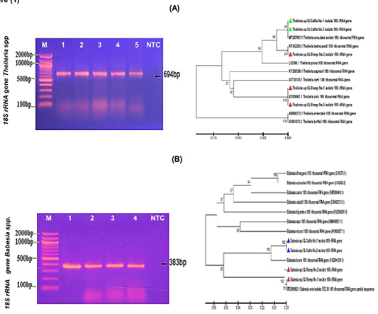
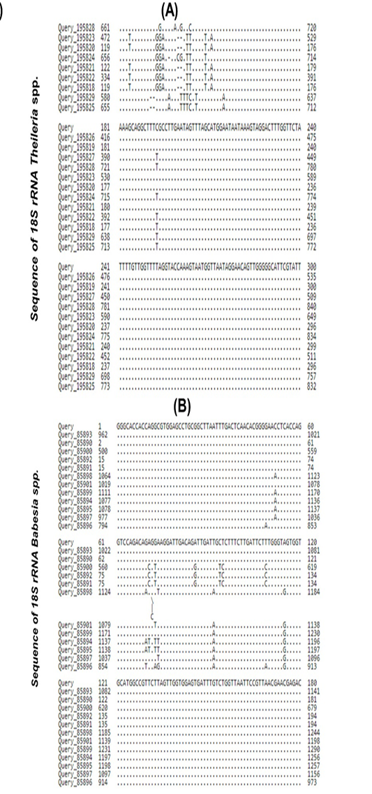
This study utilized the PCR technique, which relies on the 18S rRNA gene, to detect Theileria and Babesia species in blood samples from cattle and sheep. The results of the investigation verified the existence of infections with Theileria and Babesia spp. specifically in the cattle. The genomic DNA extracted from the blood samples of sheep and cattle was subjected to agarose gel electrophoresis using a 1% concentration. This was done to identify the presence of the 18S rRNA gene in Theileria spp. As a result, the samples from sheep and cattle tested positive for Theileria spp. at 694bp, as shown in Figure 1A. The polymerase chain reaction (PCR) was used to identify the presence of Babesia spp. in the blood samples of sheep and cattle. Specifically, the PCR product size was determined by targeting the 18S rRNA gene, resulting in a fragment of 383 base pairs (bp) (Figure 1B). The DNA sequences of Theileria and Babesia species were analyzed utilizing phylogenetic tree and ClustalW alignment analysis with the MEGA 6.0 version. The local species isolates were subjected to NCBI-Blast analysis and compared to isolates from various countries. The results suggested a strong resemblance in the multiple alignment analysis and mutation of the nucleotide sequences in the 18S ribosomal rRNA gene (Figure 2). The genetic link between local and global Theileria and Babesia spp. was determined by comparing the 18S rRNA gene sequences using NCBI-BLAST homology analysis. Sheep isolates No. 1 and No. 3 from NCBI-Blast are genetically identical to Theileria ovis (AY508461.1), a strain found in Turkish sheep. The sheep isolate No. 2 from NCBI-Blast is genetically identical to Theileria lestoquardi (KP342263.1), a strain found in Iraqi sheep. Cattle isolates No. 1 and No. 2 from NCBI-Blast are genetically identical to Theileria annulata (MF287951.1), a strain found in Bengal cattle (Figure 2 and Table 1). The homology sequence identity between Babesia spp. sheep isolates (No.1 and No. 2) and Babesia ovis (MG569902.1), a Turkey isolate, is 100%
according to NCBI-Blast. Similarly, the homology sequence identity between Babesia spp. cattle isolates (No.1 and No. 2) and Babesia bovis (HQ264126.1), a USA isolate, is also 100% according to NCBI-Blast (Table 2). The analysis of the phylogenetic tree revealed that the sheep isolates (No. 1 and No. 3) of Theileria spp. were genetically similar to the NCBI-Blast Theileria ovis (AY508461.1), while the sheep isolate (No. 2) of Theileria spp. was genetically similar to the NCBI-Blast Theileria lestoquardi (KP342263.1).
Table 1: NCBI-BLAST homology sequence identity between local Theileria spp. isolates and closely related Theileria spp. isolates in NCBI-BLAST.
Isolate No.
Genbank Accession number
NCBI-BLAST Homology Sequence Identity
Identical Theileria sp.
Genbank Accession number
Identity (%)
Theileria sp. Sheep No.1
OQ818311
Theileria ovis
AY508461.1
100%
Theileria sp. Sheep No.2
OQ818312
Theileria lestoquardi
KP342263.1
100%
Theileria sp. Sheep No.3
OQ818313
Theileria ovis
AY508461.1
100%
Theileria sp. Cattle No.1
OR438632
Theileria annulata
MF287951.1
100%
Theileria sp. Cattle No.2
OR438633
Theileria annulata
MF287951.1
100%
Table 2: NCBI-BLAST homology sequence identity between local Babesia spp. isolates and closely related Babesia spp. isolates in NCBI-BLAST.
Isolate No.
Genbank Accession number
NCBI-BLAST Homology Sequence Identity
Identical Babesia sp.
Genbank Accession number
Identity (%)
Babesia sp. Sheep No.1
OQ818624
Babesia ovis
MG569902.1
100%
Babesia sp. Sheep No.2
OQ818625
Babesia ovis
MG569902.1
100%
Babesia sp. Cattle No.1
OQ818626
Babesia bovis
HQ264126.1
100%
Babesia sp. Cattle No.2
OQ818627
Babesia bovis
HQ264126.1
100%
Likewise, the cattle isolates (No. 1 and No. 2) of Theileria spp. were genetically similar to the NCBI-Blast Theileria annulata (MF287951.1), with a total genetic change ranging from 0.005% to 0.015% (Figure 1). The Babesia spp. sheep isolates (No.1 and No. 2) were found to have a close genetic relationship with NCBI-Blast Babesia ovis (MG569902.1). Similarly, the Babesia spp. cattle isolates
(No.1 and No. 2) were found to be closely related to NCBI-Blast Babesia bovis (HQ264126.1). This genetic similarity was observed to have a total genetic change ranging from 0.01% to 0.06%, as depicted in Figure 2. Ultimately, the isolated specimens of locally identified species were sent to the NCBI-GenBank, and specific GenBank accession numbers were assigned to each isolate of Theileria and Babesia species.
Prior research on Theileria spp. has demonstrated that there are six distinct species of Theileria that are capable of infecting various animal species. The key species identified in cattle are T. annulata, which is responsible for causing tropical theileriosis, and T. parva, which causes East Coast fever ; . The conventional PCR technique was employed to analyze blood samples from cattle, leading to the identification of Theileria spp. at 694bp. The size of the PCR product was determined based on the partial sequence of the 18S ribosomal rRNA gene of Theileria spp., which was previously confirmed by studies conducted by ; ; . Furthermore, our research has validated the use of DNA sequencing analysis to identify Theileria annulata in positive blood samples from cattle, aligning with previous studies that also identified Theileria annulata and Theileria parva in cows . However, our analyses have not detected the presence of Theileria parva, most likely because Theileria annulata is more prevalent in Iraqi cattle. Therefore, it is imperative to do additional research by examining various blood samples from cattle in order to gain deeper insights. Furthermore, it is crucial to examine the impact of seasons and the geographical distribution of cattle in order to ascertain the degree to which environmental factors can affect the transmission and occurrence of certain illnesses in cattle. This study differentiated between the sheep species Theileria ovis and Theileria lestoquardi by analyzing the phylogenetic tree of Theileria spp. This finding aligns with a previous study conducted by ; , which used a nested PCR–restriction fragment length polymorphism (RFLP) approach. Similarly, this study discovered that Theileria annulata did not induce any infections in sheep. This finding is consistent with the study conducted by Zaeemi et al. (2011), which suggested that the identification of Theileria annulata in sheep using RFLP-based methods could be more efficient depending on aspects such as sample size, season, and geographical location. In addition, this study detected Babesia spp. at a length of 383 base pairs (bp) in both cattle and sheep. This finding is consistent with prior studies conducted by ; for both species. Furthermore, the veracity of this discovery was validated through the examination of the DNA sequence and the study of the phylogenetic tree of Babesia species. Consequently, the presence of Babesia ovis was detected in the samples taken from the infected sheep, whereas Babesia bovis was identified in the infected cattle. These findings are consistent with previous research conducted on Babesia species in sheep and cattle, as reported by ; . Notwithstanding these studies and research, this study found no further species of Babesia. Future investigations on the infections of cattle and sheep with Babesia spp. should take into account additional variables, including sample size, seasonal variations, and alternative methodologies. Moreover, this study indicates that T. annulata is the prevailing species causing infection in cattle, while T. ovis and T. lestoquardi are more commonly found in sheep. Moreover, Babesia bovis and Babesia ovis are the predominant pathogenic species found in cattle and sheep. Hence, further examination employing diverse methodologies should consider distinct seasons and diverse environmental conditions in order to identify all the species of Theileria and Babesia spp.
CONCLUSIONS AND RECOMMENDATIONS
Overall, the present study reveals that Theileria ovis, Theileria lestoquardi, and Babesia ovis predominantly infect sheep, while Theileria annulata and Babesia bovis primarily infect cattle during this period. Although there are distinct differences between Theileria and Babesia species, they also share considerable similarities with each other and with other related taxa. Therefore, future research should adopt innovative methodologies to accurately identify the species of these parasites.
ACKNOWLEDGEMENTS
Many thanks to The Research team who Worked with me.
NOVELTY STATEMENTS
This study is one of the rare studies in the genetic diagnosis
of these pathogens.
AUTHOR’S CONTRIBUTIONS
All the research team for this work have equal roles.
Conflict of Interest
The authors have declared no conflict of interest.
Figures
Figure 1:.
(A) This figure illustrates the PCR product of the 18S rRNA gene in Theileria spp. of sheep and cattle blood samples at 694bp. M: DNA marker ladder (2000-100bp), and Lane (1-3) showed some positive samples of Theileria spp. from Sheep and Lane (4-5) positive samples of Theileria spp. from Cattle, and lane (NTC) Non-template negative control. Besides, the analysis of the phylogenetic tree of Theileria spp. is based on the 18S ribosomal rRNA gene partial sequence and genetic identification. The phylogenetic tree was constructed using the Unweighted Pair Group method with Arithmetic Mean (UPGMA tree) in the MEGA 6.0 version. Theileria spp. Sheep isolates (No. 1 and No. 3) were found to be closely related to NCBI-Blast Theileria ovis (AY508461.1), and Theileria spp. sheep isolates (No. 2) were identified to be closely related to NCBI-Blast Theileria lestoquardi (KP342263.1). Theileria spp. Cattle isolates (No. 1 and No. 2) were found to be closely related to NCBI-Blast Theileria annulata (MF287951.1) at a total genetic change (0.005-0.015%).(B): This figure illustrates the PCR product run on 1% Agarose gel electrophoresis and detected the 18S rRNA gene in Babesia spp. of sheep and cattle blood samples M: DNA marker ladder (2000-100bp), and Lane (1-2) showed some positive samples of Babesia spp. from Sheep and Lane (3-4) some positive samples of Babesia spp. from Cattle at 383bp PCR product size and lane (NTC) Non-template negative control. Besides, the analysis of the phylogenetic tree is based on 18S ribosomal rRNA gene partial sequence in the local Babesia species sheep and cattle isolates that were used for Babesia species genetic identification. The phylogenetic tree was analyzed using the Unweighted Pair Group method with Arithmetic Mean (UPGMA tree) in the MEGA 6.0 version. Babesia spp. sheep isolates (No.1 and No.2) were found to be closely related to NCBI-Blast Babesia ovis (MG569902.1) and Babesia spp. cattle isolates (No.1 and No. 2) were found to be closely related to NCBI-Blast Babesia bovis (HQ264126.1) at a total genetic change (0.01-0.06%).
Figure 2:.
(A) Multiple sequence alignment analysis for 18S ribosomal rRNA gene partial sequence in the local Theileria spp. isolates and the GenBank located Theileria spp. isolates based on the NCBI-BLAST Alignment tool. The figure shows similarity in the multiple alignment analysis (*) and mutation in the nucleotide sequences of the 18S ribosomal rRNA gene. (B): Multiple sequence alignment analysis for 18S ribosomal rRNA gene partial sequence in the local Babesia spp. isolates and the GenBank located Babesia spp. isolates based on the NCBI-BLAST Alignment tool. The figure shows similarity in the multiple alignment analysis (*) and mutation in the nucleotide sequences of the 18S ribosomal rRNA gene.
References
Ahmed J, Yin H, Bakheit M, Liu Z, Mehlhorn H, Seitzer U (2011). Small ruminant theileriosis. Prog. Parasitol., 135-153. https://doi.org/10.1007/978-3-642-21396-0_8[CrossRef]
Al-Hosary AA, ElSify A, Salama AA, Nayel M, Elkhtam A, Elmajdoub LO, Rizk MA, Hawash MM, Al-Wabel MA, Almuzaini AM, Ahmed LS, Paramasivam A, Mickymaray S, Omar MA (2021). Phylogenetic study of Theileria ovis and Theileria lestoquardi in sheep from Egypt: Molecular evidence and genetic characterization. Vet. World, 14(3): 634-639. https://doi.org/10.14202/vetworld.2021.634-639[CrossRef]
Almazán C, Scimeca RC, Reichard MV, Mosqueda J (2022). Babesiosis and theileriosis in North America. Pathogens, 11(2): 168. https://doi.org/10.3390/pathogens11020168[CrossRef]
Altay K, Dumanli N, Aktas M (2012). A study on ovine tick-borne hemoprotozoan parasites (Theileria and Babesia) in the East Black Sea Region of Turkey. Parasitol. Res., 111: 149-153. https://doi.org/10.1007/s00436-011-2811-8[CrossRef]
Alvarez JA, Rojas C, Figueroa JV (2019). Diagnostic tools for the identification of Babesia sp. in persistently infected cattle. Pathogens, 8(3): 143. https://doi.org/10.3390/pathogens8030143[CrossRef]
Calleja‐Bueno L, Sainz Á, García‐Sancho M, González‐Martín JV, Díaz‐Regañón D, Rodríguez‐Franco F, Agulla B, Tormo B, Villaescusa A (2022). First detection of Anaplasma phagocytophilum and Babesia divergens and high infection rates of Anaplasma marginale and Babesia bigemina in cattle in extensive grazing systems of Central Spain. Transboundary Emerg. Dis., 69(4): e1090-e1100. https://doi.org/10.1111/tbed.14394[CrossRef]
Elsworth B, Duraisingh MT (2021). A framework for signaling throughout the life cycle of Babesia species. Mol. Microbiol., 115(5): 882-890. https://doi.org/10.1111/mmi.14650[CrossRef]
Esmaeilnejad, B, Tavassoli M, Asri-Rezaei S, Dalir-Naghadeh B, Mardani K, Jalilzadeh-Amin G, Golabi M, Arjmand J (2014). PCR‐based detection of Babesia ovis in Rhipicephalus bursa and small ruminants. J. Parasitol. Res., (1): 294704. https://doi.org/10.1155/2014/294704[CrossRef]
Friedhoff KT (2018). Transmission of Babesia. In Babesiosis of domestic animals and man: 23-52. CRC Press. https://doi.org/10.1201/9781351070027-2[CrossRef]
Ganaie Z, Shahardar R, Maqboo I, Bulbul K, Allaie I, Wani Z (2019). An overview of bovine theileriosis. Int. J. Vet. Sci. Anim. Husbandry, 4(1): 09-13.
Ganzinelli S, Rodriguez A, Schnittger L, Florin-Christensen M (2018). Babesia in domestic ruminants. Parasitic Protozoa of farm animals and pets: 215-239. https://doi.org/10.1007/978-3-319-70132-5_9[CrossRef]
Gray JS, Estrada-Peña A, Zintl A (2019). Vectors of babesiosis. Annu. Rev. Entomol., 64: 149-165. https://doi.org/10.1146/annurev-ento-011118-111932[CrossRef]
Gul N, Ayaz S, Gul I, Adnan M, Shams S, Akbar N (2015). Tropical theileriosis and east coast fever in cattle: present, past and future perspective. Int. J. Curr. Microbiol. Appl. Sci., 4(8):1000-1018.
Jalali SM, Khaki Z, Kazemi B, Rahbari S, Shayan P, Bandehpour M, Yasini SP (2014). Molecular detection and identification of Theileria species by PCR-RFLP method in sheep from Ahvaz, Southern Iran. Iranian J. Parasitol., 9(1): 99-106.
Karasov M., Tóthová C, Grelová S, Fialkovičová M (2022). The etiology, incidence, pathogenesis, diagnostics, and treatment of canine babesiosis caused by Babesia gibsoni infection. Animals, 12(6): 739. https://doi.org/10.3390/ani12060739[CrossRef]
Kundave VR (2014) Qualitative and quantitative assessment of Theileria annulata in cattle and buffaloes Polymerase Chain Reaction.
Khamesipour F, Doosti A, Koohi A, Chehelgerdi M, Mokhtari-Farsani A, Chengula AA (2015). Determination of the presence of Babesia species in blood samples of cattle, camel and sheep in Iran by PCR. Arch. Biol. Sci., 67(1): 83-90. https://doi.org/10.2298/ABS140410009K[CrossRef]
Kho KL, Amarajothi ADG, Koh FX, Panchadcharam C, Nizam QNH, Tay ST (2017). The first molecular survey of theileriosis in Malaysian cattle, sheep, and goats. Vet. Parasitol. Reg. Stud. Rep., 10: 149-153. https://doi.org/10.1016/j.vprsr.2017.08.003[CrossRef]
Kuttler KL (2018). World-wide impact of babesiosis. In Babesiosis of domestic animals and man:1-22. CRC Press. https://doi.org/10.1201/9781351070027-1[CrossRef]
Maharana BR, Tewari AK, Saravanan BC, Sudhakar NR (2016). Important hemoprotozoan diseases of livestock: Challenges in current diagnostics and therapeutics: An update. Vet. world, 9(5): 487. https://doi.org/10.14202/vetworld.2016.487-495[CrossRef]
Martínez-Garcí, G, Santamaría-Espinosa RM, Lira-Amaya JJ, Figueroa JV (2021). Challenges in tick-borne pathogen detection: the case for Babesia spp. identification in the tick vector. Pathogens, 10(2): 92. https://doi.org/10.3390/pathogens10020092[CrossRef]
Morrison W (2015). The aetiology, pathogenesis and control of theileriosis in domestic animals. Rev. Sci. Tech., 34(2): 599-611. https://doi.org/10.20506/rst.34.2.2383[CrossRef]
Naik BS, Maiti SK, Raghuvanshi PDS (2016). Prevalence of tropical theileriosis in cattle in Chhattisgarh state. J. Anim. Res., 6(6): 1043-1045. https://doi.org/10.5958/2277-940X.2016.00151.0[CrossRef]
Nayel M, El-Dakhly KM, Aboulaila M, Elsify A, Hassan H, Ibrahim E, Salama A, Yanai T (2012). The use of different diagnostic tools for Babesia and Theileria parasites in cattle in Menofia, Egypt. Parasitol. Res., 111: 1019-1024. https://doi.org/10.1007/s00436-012-2926-6[CrossRef]
Nene V, Morrison WI (2016). Approaches to vaccination against Theileria parva and Theileria annulata. Parasite Immunol., 38(12): 724-734. https://doi.org/10.1111/pim.12388[CrossRef]
Ngetich EC (2019). Prevalence and The Effect of Parasites on Haematological Parameters of Livestock in The Upper Kerio Valley, Kenya (Doctoral dissertation, University of Eldoret).
Nourollahi-Fard SR, Khalili M, Ghalekhani N (2015). Detection of Theileria annulata in blood samples of native cattle by PCR and smear method in Southeast of Iran. Journal of Parasitic Diseases, 39: 249-252. https://doi.org/10.1007/s12639-013-0333-2[CrossRef]
Oakes VJ, Yabsley MJ, Schwartz D, LeRoith T, Bissett C, Broaddus C, Schlater JL, Todd SM, Boes KM, Brookhart M, Lahmers KK (2019). Theileria orientalis Ikeda genotype in cattle, Virginia, USA. Emerging Infectious Diseases, 25(9): 1653. https://doi.org/10.3201/eid2509.190088[CrossRef]
Ranjbar-Bahadori S, Eckert B, Omidian Z, Shirazi NS, Shayan P (2012). Babesia ovis as the main causative agent of sheep babesiosis in Iran. Parasitol. Res., 110(4): 1531-1536. https://doi.org/10.1007/s00436-011-2658-z[CrossRef]
Saeed S, Jahangir M, Fatima M, Shaikh RS, Khattak RM, Ali M, Iqbal F (2015). PCR based detection of Theileria lestoquardi in apparently healthy sheep and goats from two districts in Khyber Pukhtoon Khwa (Pakistan). Trop. Biomed., 32(2): 225-232.
Shahzad W, Haider NOOR, Mansur-ud-Din A, Munir R, Saghar MS, Mushtaq MH, Ahmad N, Akbar G, Mehmood, F (2013). Prevalence and molecular diagnosis of Babesia ovis and Theileria ovis in Lohi sheep at livestock experiment station (LES), Bahadurnagar, Okara, Pakistan. Iran. J. Parasitol., 8(4): 570-578.
Sivakumar T, Hayashida K, Sugimoto C, Yokoyama N (2014). Evolution and genetic diversity of Theileria. Infect. Genet. Evol., 27: 250-263.
Tavassoli M, Tabatabaei M, Mohammadi M, Esmaeilnejad B, Mohamadpour H (2013). PCR-based detection of Babesia spp. infection in collected ticks from cattle in west and north-west of Iran. Journal of arthropod-borne diseases, 7(2): 132.
Weiland G, Reiter I (2018). Methods for the measurement of the serological response to Babesia. Babesiosis of domestic animals and man. :143-162. https://doi.org/10.1201/9781351070027-9[CrossRef]
Wright I, Goodger B (2018). Pathogenesis of babesiosis. Babesiosis of domestic animals and man. 99-118. https://doi.org/10.1201/9781351070027-6[CrossRef]
Zaeemi M, Haddadzadeh H, Khazraiinia P, Kazemi B, Bandehpour M (2011). Identification of different Theileria species (Theileria lestoquardi, Theileria ovis, and Theileria annulata) in naturally infected sheep using nested PCR–RFLP. Parasitol. Res., 108: 837-843. https://doi.org/10.1007/s00436-010-2119-0[CrossRef]
Zeb J, Song B, Aziz MU, Hussain S, Zarin R, Sparagano O (2022). Diversity and distribution of Theileria species and their vectors in ruminants from India, Pakistan and Bangladesh. Diversity, 14(2): 82. https://doi.org/10.3390/d14020082[CrossRef]
Zhou M, Cao S, Sevinc F, Sevinc M, Ceylan O, Moumouni PFA, Jirapattharasate C, Liu M, Wang G, Iguchi A, Vudriko P, Suzuki H, Xuan X (2016). Molecular detection and genetic identification of Babesia bigemina, Theileria annulata, Theileria orientalis and Anaplasma marginale in Turkey. Ticks Tick-Borne Dis., 7(1): 126-134. https://doi.org/10.1016/j.ttbdis.2015.09.008[CrossRef]
Zhu B, Liu C, Cao J, Li X, Wei G (2010). Phylogenetic analysis of Actias selene based on 18S ribosomal RNA and mitochondrial 16S rRNA genes. Chin. Bull. Entomol., 47(2): 285-292.