The clinic pathological examination of the animals of the groups under the study revealed significant changes of some parameters in both infected groups in comparison with the Theileria negative group.
Results of erythrogram revealed significant macrocytic hypochromic anemia in the infected groups compared with Theileria negative group.
The leukogram of infected groups showed significant leukopenia due to neutropenia, lymphopenia, monocytopenia and eosinopenia, compared with the Theileria negative group.
The kidney function tests revealed significant increase of BUN and creatinine; azotemia of both infected groups compared with Theileria negative group.
Selected from the article's own text.
Abstract
Mahmoud Eliwa1, Khaled Mohamed Ahmed Mahran2*, Waheed Ali Mousa3, Naglaa Hagag1, Mohamed Ibrahim Shaalan4, Mostafa Mahmoud Bashandy2
1Genome Research Unit, Animal Health Research Institute (AHRI), Dokki, Egypt; 2Department of Clinical Pathology, Faculty of Veterinary Medicine, Cairo University, Giza, Egypt; 3Department of Parasitology, Faculty of Veterinary Medicine, Cairo University, Giza, Egypt; 4Department of Pathology, Faculty of Veterinary Medicine, Cairo University, Giza, Egypt.
Abstract | Ovine theileriosis is a hemoprotazoan infection transmitted by tick bites which cause severe economic loss. This study aimed to survey Theileria infection of sheep in different localities of Egypt (Cairo, Giza and Al Monofia) by using microscopic and molecular detection associated with clinicopathological and pathological investigations. A total of 152 sheep blood samples were selected from farms and veterinary clinics. Microscopic examination of blood smears revealed that the incidence of Theileria infection was 21.7% while the incidence was 36.8% using PCR assay (universal Theileria spp. Primer). The use of species-specific primers showed a result of 53.6% single infection; Theileria ovis, and 46.4% mixed infection; T. ovis and Theileria lestoquardi. According to the results of PCR, animals were divided into 3 groups: Theileria negative group, T. ovis group and mixed T. ovis and T. lestoquardi group. The examination of both infected groups revealed non-significant changes between them. The hemogram revealed significant macrocytic hypochromic anemia, leukopenia, neutropenia, lymphopenia, monocytopenia, eosinopenia and thrombocytopenia in Theileria infected groups compared with Theileria negative group. Biochemical analysis revealed significant hypoproteinemia, hypoalbuminemia, total and indirect hyperbilirubinemia with elevations of AST and GGT activities with increase in concentration of BUN and creatinine while no significant changes in A:G ratio values and direct bilirubin concentration in Theileria infected groups compared with Theileria negative group. The pathological investigation revealed lymphocytic depletion and necrosis with hemorrhages in lymph node and spleen. Sequencing and phylogenetic analysis were performed by targeting the 18S rRNA gene of Theileria spp. In conclusion, according to our knowledge, this is the first report of phylogeny of T. lestoquardi infected sheep in Egypt.
Ovine theileriosis is one of the most important hemoprotazoan infections transmitted by tick bites which hinder the development and productivity of livestock industry, and result in severe economic losses as reduced productivity, high cost of treatment and the death of infected animals ; . Theileria is a genus belonging to Theileriidae family in Piroplasmida order. This order belongs to the Apicomplexa phylum .
The ovine theileriosis is a worldwide disease that is reported in different geographical areas including the Middle East, East and North Africa, India, China, Central Asia, and Eastern and Southern Europe . In Egypt, the Theileria was first discovered in 1914 . The incidence of ovine theileriosis by microscopic examination was 33.75% in different 4 governorates (El-Fayoum, Giza, Cairo and El-Gharbia) (Radwan and El–Kelesh, 2009), 77.93% in Halayeb and Shalatin and 15.56% in Giza .
The predominant clinical signs of ovine theileriosis are pyrexia, loss of appetite, gross enlargement of prescapular lymph nodes, pale mucous membranes, lacrimation and presence of ticks or tick bites on various parts of the body ; .
Diagnosis of theileriosis is mainly based on clinical signs in infected animals, and microscopic examination of stained thin blood and lymph node smears. In addition, Theileria species can be differentiated by using serological assays as enzyme-linked immunosorbent assay (ELISA), and molecular techniques such as polymerase chain reaction (PCR) ; . PCR product may be sequenced to confirm the identity of the parasite ; .
Hematological and biochemical alterations during Theileria infection in sheep were reported in several studies as result of multiple organs affection causing numerous gross and microscopic pathological changes . These alterations provide some information about the severity and pathogenicity of the disease, and guide for the differential diagnosis and prognosis of the disease condition ; ; .
This study aims to survey the prevalence of ovine theileriosis in certain areas of Egypt (Cairo, Giza and Al Monofia), and to demonstrate the hematological, biochemical and pathological changes during Theileria infection with the identification of Theileria species by molecular techniques such as PCR, sequencing and phylogenetic analysis.
MATERIALS AND METHODS
Animals subjected to study
A total of 152 sheep were subjected to general examination and sampling in different localities of Cairo, Giza and Al Monofia (Table 1). The animals were randomly selected from farms, slaughterhouses and veterinary clinics. The animals were divided into 3 groups according to PCR result: negative group (96 sheep), T. ovis infected group (30 sheep) and mixed infection with T. ovis and T. lestoquardi group (26 sheep). This study was approved (VetCU10102019091) from the Institutional Animal Care and Use Committee (IACUC) of Faculty of Veterinary Medicine, Cairo University, Egypt.
Clinical examination of animals
All sheep, under the study, were subjected to the general clinical examination. Moreover, they were examined for the presence of ticks or tick bites, anemic mucous membrane and enlarged lymph nodes were also considered.
Table 1: Numbers of examined sheep according to different geographical localities.
Location
Number of sheep
1. Badr center (Al Monofia)
29
2. Bahtim and Marg (Cairo)
47
3. Kafr Tohormous and Faisal (Giza)
29
4. Al Oubour (Cairo)
13
5. Al-Saff (Giza)
34
Total
152
Samples collection
Blood and serum collection
The blood samples were collected from jugular vein by a sterile needle. The samples were divided into sterile air-vacuumed tubes with potassium salt of ethylene diamine tetra acetic acid (EDTA) as anticoagulant for hematological and molecular examination and into plane tubes for serum collection for biochemical examination.
Lymph node biopsy
The prescapular lymph nodes biopsies were aspirated by fine needle aspiration after sterilization of sampling site .
Organ tissue specimens
Swollen lymph nodes and enlarged spleen were collected just after slaughter of the animals in the slaughterhouse and preserved in 10% neutral buffered formalin, for histopathological examination .
Microscopic smear examination
Blood and lymph node smears were prepared on clean slide, air dried, fixed by methanol and stained with Field stain . The blood smears were examined microscopically to detect intra-erythrocytic merozoites and schizonts in lymphocytes, while the lymph node smears were examined to detect the schizonts (Koch’s blue bodies) inside the lymphocytes.
Polymerase chain reaction (PCR)
The DNA of Theileria was extracted from collected whole blood by using Thermo Scientific GeneJET Genomic® extraction kit (Life Technologies Inc., Carlsbad, California, USA) according to the manufacturer’s instructions. The used primers are listed in Table 2 as universal Theileria spp. and specific primers; T. lestoquardi and T. ovis . A universal Theileria spp. primer was used for all collected blood samples while the specific primers were only applied on the positive samples resulted from universal Theileria spp. primer. The PCR amplifications were performed in total volume of 25 µl containing 6.5 µL of template DNA, 12.5 µl of Thermo Scientific® 2× PCR Master mix (Life Technologies Inc., Carlsbad, California, USA), and 3 µl of each forward and reverse primer (20 pmol/µl). The PCR reactions were carried out in a thermocycler (BIO-RAD®, Singapore). The PCR cycling program were done according to Riaz and Tasawar (2017) as; initial denaturation at 94°C for 15 min for 1 cycle followed by denaturation, annealing and extension for 35 cycles at temperature (94, 60 and 72°C, respectively). At last final extension at 72°C for 7 min in 1 cycle. The PCR products were checked on 1.5% agarose gel stained with ethidium bromide, visualized and photographed using a gel document system (BIO-RAD®, Singapore).
Clinicopathological examination
Hematological studies
Hematological studies were performed by using Exigo vet® hematology analyzer on whole blood samples. The examination included RBCs count, hematocrit (Hct), hemoglobin (Hb) concentration, red cell indices [mean corpuscular volume (MCV) and mean corpuscular hemoglobin concentration (MCHC)], total leukocyte count (TLC) and platelet count. Differential leukocyte count (DLC) were performed manually by the method described by Weiss and Wardrop (2010).
Biochemical analysis
The biochemical examination was performed on serum samples by using commercial biochemical diagnostic kits provided by Vitro Scient®, Egypt. The biochemical parameters included total proteins , albumin , globulin, A/G ratio, aspartate aminotransferase (AST) , gamma-glutamyl transpeptidase (GGT) (Persijn and van der Slike, 1982; , total, direct and indirect bilirubin , blood urea nitrogen (BUN) and creatinine .
Histopathological examination
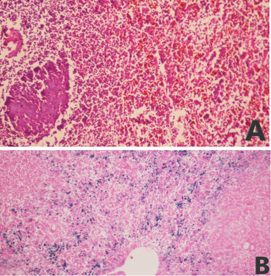
The collected tissues were routinely processed and stained with hematoxylin and eosin (H and E) for microscopic examination . Additionally, spleen smears were stained with Prussian blue stain to demonstrate the hemosiderin pigment deposition .
Sequencing and phylogenetic analysis
The gene-specific PCR amplicons of the 18S rRNA gene of T. ovis and T. lestoquardi were size-separated using agarose gel electrophoresis, excised and purified from gels using the QIAquick® Gel Extraction Kit (Qiagen, Germany). Further, purified products from gel were used directly for cycle sequencing reactions using the BigDye® Terminator v3.1 Cycle Sequencing Kit (Applied Biosystems, USA). Products of cycle sequencing reactions were purified using DyeEx® Spin Kit (Qiagen, Germany) and sequenced on an ABI 3500XL® Genetic Analyzer (Life Technologies, USA). The obtained sequences of T. lestoquardi and T. ovis were uploaded at GenBank and given the accession numbers; MT002826 and MT002827, respectively. The 18S rRNA genes of MT002826 and MT002827 sequences were assembled, aligned and edited using the Bioedit® software, version 7.2.5 . A Basic Local Alignment Search Tool (BLAST) was used on the National Centre for Biotechnology Information (NCBI) platform. Phylogenetic trees were constructed by using molecular evolutionary genetics analysis (MEGA®) software version 10.1.7 on the bases of maximum likelihood methodology .
Table 2: Primers design of different Theileria primers sets.
Primer
Target gene
Primer specificity
Product size (bp)
Universal spp.
18S rRNA
F.5'-AGTTTCTGACCTATCAG-3'
R.5'-TTGCCTTAAACTTCCTTG-3'
1098
Specific T.ovis
18S rRNA
F. 5'-TCGAGACCTTCGGGT-3
R.5'-TCCGGACATTGTAAAACAAA-3'
520
Specific T.lestoquardi
18S rRNA
F. 5'-GTGCCGCAAGTGAGTCA-3'
R.5'GGACTGATGAGAAGACGATGAG3'
785
Statistical analysis
The data were expressed as mean ± standard deviation (SD). Analysis of variance was performed by one-way ANOVA using SPSS version 25.0 for windows (Statistical Package for the Social Sciences; SPSS Inc., Chicago, USA).
RESULTS
Clinical signs
Clinical manifestations of ovine theileriosis in infected sheep and their incidence were listed in Table 3.
Table 3: The incidence of clinical signs of theileriosis in sheep.
Clinical signs
Number of sheep
Fever
39
Cachexia and emaciation
33
Lacrimation and conjunctivitis
28
Pale mucous membrane
26
Tick bite wound
10
Prevalence of theileria infection
The microscopic examination of blood smears revealed the presence of intracellular forms of Theileria piroplasms and schizonts in 33 out of 152 samples (21.7%) while its incidence by using PCR (universal Theileria spp. Primer) was 56 out of 152 (36.8%) of examined animals. Moreover, the use of microscopic examination of blood smears and PCR assay revealed different prevalence in the different localities from which the samples were collected, Table 4.
Table 4: The prevalence of ovine theileriosis in the different localities.
Location
Number of sheep
Diagnostic techniques
Blood smears
PCR
1. Bahtim and marg (Cairo)
47
2
4.3%
14
29.8%
2. Al Oubour (Cairo)
13
0
0.0%
0
0.0%
3. Kafr tohormous and faisal (Giza)
29
12
41.4%
13
44.8%
4. Al-Saff (Giza)
34
18
52.9%
28
82.4%
5. Badr center (Al Monofia)
29
1
3.4%
1
3.4%
Total
152
33
21.7%
56
36.8%
Microscopic examination
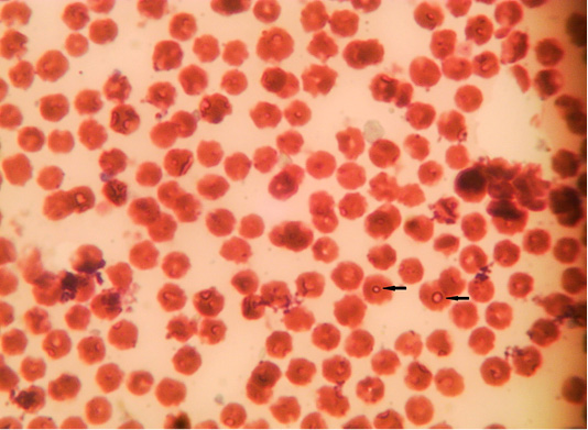
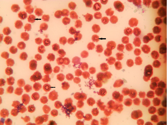
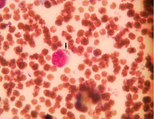
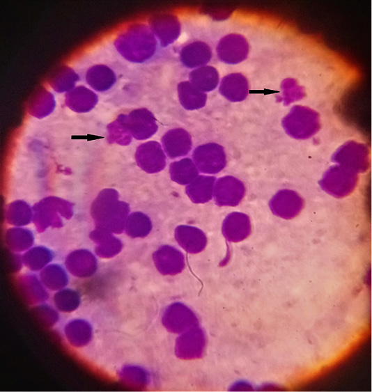
The microscopic examination of blood smear revealed pleomorphic (round, oval, rod and comma) piroplasms (Figures 1 and 2). They were observed as individual (one piroplasm per erythrocyte), double, triple and tetra-forms. Moreover, the microschizonts were observed in the lymphocytes of blood smear (Figure 3) while the macroschizonts (Koch’s blue bodies) were demonstrated in the lymphocytes of lymph nodes smears (Figure 4). The infected RBCs showed crenation, spherocytosis and acanthocytosis.
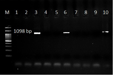
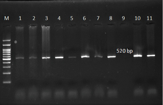
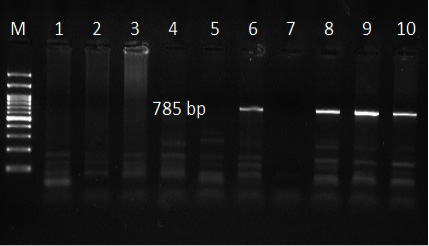
The use of specific primers assured the presence of T. ovis and T. lestoquardi in the different localities, Table 5. However, T. lestoquardi was only identified in association of T. ovis (mixed infection) in Al-Saff (Giza) group. The product size of universal Theileria, T. lestoquardi and T. ovis were 1098bp (Figure 5), 785 bp (Figure 7) and 520 bp (Figure 6), respectively.
Table 5: Prevalence of Theileria infections with different species.
There was significant decrease in RBCs count, hematocrit, Hb concentration, MCHC, TLC, neutrophils, lymphocytes, monocytes, eosinophils, and platelets with significant increase of MCV in infected groups compared with Theileria negative group (Table 6). There were no significant differences between T. ovis group and mixed Theileria group.
Significant increases in AST, GGT, total bilirubin, indirect bilirubin, urea, and creatinine with significant decreases in total proteins and albumin were recorded in infected groups in comparison with Theileria negative group. Moreover, there was no significant change in the values of A:G ratio and direct bilirubin (Table 7). There were no significant differences between T. ovis group and mixed Theileria group.
Table 6: Hematological parameters of Theileria infected and control groups.
Parameter
Unit
Theileria negative group
Theileria ovis group
Mixed Theileria group
RBCs count
×106cell/μl
9.84±0.88a
6.72±0.76b
6.62±0.33b
Hct
%
29.25±1.12a
22.97±1.94b
21.81±1.26b
Hb
g/dl
10.21±0.91a
7.11±0.54b
6.88±0.39b
MCV
fl
29.72±3.17a
34.27±2.51b
33.01±2.60b
MCHC
%
33.86±1.54a
30.83±1.26b
31.44±1.08b
TLC
×103 cell/μl
14.98±1.32a
9.51±0.84b
6.65±0.26b
Neutrophils
×103 cell/μl
5.05±0.27a
4.31±0.19b
3.58±0.14b
Lymphocytes
×103 cell/μl
8.08±0.81a
4.29±0.25b
2.41±0.19b
Monocytes
×103 cell/μl
1.493±0.088a
0.755±0.033b
0.528±0.027b
Eosinophils
×103 cell/μl
0.356±0.056a
0.151±0.016b
0.124±0.026b
Platelets
×103 cell/μl
286.58±13.55a
202.11±15.81b
188.47±9.25b
Different superscripts in the same raw refer to significant changes (p<0.05).
Table 7: Biochemical parameters of Theileria infected and control groups.
Group
Unit
Theileria negative group
Theileria ovis group
Mixed Theileria group
Total proteins
g/dl
7.98±0.47a
6.48±0.69b
6.73±0.41b
Albumin
g/dl
3.82±0.15a
3.29±0.33b
3.25±0.28b
Globulin
g/dl
4.16±0.38a
3.19±0.17b
3.48±0.72b
A:G ratio
1.51±0.099a
1.68±0.076a
1.42±0.088a
AST
U/L
26.21±3.96a
45.63±2.52b
65.25±5.56b
GGT
U/L
24.13±1.57a
41.99±1.49b
40.33±1.01b
Total bilirubin
mg/dl
0.930±0.052a
1.309±0.065b
1.118±0.021b
Direct bilirubin
mg/dl
0.608±0.054a
0.573±0.038a
0.618±0.012a
Indirect bilirubin
mg/dl
0.624±0.061a
1.002±0.035b
0.997±0.021b
Urea
mg/dl
23.16±1.74a
46.92±1.27b
50.53±1.10b
Creatinine
mg/dl
0.983±0.178a
1.609±0.209b
1.346±0.124b
Different superscripts in the same raw refer to significant changes (p<0.05).
Macroscopic and histopathological changes
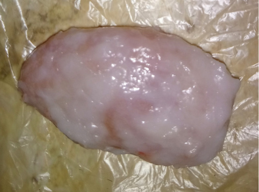
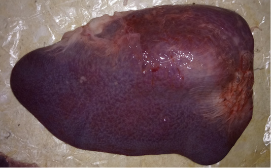
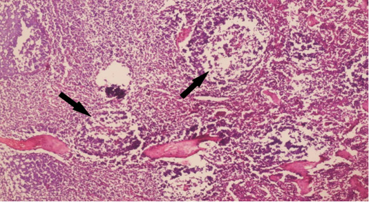
The examined organs (spleen and lymph nodes) showed severe enlargement with sub-capsular hemorrhages (Figures 8 and 9). Meanwhile, the microscopic examination of spleen revealed diffuse depletion of the lymphoid tissue, degenerative changes in the septal connective tissue and multiple necrotic foci with diffuse hemorrhages accompanied with deposition of hemosiderin pigment (Figure 10). Moreover, depletion and necrosis of lymphoid follicles were observed in the lymph nodes (Figure 11).
Phylogenetic analysis
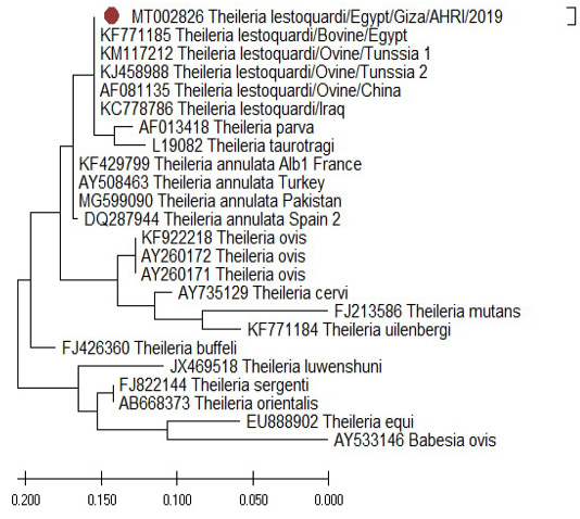
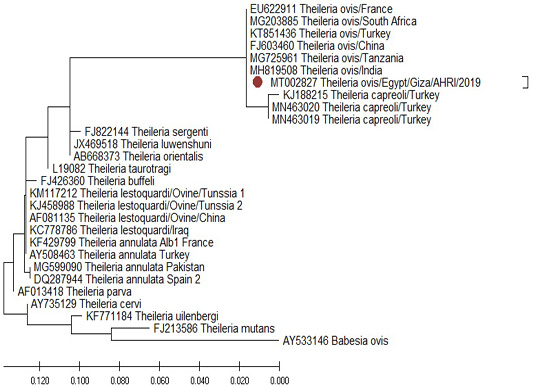
The comparison of the18S rRNA T. Lestoquardi partial sequence (MT002826) revealed 100% homology with T. lestoquardi from bovine/Egypt (KF771185), ovine/Tunisia (KM117212 and KJ458988), ovine/China (AF081135) and ovine/Iraq (KC778786). Also, there was a 99% homology with the bovine T. annulata from France (KF429799), Turkey (AY508463), Pakistan (MG599090) and Spain (DQ287944) (Figure 12) while the 18S rRNA T. ovis partial sequence (MT002827) revealed 100% homology with the ovine T. ovis from South Africa (MG203885), France (EU622911), Turkey (KT851436), China (FJ603460), Tanzania (MG725961) and India (MH819508) (Figure 13).
Ovine theileriosis is an important hemoprotazoan disease of sheep causing severe economic losses as reduced productivity and sometimes cause high mortality of infected animals . In this study, the ovine theileriosis infected animals showed the common clinical signs of the disease as pyrexia, cachexia, emaciation, lacrimation, conjunctivitis, pale mucous membrane and tick bite wounds.
Microscopic examinations of blood smears, prepared from the animals of this study, revealed that the incidences of Theileria species were 4.3%, 0.0%, 41.4%, 52.9% and 3.4% in Bahtim and Marg, Al Oubour, Kafr Tohormous and Faisal, Al-Saff, and Badr center; respectively, with an overall incidence of 21.7% while by using PCR (universal Theileria spp. primer) the incidences were 29.8%, 0.0%, 44.8%, 82.4% and 3.4% in the same localities with an overall incidence of 36.8%. These results are approximately similar to the results of Hegab et al. (2016) who reported a prevalence of 17.08% by using microscopic examination of blood smears and 40% by using PCR in the resident sheep samples collected from different localities of Egypt.
According to the results of PCR (by using universal and spp. specific primers), the animals of this study, were divided into 3 groups: Theileria negative group, T. ovis group and mixed T. ovis and T. lestoquardi group. The clinic pathological examination of the animals of the groups under the study revealed significant changes of some parameters in both infected groups in comparison with the Theileria negative group. Moreover, the significance was more prominent in the mixed group than the T. ovis group due to there is co-infection with T. lestoquardi and T. ovis.
The microscopic examination of blood smears of infected animals showed pleomorphic intra-erythrocytic piroplasms and microschizont inside lymphocytes while the lymph node smears showed macroschizont (Koch’s blue bodies) inside the lymphocytes. These microscopic findings are similar to Çöl and Uslu (2006) and Hegab et al. (2016).
Results of erythrogram revealed significant macrocytic hypochromic anemia in the infected groups compared with Theileria negative group. The hemolytic anemia occurs due to presence of parasites inside the RBCs causes hemolysis result from erythrocytic fragility and stimulation of erythrophagocytosis in reticuloendothelial system . A permanent tick blood sucking causing chronic blood loss . These results agreed with the results recorded by Al-Fetly (2012), Razavi et al. (2015) and Sohail et al. (2018).
The leukogram of infected groups showed significant leukopenia due to neutropenia, lymphopenia, monocytopenia and eosinopenia, compared with the Theileria negative group. This leukopenia may be explained as a results of destruction of infected lymphocytes due to presence of schizogony of parasite inside them while the decrease of other cells may be due to the suppression of bone marrow and leukopoiesis as a result of parasitic products and increase TNF-α production . Moreover, Weiss and Wardrop (2010) reported that the eosinopenia and pancytopenia in Theileria infected animals may be due to parasitic toxins or different causes of necrosis, fibrosis, or suppression in bone marrow. These results were in agreement with Çöl and Uslu, (2006), Yaghfoori et al. (2017), Minnat and Abdulwadood (2012) and Khan et al. (2017) while leukocytosis in Theileria infected sheep were demonstrated in some studies that results due to stage of infection and proliferation of lymphocytes in the lymphoid organs as a defensive response to parasitic infection ; ; .
Significant thrombocytopenia was also reported in the infected groups compared with Theileria negative group. The thrombocytopenia in Theileria infection occurred due to increased platelets destruction, consumption and degranulation in peripheral circulation and/or due to the pancytopenia resulting from production of TNF-α or bone marrow suppression as result of parasitic toxins .
The liver affection during ovine theileriosis was proved by significant hypoproteinemia, hypoalbuminemia, total and indirect hyperbilirubinemia. The AST and GGT activities were significant increased. Moreover, the values of A:G ratio and direct bilirubin have no significant changes in both infected groups compared with the Theileria negative group. Hypoproteinemia and hypoalbuminemia in Theileria infection might be resulted from synthesis reduction in liver due to liver failure, extravascular release proteinaceous fluids from diseased lymph nodes and/or reduction in protein intake due to anorexia during infection ; . The increased liver enzymes activities were explained by the leakage from liver damages as necrosis and degeneration of the hepatocytes, severe damage of the hepatobiliary system and increased sinusoidal spaces ; ; . The total and indirect hyperbilirubinemia were due to hemolysis of infected RBCs inside the reticuloendothelial system causing hemolytic anemia and liver failure ; ; .
The kidney function tests revealed significant increase of BUN and creatinine; azotemia of both infected groups compared with Theileria negative group. This azotemia may be related to the deposition of immune complexes of Theileria spp. in the renal tissue causing focal or diffuse coagulative necrosis and/or increased protein catabolism due to inappetence during Theileria infection ; ; .
The histopathological changes included depletion and necrosis with hemorrhages of lymphoid follicles of lymph nodes and diffuse depletion of the lymphoid tissue with degenerative changes and hemorrhages in the spleen. The lymphocytic depletion and necrosis may explain the recorded lymphopenia in the Theileria infected groups .
According to previous studies, a conserved gene of 18S rRNA is specific target for Theileria species detection by using PCR assay as described by as a sensitive diagnostic tool for ovine theileriosis. In this study, different primer sets were used for Theileria spp.; a universal primer and species-specific primers for T. ovis and T. lestoquardi . Different Theileria species as T. ovis and T. lestoquardi were identified by using specific primer sets. From positive samples of universal primers 36.8% were identified as 53.6% T. ovis and 46.4% mixed infection with T. ovis and T. leastoquardi.
Sequencing and phylogenetic analysis were performed using a blood sample collected from Al-Saff (Giza) that was positive to T. lestoquardi and T. ovis. The 18S rRNA gene of T. lestoquardi and T. ovis were partially sequenced (GenBank Accession Numbers MT002826 and MT002827). The isolate of T. leastoquardi (MT002826) revealed 100% homology with T. lestoquardi from bovine/Egypt (KF771185), ovine/Tunisia (KM117212 and KJ458988), ovine/China (AF081135) and ovine/Iraq (KC778786). This isolate showed 99% homology with T. annulata from France (KF429799), Turkey (AY508463), Pakistan (MG599090) and Spain (DQ287944). Our isolate was clustered in a single clade with T. lestoquardi, T. annulata, T. parva and T. taurotragi. This result is consistent with a previous report about T. lestoquardi from ovine source in Tunisia and T. lestoquardi from buffaloes source in Egypt . The isolate of T. ovis (MT002827) revealed 100% homology with T. ovis from South Africa (MG203885), France (EU622911), Turkey (KT851436), China (FJ603460), Tanzania (MG725961) and India (MH819508).
CONCLUSION
The infected sheep showed several clinical manifestations as pyrexia, cachexia, emaciation, lacrimation, conjunctivitis, pale mucous membrane and tick bite wounds. Hematological examination of the ovine theileriosis showed macrocytic hypochromic anemia with pancytopenia affecting the three lineages of hematopoiesis. Moreover, the disease causes multiple organ affections reflected in biochemical results as hypoproteinemia, hypoalbuminemia, hyperbilirubinemia, azotemia with increase of liver enzymes activities. according to our knowledge, this is the first report of phylogeny of T. lestoquardi in sheep in Egypt.
ACKNOWLEDGEMENTS
The authors would like to acknowledge the veterinarians in veterinary clinics, sheep farms and slaughterhouse who helped in sample collection. We also thank the staff of Genome Research Unit and Clinical Pathology unit in Animal Health Research Institute (AHRI) and Department of Clinical Pathology, Faculty of Veterinary Medicine for supportive technical assistance.
Author’s Contribution
Mostafa Bashandy, Khaled Mahran and Waheed Mousa: Conceptualization.
Mahmoud Eliwa: Samples collection.
Mahmoud Eliwa and Khaled Mahran: Cinicopathologic investigations.
Mahmoud Eliwa and Naglaa Hagag: Molecular studies.
Mohamed Shaalan: Histopathological examination.
Khaled Mahran: Data analysis.
Mahmoud Eliwa and Khaled Mahran: Writing the manuscript.
All authors read and approved the final version.
FUNDING
This research did not receive any specific grant from funding agencies in the public, commercial, or not-for-profit sectors.
CONFLICT OF INTEREST
The authors have declared no conflict of interest.
References
• Akhter W, Aslam A, Rehman MU, Rehman HU, Rashid I, Akhtar R (2017). Comparative pathological changes in sheep infected with Theileria annulata and non-infected control. J. Hellenic Vet. Med. Soc., 68(4): 629–634. https://doi.org/10.12681/jhvms.16064[CrossRef]
• Al-Fetly DRH (2012). Detection of Theileria spp. in blood samples and estimation of haematological and biochemical changes in sheep in Al-Diwaniya. Kufa J. Vet. Sci., 3(2): 45–53.
• AL-Hosary AAT, Ahmed L, Seitzer U (2015). First report of molecular identification and characterization of Theileria spp. from water buffaloes (Bubalus bubalis) in Egypt. Adv. Anim. Vet. Sci., 3(12): 629–633. https://doi.org/10.14737/journal.aavs/2015/3.12.629.633[Full text][CrossRef]
• Allsopp BA, Baylis HA, Allsopp MT, Cavalier-Smith T, Bishop RP, Carrington DM, Sohanpal B, Spooner P (1993). Discrimination between six species of Theileria using oligonucleotide probes which detect small subunit ribosomal RNA sequences. Parasitology, 107: 157–165. https://doi.org/10.1017/S0031182000067263[CrossRef]
• Almeria S, Castellà J, Ferrer D, Ortuño A, Estrada-peña A, Gutiérrez JF (2001). Bovine piroplasms in Minorca (Balearic Islands, Spain): A comparison of PCR-based and light microscopy detection. Vet. Parasitol., 99: 249–259. https://doi.org/10.1016/S0304-4017(01)00464-2[CrossRef]
• Altay K, Dumanli N, Patricia J, Holman B, Aktas M (2005). Detection of Theileria ovis in naturally infected sheep by nested PCR. Vet. Parasitol., 127: 99–104. https://doi.org/10.1016/j.vetpar.2004.09.012[CrossRef]
• Baghshani H, Razmi GR, Yaghfouri S, Dezaki AA (2012). Investigation of selected biochemical parameters in sheep naturally infected with theileriosis. Comp. Clin. Path., 21: 1417–1420. https://doi.org/10.1007/s00580-011-1308-2[CrossRef]
• Bakheit MA, Seitzer U, Ahmed JS (2006). A new recombinant protein-based ELISA for the diagnosis of malignant theileriosis of sheep and goats. Parasitol. Res., 98: 145–149. https://doi.org/10.1007/s00436-005-0034-6[CrossRef]
• Cluzel C, Javard R, Grimes C (2016). Pathology in practice. Javma J. Am. Vet. Med. A., 249(10): 1153–1155. https://doi.org/10.2460/javma.249.10.1153[CrossRef]
• Çöl R, Uslu U (2006). Haematological and coagulation profiles during severe tropical theileriosis in cattle. Turk. J. Vet. Anim. Sci., 30: 577–582.
• Doumas B, Watson W, Biggs H (1971). Albumin standards and the measurement of serum albumin with bromcresol green. Int. J. Clin. Chem. Diagn. Lab. Med., 31: 87-96. https://doi.org/10.1016/0009-8981(71)90365-2[CrossRef]
• Doumas BT, Bayse DD, Carter RJ, Peters T, Schaffer R (1981). A Candidate reference method for determination of total protein in serum I. Development and validation. Clin. Chem., 27(10): 1642–1650. https://doi.org/10.1093/clinchem/27.10.1642[CrossRef]
• Elbasheir M, Elhussein AM (2013). Theleria lestoqurdi on some haematological and biochemical parameters in desert ewes during pregnancy. J. Vet. Med. Anim. Prod., 4(2): 88–95.
• Elsify A, Sivakumar T, Nayel M, Salama A, Elkhtam A, Rizk M, Mosaab O, Sultan K, Elsayed S, Igarashi I, Yokoyama N (2015). An epidemiological survey of bovine Babesia and Theileria parasites in cattle, buffaloes, and sheep in Egypt. Parasitol. Int., 64(1): 79–85. https://doi.org/10.1016/j.parint.2014.10.002[CrossRef]
• Fadel NGM, El-Refai MA, Wahba AA, Shaker MHM, Elebeedy AA, Hafez MA, Harba SAR (2010). Survey on parasitic diseases of sheep and goats and the related clinicopathological changes in east –south of Egypt. Egypt. J. Appl. Sci., 25(5): 298–313.
• Gunes N, Arsoy D, Aktas M, Ozubek S, Ozuicli M, Aydin L, Selcuk O (2016). Changes in some biochemical parameters in small ruminant theileriosis. Bulg. J. Agric. Sci., 22(2): 303–307.
• Hall TA (1999). BioEdit: A user-friendly biological sequence alignment editor and analysis program for Windows 95/98/NT. Nucl. Acids Symp. Ser., 41: 95–98.
• Hasegawa M, Kishino H, Yano T (1985). Dating of the human-ape splitting by a molecular clock of mitochondrial DNA. J. Mol. Evol., 22(2): 160–174. https://doi.org/10.1007/BF02101694[CrossRef]
• Hegab AA, Fahmy MM, Mahdy OA, Wahba AA (2016). Parasitological and molecular identification of Theileria Species by PCR-RFLP method in sheep, Egypt. Int. J. Adv. Res. Biol. Sci., 3(7): 48–55.
• Jaffé M (1986). About the precipitate that picric acid produces in normal urine and about a new reaction of creatinine. Hoppe Seylers Z. Physiol. Chem., 10: 391–400.
• Jendrassik L, Grof P (1938). Colorimetric method of determination of bilirubin. Biochem. Z., 297: 81–82.
• Khan A, Ashfaq K, ud Din I, ul Haq R, Jamil M, Ullah B, Ullah S, ur Rehman H, Ullah F (2017). Bovine theileriosis: Prevalence, estimation of hematological profile and chemotherapy in cattle in Dera Ismail Khan, Khyber Pakhtunkhwa Province, Pakistan. Am. Sci. Res. J. Eng. Technol. Sci., 32(1): 8–17.
• Kirvar E, BIlhan T, Katzer F, Wilkie G, Hooshmand-Rad P, Brown D (1998). Detection of Theileria lestoquardi (hirci) in ticks, sheep, and goats using polymerase chain reaction. Ann. N. Y. Acad. Sci., 849: 52–62. https://doi.org/10.1111/j.1749-6632.1998.tb11033.x[CrossRef]
• Kumar S, Stecher G, Li M, Knyaz C, Tamura K (2018). MEGA X: Molecular evolutionary genetics analysis across computing platforms. Mol. Biol. Evol., 35(6): 1547–1549. https://doi.org/10.1093/molbev/msy096[CrossRef]
• Levine ND (1985). Apicomplexa: The piroplasms. Vet. Protozool., pp. 291–328.
• Littlewood W (1915). Annual report for 1914. Ministry of agriculture Egypt, Vet. Ser., 39(5).
• Liu Q, Zhou YQ, He GS, Oosthuizen MC, Zhou DN, Zhao JL (2010). Molecular phylogenetic studies on Theileria spp. isolates (China) based on small subunit ribosomal RNA gene sequences. Trop. Anim. Health Prod., 42: 109–114. https://doi.org/10.1016/j.ympev.2009.09.034[CrossRef]
• Mahmoud MHS, Al-dhalimy AMB, Al-dujaily AH (2019). Study of hematological and biochemical changes in sheep and goats infected with theileriosis at Al-Najaf province, Iraq. Biochem. Cell. Arch., 19(1): 803–806.
• Minnat TR, Abdulwadood I (2012). Study of clinical, hematological and serological diagnosis of ovine theileriosis in Basrah province. Basra J. Vet. Res., 11(4): 1–18.
• Persijn J, van der Slike W (1982). A new method for the determination of gamma glutamyletrans-peptidase in serum. J. Clin. Chem. Clin. Biochem., 14: 421–427.
• Radwan IGH, El–Kelesh EA (2009). Identification of Theileria species in sheep andgoats by ploymerase chain reaction (PCR). Kafrelsheikh Vet. Med. Journal, 3rd Sci. Conf., pp. 460-473. https://doi.org/10.21608/kvmj.2009.108505[CrossRef]
• Razavi SM, Espandarnia A, Rakhshandehroo E, Ghane M, Nazifi S (2015). Malignant ovine theileriosis: Serum concentrations of some inflammatory components and adenosine deaminase activity. Comp. Clin. Path., 24: 639–643. https://doi.org/10.1007/s00580-014-1959-x[CrossRef]
• Razmi G, Yaghfoori S, Mohri M, Haghparast A, Tajeri S, Republic I (2019). The haematological, proinflammatory cytokines and IgG changes during an ovine experimental theileriosis. Onderstepoort J. Vet. Res., 86(1): a1629. https://doi.org/10.4102/ojvr.v86i1.1629[CrossRef]
• Reitman S, Frankel S (1957). A colorimetric method for the determination of serum glutamic oxalacetic and glutamic pyruvic transaminases. Am. J. Clin. Pathol., 28: 56–63. https://doi.org/10.1093/ajcp/28.1.56[CrossRef]
• Riaz M, Tasawar Z (2017). A study on molecular surveillance of Theileria spp. infection and its impact on hematological and biochemical changes in naturally infected small ruminants at Multan, Pakistan. Pure Appl. Biol., 6(4): 1427–1435. https://doi.org/10.19045/bspab.2017.600154[CrossRef]
• Rjeibi MR, Darghouth MA, Rekik M, Amor B, Sassi L, Gharbi M (2016). First molecular identification and genetic characterization of Theileria lestoquardi in sheep of the Maghreb region. Transbound. Emerg. Dis., 63: 278–284. https://doi.org/10.1111/tbed.12271[CrossRef]
• Sampson EJ, Baird MA, Burtis CA, Smith EM, Witte DL, Bayse DD (1980). A coupled-enzyme equilibrium method for measuring urea in serum: Optimization and evaluation of the AACC study group on urea candidate reference method. Clin. Chem., 26(7): 816–826. https://doi.org/10.1093/clinchem/26.7.0816[CrossRef]
• Sohail A, Khan MY, Burni T, Khalil IH, Mehar S, Shah A (2018). A Study on molecular surveillance of Theileria spp. infection and its impact on hematological and biochemical changes in naturally infected small ruminants at Multan, Pakistan. ARPN J. Agric. Biol. Sci., 13(1): 1–7.
• Soulsby E (1982). Helminths, arthropods and protozoa of domesticated animals (7th ed.). Baillier Tindall and Cassel Ltd.
• Suvarna KS, Layton C, Bancroft JD (2012). Bancroft’s theory and practice of histological techniques E-book (7th ed.). Elsevier Health Sciences.
• Szasz G (1969). A kinetic photometric method for serum gamma-glutamyletranspeptidase. Clin. Chem., 15: 124–136. https://doi.org/10.1093/clinchem/15.2.124[CrossRef]
• Weiss DJ, Wardrop KJ (2010). Schalm’s veterinary hematology (eds. Weiss DJ and Wardrop KJ); 6th ed.). Blackwell Publishing Ltd.
• Yaghfoori S, Mohri M, Razmi G (2017). Experimental Theileria lestoquardi infection in sheep: Biochemical and hematological changes. Acta. Tropica., 173: 55–61. https://doi.org/10.1016/j.actatropica.2017.05.029[CrossRef]
• Youssef SY, Yasien S, Mousa WMA, Nasr SM, El-Kelesh EAM, Mahran KM, Abd-El-Rahman AH (2015). Vector identification and clinical, hematological, biochemical, and parasitological characteristics of camel (Camelus dromedarius) theileriosis in Egypt. Trop. Anim. Health Prod., 47: 649–656. https://doi.org/10.1007/s11250-015-0771-1[CrossRef]
• Zhang X, Liu Z, Yang J, Chen Z, Guan G, Ren Q, Liu A, Luo J, Yin H, Li Y (2014). Multiplex PCR for diagnosis of Theileria uilenbergi, Theileria luwenshuni, and Theileria ovis in small ruminants. Parasitol. Res., 113(2): 527–531. https://doi.org/10.1007/s00436-013-3684-9[CrossRef]
• Zhao L, Wang J, Ding Y, Li K, He B, Li F, Zhang L, Li X, Liu Y (2019). Theileria ovis (Piroplasmida: Theileriidae) detected in melophagus ovinus (Diptera: Hippoboscoidea) and ornithodoros lahorensis (Ixodida: Argasidae) removed from sheep in Xinjiang, China. J. Med. Entomol., 1915(10): 1–5. https://doi.org/10.1093/jme/tjz193[CrossRef]