
Pseudomonas aeruginosa is a common disease causing bacterium that causes respiratory tract infections, urinary tract infections and sepsis syndrome. It is found ubiquitously in nature and can cause diseases in immunocompromised patients. Current study was conducted to estimate genetic diversity and analyze the existence of extended spectrum beta lactamases in P. aeruginosa recovered from drinking water in Peshawar, Pakistan. Drinking water samples were gathered from distinct areas of Peshawar during 2021-22 and examined for existence of P. aeruginosa. Fifty isolates of P. aeruginosa were recovered from drinking water samples. Antibiotic sensitivity was conducted against nine different antibiotics i.e., meropenem (10 µg), piperacillin/tazobactum (110 µg), gentamicin (10 µg), minocycline (30 µg), ofloxacin (5 µg), clarithromycin (15 µg), cefixime (5 µg), ceftriaxone (30 µg) and amikacin (30 µg). Genetic diversity was analyzed through ERIC-PCR. The occurrence of four extended spectrum beta lactamases (bla-TEM, bla-SHV, blaOXA-10 and bla-CTX-M) was analyzed in the isolates. Antibiotic sensitivity was 0, 2, 4, 6, 10, 64, 74 and 100% towards meropenem, piperacillin/tazobactum, ofloxacin, amikacin, ceftriaxone, gentamicin, minocycline, cefixime and clarithromycin respectively. Prevalence of bla-TEM gene was 18% and blaOXA-10, bla-SHV and blaCTX-M were not detected in any of the isolates. Genetic distances among the isolates ranged from 0 to 100 percent. Based on ERIC PCR profiling, out of 50 isolates, 14 isolates were clustered into 6 distinct clones and 36 isolates showed unique ERIC profiling according to similarity coefficients of ≥85%. These results highlight the prevalence of diverse P. aeruginosa strains in drinking water of Peshawar. These bacteria also are resistant to some of the last resort antibiotics and possess antimicrobial resistance genes. These findings are alarming and proper water treatment and hygienic principles are required with regard to supplying clean drinking water.
Research Article · Volume 58, Issue 6, 30 September 2026 · Open Access
Genetic Diversity and Molecular Characterization of Extended Spectrum Beta Lactamases in Pseudomonas Aeruginosa Isolated from Drinking Water in Peshawar, Pakistan
· ·
Affiliations & Notes ⌄
- 1Centre of Biotechnology and Microbiology, University of Peshawar, Peshawar, Pakistan * Corresponding author: kafeelpbg@gmail.com
Article Info ⌄
- Journal:
- Pakistan Journal of Zoology
- Published:
- 30 September 2026

From the article
- Antibiotic resistance among P. aeruginosa is increasing due to its capacity to be intrinsically resistant or develop resistance through pathways like reduced membrane permeability, AmpC β-lactamase production, multidrug efflux pumps, and β-lactamase production, including production of ESBLs and metallo-β-lactamases.
- In the current study, among 50 isolates, 18% (n=9) were positive for the bla-TEM gene, but none were positive for bla-SHV, bla-OXA-10, or bla-CTX-M. All isolates were phenotypically negative for the production of ESBLs.
- In this study, the OprL gene amplification was used as standard for molecular confirmation all 50 isolates of P. aeruginosa.
- The current study confirmed the occurrence of ESBL coding genetic determinants in Pseudomonas aeruginosa recovered from drinking water of different districts of Peshawar, Pakistan.
Selected from the article's own text.
Abstract
INTRODUCTION
Pseudomonas aeruginosa is widely distributed in nature and could be found in diverse forms in soil and water. Clinical isolates of P. aeruginosa demonstrate virulence by producing proteases, invading epithelial cells and exhibiting low susceptibility to a broad spectrum of antibiotics. P. aeruginosa could be found in various environments . These external reservoirs could serve as potential sources of infections in human. P. aeruginosa has been frequently reported to exist in main water supplies and aquatic settings which could harbor antibiotic resistance genes. The release of antimicrobials into water bodies may lead to greater chances of bacterial resistance development in P. aeruginosa ; . Production of extended spectrum beta-lactamases (ESBLs) is one prominent resistance mechanism to inactivate beta-lactam antibiotics . Several forms of beta lactamases have been found in P. aeruginosa. The OXA type enzymes which cleave oxacillin, are the most prevalent type that can be found in P. aeruginosa. These enzymes show highest hydrolytic activity against cloxacillin and oxacillin rather than benzyl penicillin . SHV-type beta-lactamases are named after the presence of sulfhydryl moiety and in comparison to other beta-lactamases they are frequently isolated from hospital samples . CTX-M type beta-lactamases were initially identified in Munich which is why they are called CTX-M, and are termed for their activity against cefotaxime. Variants of blaCTX-M like CTX-M-1, 2, 8, 9 and 25 have been identified that show homology at the level of amino acids . It has been documented that P. aeruginosa possesses CTX-M-1, 2 and 43 variants . TEM-type beta-lactamases exhibit the highest hydrolyzing capacity against ampicillin and low efficacy against extended-spectrum cephalosporins. Over 100 distinct TEM variants, predominantly classified as extended-spectrum beta-lactamases (ESBLs), have been identified. Different TEM variants have been reported in P. aeruginosa like TEM-1, 2, 4, 21, 24, 42 and 116 .
Molecular typing of bacteria is an important component of epidemiological studies that play crucial role in tracking the spread and origin of a bacterial infection. Various molecular techniques are used for estimating genetic relationships among bacteria. One such method is the enterobacterial repetitive intergenic consensus polymerase chain reaction (ERIC-PCR) that is cost-effective technique to assess similarities and differences among bacterial strains in a low budget set up .
The contamination of drinking water by microorganisms like P. aeruginosa is a significant health concern. Pathogenic microorganisms, despite being present in lower proportions in the environment, could still induce illnesses in individuals who are otherwise healthy. Pathogenic microorganisms in drinking water represent one of the most important potential health risks in potable water . No or little information is available on the molecular characteristics of potential pathogenic bacteria prevailing in drinking water in Peshawar, Pakistan. Specifically, there has been no investigation into the prevalence of beta-lactamase genes and the genetic diversity of Pseudomonas aeruginosa contaminating drinking water in this region. Current study was conducted to estimate genetic diversity and analyze the occurrence of extended spectrum beta lactamases in P. aeruginosa recovered from drinking water in Peshawar, Pakistan.
MATERIALS AND METHODS
Isolation, culturing and identification of bacteria
A total of fifty water samples were collected from various locations in Peshawar during 2021-22 using sterile Falcon tubes and samples subsequently processed under controlled laboratory conditions. MacConkey agar medium (Oxoid, UK; 51 g/L) was prepared for bacterial isolation, and the collected samples were inoculated onto the prepared media. The identification of Pseudomonas aeruginosa isolates was confirmed using staining techniques and a series of biochemical tests, including catalase, oxidase, citrate utilization, triple sugar iron (TSI) test, methyl red (MR), Voges–Proskauer (VP), motility, gelatin hydrolysis, and casein hydrolysis as reported previously .
Molecular identification of P. aeruginosa
For molecular confirmation of P. aeruginosa isolates, the oprL gene was amplified using forward and reverse primers described in previous studies . The PCR reaction mix was prepared with 4 μL of 5× FIREPol Master Mix (Cat. No: 04-12-00115), 1 μL each of the forward and reverse primers, 1 μL of DNA template, and 13 μL of molecular-grade water, bringing the total reaction volume to 20 μL. The PCR products were then analyzed by running them on a 1% agarose gel. The primer sequences and the specific PCR conditions used for oprL gene amplification are provided in Table I.
Antibiotic susceptibility
Antibiotic sensitivity analysis was carried out using the disc diffusion method . Mueller Hinton Agar (MHA) medium (Oxoid, UK; 38 g/L of distilled water) was prepared, and fresh bacterial culture was inoculated onto the MHA surface using a sterile swab, evenly streaking the plate. Antibiotic discs were then placed onto the bacterial lawn, and the zones of inhibition were measured. The results were interpreted based on the Clinical Laboratory Standards Institute (CLSI) guidelines and reported procedures to assess antibiotic susceptibility and resistance .
Phenotypic detection of β-lactamase production
Pseudomonas aeruginosa isolates were phenotypically identified using the double disc synergy test . The β-lactam antibiotic discs were placed onto the agar plate with amoxicillin-clavulanic acid (AMC 30 μg) in the middle and cefotaxime (CTX 30 μg), ceftazidime (CAZ 30 μg), cefepime (FEP 30 μg), and aztreonam (ATM 30 μg) surrounding it. The enlargement of the inhibition zone surrounding one of the cephalosporin discs or aztreonam towards the amoxicillin-clavulanic acid disc was indicative of a positive ESBL outcome.
Analysis of ESBLs genes
Specific primers, as previously described, were employed to detect ESBL genes in isolates of P. aeruginosa . Four genes blaSHV, blaCTX-M, blaOXA-10, and blaTEM linked to ESBLs were examined. A PCR reaction mixture was prepared with a total volume of 20 μl. The amplified products were examined using 1% agarose
Table I. Primer sequences and reaction condition for amplification of OprL resistance genes.
|
Gene |
Primer sequences 5`→3` |
Fragment size (bp) |
PCR profile (40 cycles) |
|
OprL |
F-ATGGAAATGCTGAAATTCGC R-CTTCTTCAGCTCGACGCGACG |
504 |
Initial denaturation at 96 oC for 5 min, followed by 40 cycle, each of Denaturation at 96 oC for 1 min, Annealing at 55 oC for 1 min, Extension at 72 oC for 1 min, Final extension at 72 oC for 10 min. |
|
bla-SHV |
F’AAGATCCACTATCGCCAGCAG R’ATTCAGTTCCGTTTCCCAGCGG |
230 |
Initial denaturation at 95 oC for 5 min followed by 30 cycles each of denaturation at 94 oC for 1 min, annealing at 60 oC for 1 min, extension at 72 oC for 1 min, final extension at 72 oC for 5 min. |
|
bla-CTX-M |
F’ CGCTTTGCGATGTGCAG R’ ACCGCGATATCGTTGGT |
552 |
Initial denaturation at 94 oC for 1 min followed by 30 cycles each of denaturation at 55 oC for 1 min, annealing at 72 oC for 1 min, extension at 72 oC for 5 min, final extension at 72 oC for 5 min. |
|
blaOXA-10 |
F’ TATCGCGTGTCTTTCGAGTA R’ TTAGCCACCAATGATGCC |
760 |
Initial denaturation at 94 oC for 1 min followed by 30 cycles each of denaturation at 57 oC for 1 min, annealing at 72 oC for 1 min, extension at 72 oC for 1 min, final extension at 72 oC for 5 min. |
|
bla TEM |
F’ATGAGTATTCAACATTTCCGC R’CAATGCTTAATCAGTGAGG |
856 |
Initial denaturation at 94 oC for 1 min followed by 30 cycles each of denaturation at 55 oC for 1 min, annealing at 72 oC for 1 min, extension at 72 oC for 5 min. |
|
ERIC |
ERIC-I ATGTAAGCTCCTGGGGATTC ERIC-II AAGTAAGTGACTGGGGTGAG |
Variable |
Initial denaturation at 95 oC for 3 min, followed by 30 cycle, each of denaturation at 90 oC for 0.5 min, annealing at 40 oC for 1 min, extension at 72 oC for 1 min, final extension at 72 oC for 8 min. |
gel electrophoresis. The primer sequences and PCR conditions for amplification of ESBLs are provided in Table I.
Genetic diversity analysis
Genetic diversity among P. aeruginosa isolates was assessed through ERIC-PCR using the ERIC-R1 and ERIC-R2 primers . The resulting PCR products were analyzed by gel electrophoresis. The primer sequences and PCR conditions used for amplifying the ERIC-PCR are listed in Table I.
Statistical analysis
Bivariate data, indicating the presence (1) or absence (0) of bands amplified through ERIC-PCR, was generated. Each band was assigned a specific label, and the presence or absence of each band was verified for every isolate. The genetic dissimilarity between isolates was quantified using the Nei and Li formula, denoted as Dab=1-(Nab)/ (Na + Nb – Nab) . In this equation, Dab represents the difference between isolates A and B, Nab signifies the count of shared bands between isolates A and B, where Na represents the number of bands in isolate A, and Nb signifies the number of bands in isolate B. Bivariate data from ERIC-PCR analysis was utilized to construct a dendrogram using molecular evolutionary genetic analysis (MEGA X) software. The genetic similarity between the P. aeruginosa isolates recovered from drinking water was assessed through the help of the resulting dendrogram.
RESULTS
Antibiotic sensitivity
Variable levels of susceptibilities were observed in P. aeruginosa isolates against the test antibiotics. Susceptibility to meropenem was 100%. Susceptibility to piperacillin/tazobactam was 98% whereas 2% of the isolates exhibited intermediate susceptibility. Susceptibility to gentamicin was 90% while 10% of isolates were classified as intermediate. Against minocycline, 36% of isolates were susceptible, 22% showed intermediate response, and 42% were resistant. Susceptibility to ofloxacin was 96%, whereas 100% resistance was observed against clarithromycin. Against cefixime, 26% of the isolates were susceptible, 8% showed intermediate resistance and 66% were found resistant. Susceptibility to amikacin was 96% susceptibility while 4% of isolates showed intermediate resistancce. Resistance to amoxicillin-clavulanic acid was 100%. Susceptibility to cefotaxime was 34% while 48% of the isolates showed intermediate resistance and 18% showed complete resistance. Susceptibility to cefepime was 92% while 4% of the isolates showed intermediate resistance and 4% showed complete resistance. Against aztreonam, 72% susceptibility was recorded while 28% of the isolates showed complete resistance. The detailed antibiotic sensitivity results are presented in Table II.
Table II. Antibiotic sensitivity of P. aeruginosa isolates.
|
Antibiotics |
Conc. (µg) |
Susceptible % |
Intermediate % |
Resistant % |
|
Meropenem (MEM) |
10 |
100%(n=50) |
0%(n=0) |
0%(n=0) |
|
Pipercillin/Tazobactum (TZP) |
110 |
98%(n=48) |
2%(n=1) |
0%(n=0) |
|
Gentamicin (CN) |
10 |
90%(n=45) |
10%(n=5) |
0%(n=0) |
|
Minocycline (MH) |
30 |
36%(n=18) |
22%(n=11) |
42%(n=21) |
|
Ofloxacin (OFX) |
5 |
96%(n=48) |
4%(n=2) |
0%(n=0) |
|
Clathiromycin (CLR) |
15 |
0%(n=0) |
0%(n=0) |
100%(50) |
|
Cefixime (CFM) |
5 |
26%(n=13) |
8%(n=4) |
66%(33) |
|
Cefriaxone (CRO) |
30 |
18%(n=9) |
56%(n=28) |
6%(n=3) |
|
Amikacin (AK) |
30 |
96%(n=48) |
4%(n=2) |
0%(n=0) |
|
Amoxicillin/Clauvlanic acid (AMC) |
30 |
0%(n=0) |
0%(n=0) |
100%(n=50) |
|
Cefotaxime (CTX) |
30 |
34%(n=17) |
48%(n=24) |
18%(n=9) |
|
Ceftazidine (CAZ) |
30 |
92%(n=46) |
8%(n=4) |
0%(n=0) |
|
Cefepime (FEP) |
30 |
86%(n=43) |
10%(n=5) |
4%(n=2) |
|
Aztreonam (ATM) |
30 |
72%(n=36) |
0%(n=0) |
28%(n=14) |
Analysis of extended spectrum beta lactamases production
P. aeruginosa isolates were molecularly confirmed by amplifying the OprL gene, and all the isolates showed presence of the gene.
Phenotypically no isolates were found to produce beta lactamases using the double-disk synergy test. However, at molecular level, out of the 50 isolates, 18% (n=9) tested positive for bla-TEM gene possession while none of the isolates showed the presence of bla-SHV, bla OXA-10, or bla CTX-M genes. Among the subset of isolates carrying bla-TEM (18%, n=9), 33.33% (n=3) exhibited resistance to minocycline, 100% (n=9) were resistant to clarithromycin, 66.66% (n=6) were resistant to cefixime, 100% (n=9) were resistant to amoxicillin-clavulanic acid, 22.22% (n=2) showed resistance to cefotaxime while 44.44% (n=4) were resistant to aztreonam.
Analysis of genetic diversity through ERIC-PCR
A total of 15 distinct bands were generated through ERIC-PCR. Based on the phylogenetic tree and a similarity coefficient of ≥85%, among the 50 P. aeruginosa isolates, 14 were grouped into six distinct clones, while the remaining 36 exhibited unique ERIC profiles (Fig. 1). Clone C1 comprised isolates D1, D16, and D17; clone C2 included D38 and D39; clone C3 consisted of D36 and D37; clone C4 included D13 and D45; and clone C5 comprised D12, D49, and D50. Clone C6 included isolates D18 and D19. The remaining isolates did not cluster into any of the identified clonal groups.
DISCUSSION
Infections due to ESBL-producing P. aeruginosa are on the rise worldwide leading to increased morbidity, mortality and cost of healthcare especially in areas of poor hygiene and sanitation . As stipulated by the EU Directive 98/83/EC on the quality of water for human consumption, there should be no P. aeruginosa present in drinking water . However, in this study all 50 water samples (100%) were found to be contaminated with P. aeruginosa. Antibiotic resistance among P. aeruginosa is increasing due to its capacity to be intrinsically resistant or develop resistance through pathways like reduced membrane permeability, AmpC β-lactamase production, multidrug efflux pumps, and β-lactamase production, including production of ESBLs and metallo-β-lactamases. Carbapenems are the drug of choice for infections caused by both Gram-negative and Gram-positive bacteria . In the current study, meropenem was the best-performing antibiotic, with 100% susceptibility among the isolates. This observation is consistent with a study carried out in Dhaka on environmental reservoirs, which showed 100% susceptibility to meropenem . Penicillins, by inhibiting bacterial cell wall formation, are commonly made ineffective by β-lactamase production and low membrane permeability. High penicillin resistance has been extensively documented . In this study, all the isolates (100%) were resistant to amoxicillin-clavulanic acid. Likewise, a study recently done in Peshawar, Pakistan found 98% resistance to ampicillin among clinical P. aeruginosa isolates . Cephalosporins are widely used for the treatment of P. aeruginosa infections, although resistance is rising. In this study, 86% of isolates were susceptible to cefepime, a fourth-generation cephalosporin. This is in contrast to the results of study from Karachi, Pakistan where 44% of the clinical isolates were susceptible to cefepime . Various docking studies indicate that the amino acids GLN547, GLU568, GLU550, and THR565 of the penicillin-binding protein in P. aeruginosa are responsible for sensitivity to cefepime. Regarding third-generation cephalosporins, 34% of the isolates were found susceptible to cefotaxime and 18% were susceptible to ceftriaxone. These findings are aligned with the findings of another study from Peshawar, Pakistan which reported moderate resistance to cefotaxime . Aminoglycosides have a very effective action against Gram-negative bacilli, but resistance mechanisms like enzymatic modification and decreased permeability can reduce their effectiveness. In this study, 96% of the isolates were amikacin susceptible, a figure higher than the 86% found in France . Gentamicin was found effective in the current study with 90% susceptibility. These findings are in contrast with the 79.7% resistance reported by Strateva et al. (2007) from Bulgaria. Fluoroquinolones are most commonly used for P. aeruginosa because of their broad activity. Resistance is, however, constantly on the rise, depending on geographic location, bacterial origin, and local prescribing patterns . In the current study, ofloxacin was found quite effective with 96% susceptibility. The existence of resistant microorganisms in water and subsequent transfer to human could result in the colonization of human gut which could promote the horizontal transfer of resistance genes to indigenous microbiota. This leads to the development of multidrug-resistant microorganisms, making it challenging to treat infections and prompting the introduction of newer, frequently more expensive antibiotics .
The biosynthetic genes responsible for ESBLs production, commonly carried on plasmids, are among the main determinants of antibiotic resistance. Resistance plasmids may carry multiple classes of genes for antibiotic resistance, highly restricting the choice of therapy . In the current study, among 50 isolates, 18% (n=9) were positive for the bla-TEM gene, but none were positive for bla-SHV, bla-OXA-10, or bla-CTX-M. All isolates were phenotypically negative for the production of ESBLs. In a study conducted in northwest Pakistan, 19.7% prevalence of CTX gene and 11.26% prevalence of OXA gene was reported in clinical isolates . A study from Faisalabad, Pakistan reported 95% prevalence of bla-TEM in P. aeruginosa isolates from poultry meat . Another study on non-clinical isolates of P. aeruginosa reported prevalence of different beta lactam genes as: bla-SHV (93.3%), bla-TEM (40.0%), and bla-CTX-M (20.0%) . A Brazilian study reported 32% prevalence of bla-OXA-10 gene in hospital wastewater representing the contribution of hospital effluents in spreading multidrug-resistant strains into aquatic environments . A South African study revealed a 20% prevalence of bla-CTX-M gene among isolates collected from abattoir wastewater, Mthatha River water and its surrounding dam . The TEM-1 β-lactamase enzyme remains a major factor in penicillin and first-generation cephalosporin resistance and exhibits resistance to more recently developed β-lactam drugs . A study on hospital wastewater samples in Iran indicated 60%, 40% and 60% prevalence of bla-TEM, bla-SHV and bla-CTX respectively (Baghal et al., 2021). In Rivers State, Nigeria, 16.7% of river water and aquaculture isolates were detected to carry bla-SHV . SHV-type ESBLs are able to hydrolyze an extended range of β-lactams including monobactams and carbapenems . Previous studies have reported the prevalence of ESBL genes in phenotypically negative isolates . These findings are in agreement with the current study and indicate the shortcoming of phenotypic methods and the necessity of molecular detection methods like PCR.
Molecular approaches are increasingly being integrated into the identification of bacteria in addition to conventional biochemical and morphological tests. Molecular identification strategies using DNA-based methods such as PCR amplification of specific genes related to target species are much more precise . In this study, the OprL gene amplification was used as standard for molecular confirmation all 50 isolates of P. aeruginosa. Genotypic methods provide more reliability compared to phenotypic methods by being able to cancel out variability contributed by bacterial expression differences . The genetic diversity estimation of P. aeruginosa isolates has important role in tracing infection sources and analyzing outbreaks. In the current study, ERIC-PCR proved a valuable tool for differentiating the isolates. In this study, six different clones and 36 unique isolates were identified showing a high degree of diversity among the isolates. Similarly, a Brazilian research study identified a variety of genetic profiles within clinical isolates, which is an indicator of restricted cross-transmission . Another study from Iran reported five ERIC profiles indicating low genetic variation . These results are in accordance with the perception that P. aeruginosa is continuously evolving as a result of genetic transfer, recombination and gene loss.
CONCLUSION
The current study confirmed the occurrence of ESBL coding genetic determinants in Pseudomonas aeruginosa recovered from drinking water of different districts of Peshawar, Pakistan. Owing to the enormous population size of the region, bacterial infections are now being viewed as a matter of concern that could be disseminated through contaminated drinking water. Hence, upgradation of water treatment and distribution infrastructure and stringent microbial water quality regulations with regular water testing are required to control and prevent the spread of resistant potentially pathogenic bacteria to the community.
DECLARATIONS
Acknowledgement
The facilities provided by Centre of Biotechnology and Microbiology, University of Peshawar are greatly acknowledged.
Funding
The study received no external funding.
Generative AI and AI assisted technology statement
The authors declare that no generative AI and AI assisted technology was used in the creation of this manuscript.
Statement of conflict of interest
The authors have declared no conflict of interest.
Figures
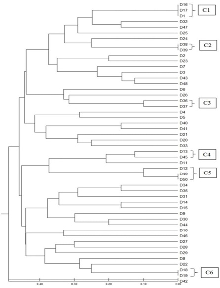
Phylogenetic tree: demonstrating the clustering of P. aeruginosa isolates. The UPGMA method and MEGA-X software was utilized to construct the phylogenetic tree. Fourteen isolates were clustered into six distinct clones: C1 (1, 16 and 17). C2 (38, 39). C3 (36, 37). C4 (13, 45). C5 (12, 49 and 50) C6 (18, 19) based on similarity coefficient ≥ 80%.
References
- Abbas, Y.A., Khudaier, B.Y. and Salih, H.A., 2013. Detection of multidrug resistant Pseudomonas spp. in clinical cases and hospital environments at Thi-Qar Province. J. Pure Sci. Sci. Educ., 3: 1377-1383.
- Abdelrahman, D.N., Taha, A.A., Dafaallah, M.M., Mohammed, A.A., El-Hussein, A.R.M., Hashim, A.I. and Altayb, H.N., 2020. β-lactamases (bla TEM, bla SHV, bla CTXM-1, bla VEB, bla OXA-1) and class C β-lactamases gene frequency in Pseudomonas aeruginosa isolated from various clinical specimens in Khartoum State, Sudan: A cross sectional study. F1000Res., 9: 147-151. https://doi.org/10.12688/f1000research.24818.2[CrossRef]
- Abdullah, R.M., Ali, S., Aslam, B. and Arshad, M.I., 2024. Molecular characterization and drug resistance pattern of pseudomonas aeruginosa isolated from poultry meat and meat products. Pak. Vet. J., 44: 34-78.
- Adelowo, O.O., Caucci, S., Banjo, O.A., Nnanna, O.C., Awotipe, E.O., Peters, F.B. and Berendonk, T.U., 2018. Extended spectrum beta-lactamase (ESBL)-producing bacteria isolated from hospital wastewaters, rivers and aquaculture sources in Nigeria. Environ. Sci. Pollut. Res., 25: 2744-2755. https://doi.org/10.1007/s11356-017-0686-7[CrossRef]
- Al-Ahmadi, G.J. and Roodsari, R.Z., 2016. Fast and specific detection of Pseudomonas aeruginosa from other pseudomonas species by PCR. Annls Burn. Fire Dis., 29: 264-270.
- Ali, S.G., Ansari, M.A., Alzohairy, M.A., Alomary, M.N., Jalal, M., AlYahya, S. and Khan, H.M., 2020. Effect of biosynthesized ZnO nanoparticles on multi-drug resistant Pseudomonas aeruginosa. Antibiotics, 9: 260-265. https://doi.org/10.3390/antibiotics9050260[CrossRef]
- Alpay-Karaoglu, S., Ozgumus, O.B., Sevim, E., Kolayli, F., Sevim, A. and Yesilgil, P., 2007. Investigation of antibiotic resistance profile and TEM-type β-lactamase gene carriage of ampicillin-resistant Escherichia coli strains isolated from drinking water. Annls Microbiol., 57: 281-288. https://doi.org/10.1007/BF03175221[CrossRef]
- Ashbolt, N.J., 2015. Microbial contamination of drinking water and human health from community water systems. Curr. Environ. Hlth. Rep., 2: 95-106. https://doi.org/10.1007/s40572-014-0037-5[CrossRef]
- Baghal-Asghari, F., Dehghani, M.H., Dehghanzadeh, R., Farajzadeh, D., Yaghmaeian, K., Mahvi, A.H. and Rajabi, A., 2021. Antibiotic resistance and antibiotic-resistance genes of Pseudomonas spp. and Escherichia coli isolated from untreated hospital wastewater. Water Sci. Technol., 84: 172-181. https://doi.org/10.2166/wst.2021.207[CrossRef]
- Bauer, A.W., Perry, D.M. and Kirby, W.M., 1959. Single-disk antibiotic-sensitivity testing of staphylococci: An analysis of technique and results. AMA Arch. Intern. Med., 104: 208-216. https://doi.org/10.1001/archinte.1959.00270080034004[CrossRef]
- Bibi, K., Khattak, M., Ahmad, N., Ishaq, M.S. and Muhammad, A., 2015. Isolation, identification and antimicrobial analysis of Pseudomonas aeruginosa from different clinical samples and to study the interactions of target and resistant proteins with cefepime and imipenem through docking. Khyber J. med. Sci., 8: 30-37
- Bhattacharya, D, Sarma, P. M., Krishnan, S., Mishra, S. and Lal, B., 2003. Evaluation of genetic diversity among Pseudomonas citronellolis strains isolated from oily sludge-contaminated sites. Appl. Environ. Microbiol., 69: 1435-1441. https://doi.org/10.1128/AEM.69.3.1435-1441.2003[CrossRef]
- Cai, B., Echols, R., Magee, G., Ferreira, J.C.A., Morgan, G., Ariyasu, M., Sawada, T. and Nagata, T.D., 2017. Prevalenve of carbapenem resustant Gram negative infections in the United States predominated by Acinetobacter baumannii and Pseudomonas aeroginosa. Open. Forum. Infect. Dis., 4: 1-7. https://doi.org/10.1093/ofid/ofx176[CrossRef]
- Cavallo, J.D., Hocquet, D., Plesiat, P., Fabre, R., Roussel, D.M., 2007. Susceptibility of Pseudomonas aeruginosa to antimicrobials: A 2004 French multicentre hospital study. J. Antimicrob. Chemother., 59: 1021–1024. https://doi.org/10.1093/jac/dkm076[CrossRef]
- Chikwendu, C.I., Ibe, S.N. and Okpokwasili, G.C., 2011. Detection of blaSHV and blaTEM beta-lactamase genes in multi-resistant Pseudomonas isolates from environmental sources. Afr. J. Microbiol. Res., 5: 2067-2074. https://doi.org/10.5897/AJMR11.149[CrossRef]
- Doi, Y., 2019. Treatment options for carbapenem-resistant gram-negative bacterial infections. Clin. Infect. Dis., 69(Supp. 7): S565-S575. https://doi.org/10.1093/cid/ciz830[CrossRef]
- Foght, J.M., Westlake, D.W., Johnson, W.M. and Ridgway, H.F., 1996. Environmental gasoline- utilizing isolates and clinical isolates of Pseudomonas aeruginosa are taxonomically indistinguishable by chemotaxonomic and molecular techniques. Microbiology, 142: 2333-2340. https://doi.org/10.1099/00221287-142-9-2333[CrossRef]
- Fróes, A.M., da Mota, F.F., Cuadrat, R.R. and Dávila, A.M., 2016. Distribution and classification of serine β-lactamases in Brazilian hospital sewage and other environmental metagenomes deposited in public databases. Front. Microbiol., 7: 1790-1794. https://doi.org/10.3389/fmicb.2016.01790[CrossRef]
- Gawad, M.A. and Gharbi, W.A., 2022. Molecular Detection of oprI and oprL Virulence Genes of Pseudomonas aeruginosa isolated from Burns and Wounds. Iraqi J. Biotechnol., 21: 1-7.
- Hosu, M.C., Vasaikar, S., Okuthe, G.E. and Apalata, T., 2021. Antibiotic resistant gene of Pseudomonas aeruginosa from non-clinical environment: Public health implications in Mthatha, Eastern Cape Province. S. Afr., 8: 67-71. https://doi.org/10.21203/rs.3.rs-31330/v1[CrossRef]
- Jalier, V., Nicolas, M.H., Fournier, G. and Phillippon, A., 1988. Extended-spectrum beta-lactamases conferring transferable resistance to newer beta-lactam agents in Enterobacteriaceae: Hospital prevalence and susceptibility patterns. Rev. Infect. Dis., 10: 867–878. https://doi.org/10.1093/clinids/10.4.867[CrossRef]
- Li, Q., Chang, W., Zhang, H., Hu, D. and Wang, X., 2019. The role of plasmids in the multiple antibiotic resistance transfer in ESBLs-producing Escherichia coli isolated from wastewater treatment plants. Front. Microbiol., 10: 633. https://doi.org/10.3389/fmicb.2019.00633[CrossRef]
- Mansoor, K., Tanvir, S.B., Shariq, A., Hussain, A., Farooqi, B.J., Ahmed, S. and Tanvir, R., 2015. Frequency and susceptibility pattern of Multidrug Resistant Pseudomonas aeruginosa in isolates of patients from a tertiary care hospital of Karachi, Pakistan. Eur. J. Biotechnol. Biol. Sci., 2: 33-36.
- Megersa, R., Mathewos, M. and Fesseha, H., 2019. Isolation and identification of Escherichia coli from dairy cow raw milk in Bishoftu town, Central Ethiopia. Arch. Vet. Anim. Sci., 1: 1-7.
- Mohamed, A. and Abdelhamid, F., 2020. Antibiotic susceptibility of Pseudomonas aeruginosa isolated from different clinical sources. Zagazig J. Pharm. Sci., 28: 10-17.
- Nei, M. and Li, W.H., 1979. Mathematical model for studying genetic variations in terms of restriction endonucleases. Proc. natl. Acad. Sci., 76: 5269-5273. https://doi.org/10.1073/pnas.76.10.5269[CrossRef]
- Nikolaidis, I., Favini-Stabile, S. and Dessen, A., 2014. Resistance to antibiotics targeted to the bacterial cell wall. Protein Sci., 23: 243-259. https://doi.org/10.1002/pro.2414[CrossRef]
- Polotto, M., Casella, T., de Lucca Oliveira, M.G., Rubio, F.G., Nogueira, M.L., de Almeida, M.T. and Nogueira, M.C., 2012. Detection of Pseudomonas aeroginosa harboring bla CTX-M-2, bla GES-1 and bla GES-5, bla IMP-1 and bla SPM-1 causing infections in Brazillian tertiary care hospital. BMC Infect. Dis., 12: 1-8. https://doi.org/10.1186/1471-2334-12-176[CrossRef]
- Pratama, I.G.W.T., Darwinata, A.E. and Tarini, N.M.A., 2023. Gene similarity examination Pseudomonas aeruginosa in Intensive Care Unit (ICU) at Prof. Dr. dr. IGNG Ngoerah hospital using the ERIC-PCR method. Intisari. Sains. Medis., 14: 608-611. https://doi.org/10.15562/ism.v14i2.1656[CrossRef]
- Raziq, A.H., 2017. Molecular study of two virulence genes of Pseudomonas aeruginosa, The Oxa 10 and Tox a with the comparisons of the relevant sequences. Med. J. Babyl., 14: 28-38.
- Ruiz-Garbajosa, P. and Cantón, R., 2017. Epidemiology of antibiotic resistance in Pseudomonas aeruginosa. Implications for empiric and definitive therapy. Rev. Esp. Quimioter., 30: 8-12
- Saleem, S. and Bokhari, H., 2020. Resistance profile of genetically distinct clinical Pseudomonas aeruginosa isolates from public hospitals in central Pakistan. J. Infect. Publ. Hlth., 13: 598-605. https://doi.org/10.1016/j.jiph.2019.08.019[CrossRef]
- Shourav, A.H., Hasan, M. and Ahmed, S., 2020. Antibiotic susceptibility pattern of Listeria spp. isolated from cattle farm environment in Bangladesh. J. Agric. Fd. Res., 2: 100082. https://doi.org/10.1016/j.jafr.2020.100082[CrossRef]
- Siqueira, V.L.D., Cardoso, R.F., Pádua, R.A.F.D., Caleffi-Ferracioli, K.R., Helbel, C., Santos, A.C.B. and Nakamura, C.V., 2013. High genetic diversity among Pseudomonas aeruginosa and Acinetobacter spp. isolated in a public hospital in Brazil. Braz. J. Pharm. Sci., 49: 49-56. https://doi.org/10.1590/S1984-82502013000100006[CrossRef]
- Strateva, T., Ouzounova-Raykova, V., Markova, B., Todorova, A., Marteva-Proevska, Y. and Mitov, I., 2007. Problematic clinical isolates of Pseudomonas aeruginosa from the university hospitals in Sofia, Bulgaria: current status of antimicrobial resistance and prevailing resistance mechanisms. J. med. Microbiol., 56: 956-963. https://doi.org/10.1099/jmm.0.46986-0[CrossRef]
- Tängdén, T., Carrara, E., Hellou, M.M., Yahav, D. and Paul, M., 2025. Introducing new antibiotics for multidrug-resistant bacteria: Obstacles and the way forward. Clin. Microbiol. Infect., 31: 354-359. https://doi.org/10.1016/j.cmi.2024.09.025[CrossRef]
- Ullah, F., Malik, S.A. and Ahmed, J., 2009. Antimicrobial susceptibility and ESBL prevalence in Pseudomonas aeruginosa isolated from burn patients in the North West of Pakistan. Burns, 35: 1020-1025. https://doi.org/10.1016/j.burns.2009.01.005[CrossRef]
- Wei, L., Wu, Q., Zhang, J., Guo, W., Gu, Q., Wu, H. and Zeng, X., 2020. Prevalence, virulence, antimicrobial resistance, and molecular characterization of Pseudomonas aeruginosa isolates from drinking water in China. Front. Microbiol., 11: 544653. https://doi.org/10.3389/fmicb.2020.544653[CrossRef]
- Zarei, O., Shokoohizadeh, L., Hossainpour, H. and Alikhani, M.Y., 2018. Molecular analysis of Pseudomonas aeruginosa isolated from clinical, environmental and cockroach sources by ERIC-PCR. BMC Res. Notes, 11: 1-7. https://doi.org/10.1186/s13104-018-3765-z[CrossRef]
Services
Article usage statistics
Loading…