
During the worldwide spread of coronavirus disease 2019 (COVID-19), outbreaks of Candida auris- multidrug-resistant (MDR) yeast have been reported. The aim of this study was to provide comprehensive detection methods for C. auris. Accurate and timely identification is important for antifungal therapy. This retrospective cohort study was conducted on several microbial cultures. Thirty-seven isolates were identified, where 56.8% were obtained from blood specimens, sputum (21.6%), bronchoalveolar lavage (16.2%), and urine (5.4%). These isolates were identified using MALDI-TOF MS compared to VITEK 2 and BioFire® Blood Culture Identification 2 (BIOFIRE BCID2). The identification included 37 C. auris isolates and 20 non-C. auris controls. MALDI-TOF MS identified 37 isolates as C. auris, whereas 20 control isolates belonging to other species were misidentified as C. auris by VITEK 2. BIOFIRE BCID2 evaluated 21 C. auris-positive blood cultures and 20 non-C. auris controls. BIOFIRE BCID2 identified all 21 isolates as C. auris derived from blood specimens. Antifungal susceptibility test, minimum inhibitory concentration (MIC), minimum inhibitory concentrations of 50% of the isolates (MIC50) and minimum inhibitory concentrations of 90% of the isolates (MIC90) were evaluated. The findings of this study demonstrate a substantial limitation of automated identification of C. auris with VITEk 2. Meanwhile, identification of isolates using BIOFIRE BCID2 and MALDI-TOF MS improved the species recognition. The observed results of fluconazole-resistance (100%) and flucytosine-resistance (91.9%) of C. auris isolates indicated that they were MDR and reinforced the urgent need for a reliable species-level identification and antifungal susceptibility testing for use in clinical practice and infection-control strategies.
Research Article · Volume 10, Issue 5, 7 October 2026 · Open Access
Evaluation of Automated Platforms for Accurate Identification of Post-COVID-19 Candida auris Isolates
· · · ·
Affiliations & Notes ⌄
- 1Microbiology Department, Faculty of Science, Ain Shams University, Cairo, Egypt
- 2Clinical Pathology Department, Ain Shams University Specialized Hospital, Ain Shams University, Cairo, Egypt. *Correspondence | Dalia A. M. Abdou, Microbiology Department, Faculty of Science, Ain Shams University, Cairo, Egypt
- 3Email: drdaliaali2013@sci.asu.edu.eg
Article Info ⌄
- Journal:
- Novel Research in Microbiology Journal
- Published:
- 7 October 2026
From the article
- Vitek 2 correctly identified the main 37 isolates of the study as C. auris but misidentified all 20 non-C. auris control as C. auris.
- BIOFIRE BCID2 demonstrated 100% agreement with MALDI-TOF MS for C. auris versus non-C. auris classification in the shared 41-culture subset and showed complete species-level agreement for microorganisms targeted by the panel.
- These findings indicate that BIOFIRE BCID2 and MALDI-TOF MS provided more specific identification than VITEK 2 in this study, particularly for distinguishing C. auris from closely related or other yeast species.
- The antifungal susceptibility results demonstrated a concerning pattern of reduced susceptibility among the C. auris isolates.
Selected from the article's own text.
Abstract
Introduction
Candida auris has become an important global health threat because of several concerning properties, including resistance to multiple classes of antifungal agents, tendency to cause outbreaks in clinical settings, ability to persist on environmental surfaces, and tolerance to several commonly used disinfectants . In 2009, C. auris was first identified in Japan and is known as a multidrug-resistant (MRD) yeast with great potential to be acquired as a nosocomial infection . In 2019, it was designated as an urgent public health threat by the Centers for Disease Control and Prevention (CDC) due to its growing risk and by the World Health Organization (WHO) as one of the most important fungal pathogens in severely ill people. A higher number of C. auris cases have been attributed to the degree of thermotolerance, ability of transmission among individuals, and lack of infection control practices ; .
Coronavirus disease 2019 (COVID-19) caused by the severe acute respiratory syndrome coronavirus 2 (SARS-CoV-2) was first recorded in Wuhan, China, in December 2019, and has risen from an epidemic to a global pandemic. This viral infection has exerted a remarkable impact on the lives of the public health systems, resulting in a large amount of disease burden and millions of cases occurring in China. The first case was identified in Egypt in February 2020 ; .
The emergence of new fungal infections appeared in the post pandemic period, with post-COVID-19 infection resulting in a new threat of fungal infectious disease . Given the pervasive emergence and spread of C. auris around the world and the global increasing concern over invasive candidiasis, in depth epidemiological aspects and mechanisms of resistance of Candida species in different geographical areas, including Egypt need further research and investigation . This is also relevant in previously infected COVID-19 patients who often have longer hospital stays, receive broader-spectrum antimicrobials and steroids, or may be immunosuppressed, with a consequently increased risk of getting invasive opportunistic infections such as C. auris . The spread and incidence of C. auris infections have been dramatically affected by the COVID-19 pandemic, which has caused an exponential surge in cases . There have been reports of a termendous growth in the number of C. auris cases after the pandemic, with values > 200% in some regions. For example, more than 300 clinical cases have been reported in 10 province- level regions in China by the end of 2023 .
The mortality associated with C. auris infections is exceptionally high, particularly in patients with predisposing risk factors . Host and pathogen factors of COVID-19 combined with inadequate hospital care have created opportunities for the emergence and spread of this fungal global threat with acquired resistance to the mainstream medical antifungals . The global spike in C. auris infections following the COVID-19 pandemic necessitates better surveillance, more knowledge on the pathogenic traits, and innovative preventive methods to counter the two widespread risks .
When identifying emerging pathogens such as C. auris, accurate identification of C. auris in the microbiology laboratories is challenging and requires a variety of advanced molecular and automated techniques. Molecular diagnostic tools, such as BioFire FilmArray panels, offer a streamlined sample-to-result approach for the detection of clinically significant Candida species directly from the positive samples ; . Automated platforms such as VITEK 2 are valuable as initial screening tools for C. auris isolates ; . As technology continues to progress rapidly, matrix-assisted laser desorption/ionization time-of flight mass spectrometry (MALDI-TOF MS) has become a top-tier method for the direct identification of Candida species from the routine cultures. It is frequently used as a benchmark approach for assessing the accuracy of other diagnostic techniques ; . In addition, healthcare providers often routinely use chromogenic medium, including CHROMagar Candida, for the selection of Candida species, which allows for their discrimination from each other using unique colonies (CDC, 2026). Additionally, C. auris’ ability to grow at 42oC due to its innate natural thermotolerance is one of the differential diagnostic criteria used during their identification procedure ; ; CDC, 2026). The addition of these rapid molecular assays to the standard and automated approaches provides an improved diagnostic spectrum for C. auris and allows rapid intervention in acute clinical situations .
The objective of this study was to critically evaluate and identify the most suitable and cost-effective methodologies and techniques used for the accurate and timely detection of C. auris recovered from diverse laboratory microbial cultures, with a particular emphasis on post-COVID-19 C. auris isolates, given their observed increased prevalence during the pandemic.
Materials and Methods
Study design
This study was conducted as a retrospective observational microbiological study including post-COVID-19 Candida auris isolates collected from routine laboratory microbial cultures at Ain Shams University Specialized Hospital (ASUSH), a tertiary care center in Cairo, Egypt. Candida auris isolates associated with several microbiological records that documented a COVID-19 diagnosis were eligible for inclusion. These isolates were considered eligible when the associated microbiological record indicated that the cultures were processed during the period of active COVID-19 diagnosis or within one month following the documented diagnosis, in accordance with previously published studies ; ; . Only microbiologically confirmed C. auris isolates were included in the study. Isolates identified as Candida species other than C. auris and those isolates where the required microbiological information was incomplete were excluded. Duplicate isolates were excluded according to the predefined study protocol to avoid repeated inclusion of the same isolate.
The study included previously recovered C. auris isolates from positive leftover laboratory cultures, with the associated microbiological records documenting a post-COVID-19 diagnosis. COVID-19 status was determined from the documented laboratory diagnosis accompanying the microbiological record, and the infection was considered as confirmed when a positive SARS-CoV-2 polymerase chain reaction (PCR) result was documented from a respiratory specimen obtained via a nasopharyngeal or an oropharyngeal swab. Isolate’s inclusion and exclusion criteria were predefined before the selection and analysis of the microbiological records and isolates.
Phenotypic screening of the recovered isolates
The obtained yeast isolates were initially screened for presumptive identification as C. auris based on their ability to grow at an elevated temperature (42 °C) and their characteristic colony morphology on a chromogenic agar medium (Brilliance™ Candida Agar) following the U.S. Centers for Disease Control and Prevention (CDC) guidelines for C. auris identification (CDC, 2026). Yeast isolates that grew at 42 °C and exhibited chromogenic features suggestive of C. auris were subjected to further identification (CDC, 2026).
Culture processing and Candida auris isolation
For all platforms, BACTEC™ culture bottles were incubated in the automated BACTEC™ FX system (BD Diagnostics, Franklin Lakes, NJ, USA) for up to five days, or until a positive growth signal was detected, according to the laboratory’s routine procedures and previously published protocols ; . Positive BACTEC™ cultures were subsequently processed for C. auris isolation and identification using the diagnostic platforms described below.
VITEK 2® system
Positive BACTEC™ culture bottles were subcultured onto Sabouraud’s Dextrose Agar (SDA) for C. auris recovery. Following growth, the isolates were subjected to identification using the VITEK® 2 Compact system (version 8.01, bioMérieux, Marcy-L’Etoile, France), following the manufacturer’s instructions ; CDC, 2024c).
MALDI-TOF mass spectrometry
Following obtained positive signals in the BACTEC™ system, the cultures were subcultured onto SDA. The recovered yeast isolates were subsequently identified by Matrix-assisted laser desorption/ionization time-of-flight mass spectrometry (MALDI-TOF MS, LT2 plus, SAI, UK) using the updated reference database BactoScreen-ID version 2.4 including C. auris spectra as a reference library .
BioFire FilmArray BCID2
For BioFire® FilmArray® BCID2 (bioMérieux, Marcy-l’Étoile, France), the positive cultures detected by the BACTEC™ FX system were processed according to the manufacturer’s instructions. Following a positive signal, 200 μl aliquots were directly withdrawn from the positive BACTEC™ culture bottles and transferred to the pre-hydrated BCID2 pouch, followed by the addition of the sample buffer. The pouch was then inserted into the FilmArray instrument (Release Version 2, Software Module Version 2.1.273.0). This system performs an automated nucleic acid extraction, multiplex PCR amplification and DNA melting where curve analysis-complete results become available within two hours. The BCID2 system includes C. auris isolates among the array of yeast targets, allowing a rapid identification via molecular means ; ; .
Inclusion of non-Candida auris controls and comparison of the identification platforms
The identification analysis included 37 eligible C. auris isolates and an individual comparison panel of 20 non-C. auris isolates, comprising six C. albicans, six C. tropicalis, four C. famata, and four Nakaseomyces glabratus (formerly C. glabrata). Non-C. auris isolates were included specifically as identification controls and were analyzed separately from the main post-COVID-19 C. auris group. VITEK 2 and MALDI-TOF MS results were compared for all the 57 isolates, whereas BIOFIRE FilmArray BCID2 results were evaluated for 41 positive blood-culture broths, comprising 21 corresponding C. auris-positive cultures and 20 non-C. auris controls. The procedure for each platform is described above.
MALDI-TOF MS was used as the comparator for species assignment. The primary comparison assessed the agreement in classification as C. auris or non-C. auris. Pairwise agreement was calculated as the number of concordant classifications divided by the number evaluated by both platforms and was expressed as a percentage. Species-level agreement was additionally assessed among the VITEK 2, MALDI-TOF MS, and BIOFIRE BCID2 microorganisms included in the panel’s target list. BIOFIRE results reported as C. glabrata were considered equivalent to N. glabratus. Because C. famata is not a BCID2 target, its four isolates were included in the assessment of C. auris detection/non-detection but were excluded from the assessment of a species-level agreement with BIOFIRE. Discordant results were described by the platform and comparator-assigned species. Performance was reported as an observed agreement within the selected collection rather than as an independently established diagnostic accuracy.
Antifungal susceptibility testing
Antifungal susceptibility testing (AFST) of all recovered isolates were performed using the VITEK 2® Compact System (bioMérieux, France) with the AST-YS08 test card according to the manufacturer’s instructions. Isolates were subcultured on SDA and incubated at 35–37 °C for 24 h prior to testing. A standardized suspension of a yeast equivalent to a 0.5 McFarland standard was prepared in sterile saline and run on the VITEK 2 system for automated minimum inhibitory concentration (MIC) determination. The panels ran included several antifungals of major therapeutic classes, namely fluconazole, voriconazole (azoles), caspofungin, micafungin (echinocandins), flucytosine and amphotericin B.
With no formal breakpoints established for C. auris, the antifungals were reported using tentative MIC breakpoints provided by the Centers for Disease Control and Prevention together with previously derived derivatisation method-based epidemiological cutoff values (dECOFFs) as well as extrapolated from other species . The CDC tentative breakpoints that implemented included Fluconazole ≥32 μg/ml, amphotericin B ≥2 μg/ml, micafungin ≥4 μg/ml, and caspofungin ≥2 μg/ml (CDC, 2024a). The dECOFFs breakpoints were used for identifying the non-wild-type isolates for voriconazole (1 μg/ml) ; . Flucytosine MICs were interpreted using the Clinical Laboratory Standards Institute (CLSI) breakpoint for other Candida spp. (≥ 32 μg/ml) (CLSI, 2017).
Minimum inhibitory concentrations of 50% of the isolates (MIC50) and Minimum inhibitory concentrations of 90% of the isolates (MIC90) were determined from MIC values ordered from the lowest to the highest, using the observations at ranks [0.50 × n] and [0.90 × n], respectively. MIC summaries for small source-specific groups were considered exploratory.
Statistical analysis
Jamovi software (version 2.6) was used for statistical analysis. Categorical data are expressed as frequencies and proportions. Antifungal MIC values (both determined values and interpreted in categories) were summarized by reporting the range, MIC50 (the lowest concentration where at least 50% of the isolates show inhibition), and MIC90 (the lowest concentration where at least 90% of the isolate display inhibition). Values falling below or above the testing limits were reported using the original inequality signs. Antifungal susceptibility classifications were presented as numbers and percentages of resistant and susceptible isolates. The distributions of resistant and susceptible isolates for the individual antifungal agents were evaluated using the chi-square goodness-of-fit test against equal expected frequencies. An agreement between the identification platforms was evaluated only among the isolates tested by the corresponding platforms and was reported as the number and percentage of concordant classifications. All tests were two-sided, and a p-value <0.05 was considered statistically significant, values below 0.001 were considered highly significant.
Results
Distribution of Candida auris isolates by culture source
A total of 37 C. auris isolates recovered from routine laboratory cultures were included in this study. Blood cultures accounted for the largest proportion of isolates (21/37, 56.8%), followed by sputum cultures (8/37, 21.6%), bronchoalveolar lavage cultures (6/37, 16.2%), and urine cultures (2/37, 5.4%) (Table 1). Collectively, the isolates recovered from the non-blood cultures represented 43.2% (16/37) of the collection.
Table 1: Distribution of included Candida auris isolates by original culture source.
|
Culture source |
Isolates, n (%) |
|
Blood |
21 (56.8) |
|
Sputum |
8 (21.6) |
|
Bronchoalveolar lavage |
6 (16.2) |
|
Urine |
2 (5.4) |
|
Total |
37 (100) |
Phenotypic characteristics of the recovered isolates
All 37 C. auris isolates (100%) expressed growth at 42°C. On Brilliance™ Candida Agar, the isolates produced colonies with colors ranging from white or pale pink to lavender. These obtained results supported the presumptive identification of C. auris, with a species identification that was subsequently established using the identification platforms described below (Figure 1).
Identification findings and inter-platform agreement
The identification analysis included 37 C. auris isolates and 20 non-C. auris controls. All 57 isolates were evaluated using VITEK 2 and MALDI-TOF MS, while BIOFIRE FilmArray BCID2 evaluated 41 corresponding positive blood-culture broths, comprising 21 C. auris-positive cultures and 20 non-C. auris controls.
All 37 isolates in the main study group were identified as C. auris by both VITEK 2 and MALDI-TOF MS, demonstrating a complete observed agreement within this group (37/37, 100%). BIOFIRE BCID2 detected C. auris in all 21 corresponding positive blood-culture broths, with concordant identification by VITEK 2 and MALDI-TOF MS for the recovered isolates. Among the 20 non-C. auris controls, MALDI-TOF MS identified six isolates as C. albicans, six as C. tropicalis, four as C. famata, and four as Nakaseomyces glabratus. VITEK 2 reported all 20 controls as C. auris, resulting in no agreement with MALDI-TOF MS within the control group (0/20, 0%). Across the combined 57-isolate collection, VITEK 2, and MALDI-TOF MS agreement was 37/57 (64.9%). All discordances involved a VITEK 2 identification of C. auris and a non-C. auris identification by MALDI-TOF MS.
Table 2: Identification results of VITEK 2 and BIOFIRE FilmArray BCID2 compared to MALDI-TOF MS.
|
MALDI-TOF classification |
VITEK 2 |
BIOFIRE BCID2 |
|
Candida auris (n = 21) |
21/21 concordant |
21/21 concordant |
|
Candida albicans (n = 6) |
6/6 reported as C. auris |
6/6 concordant |
|
Candida tropicalis (n = 6) |
6/6 reported as C. auris |
6/6 concordant |
|
Nakaseomyces glabratus (n = 4) |
4/4 reported as C. auris |
4/4 concordant as C. glabrata |
|
Candida famata (n = 4) |
4/4 reported as C. auris |
C. auris not detected* |
Where; *Species not targeted.
BIOFIRE BCID2 yielded no C. auris detections among the 20 non-C. auris controls. A species-level identifications agreed with MALDI-TOF MS for all 16 controls belonging to species targeted by BIOFIRE BCID2: C. albicans (n= 6), C. tropicalis (n= 6), and N. glabratus, reported as C. glabrata (n= 4). The four C. famata controls were evaluated for the absence of false-positive C. auris detection but were excluded from species-level agreement calculations because C. famata was not a BIOFIRE BCID2 target. Within the shared 41-culture subset, BIOFIRE BCID2 and MALDI-TOF MS displayed a complete observed agreement for C. auris versus non-C. auris classification (41/41, 100%), whereas VITEK 2 agreed with each platform for 21/41 classifications (51.2%). Restricting the BIOFIRE BCID2–MALDI-TOF MS species-level comparison to microorganisms targeted by BIOFIRE BCID2 yielded an agreement for all the 37 eligible cultures (37/37, 100%) (Table 2, Figure 2).
Antifungal susceptibility profiles and MIC distributions
Antifungal susceptibility testing was performed on all 37 C. auris isolates. The overall and culture-source-specific MIC distributions are summarized in Table 3. Fluconazole MICs were 32 µg/ml for all isolates, yielding MIC50 and MIC90 values of 32 µg/ml. All isolates therefore met the CDC tentative resistance threshold for fluconazole (37/37, 100%). Voriconazole MICs ranged from ≤0.12 to 1 µg/ml, with MIC50 and MIC90 values of 0.25 and 0.5 µg/ml, respectively. These results were reported descriptively without assigning clinical susceptibility categories. Amphotericin B MICs ranged from 0.125 to 0.75 µg/ml, with MIC50 and MIC90 values of 0.5 and 0.75 µg/ml, respectively; none of the isolates reached the CDC tentative resistance threshold of ≥2 µg/ml (Figure 3). Flucytosine MICs were ≥64 µg/ml in 34 isolates (91.9%) and ≤1 µg/ml in three isolates (8.1%), resulting in both MIC50 and MIC90 values of ≥64 µg/ml. The 34 isolates with elevated MICs exceeded the exploratory threshold of ≥32 µg/ml specified in the study methodology. This classification was not interpreted as resistance established using a validated C. auris-specific clinical breakpoint. Caspofungin MICs ranged from 0.25 to 2 µg/ml, with MIC50 and MIC90 values of 0.25 and 1 µg/ml, respectively. Three isolates (8.1%) met the CDC tentative resistance threshold of ≥2 µg/ml. Micafungin MICs ranged from 0.12 to 4 µg/ml, with MIC50 and MIC90 values of 0.12 and 4 µg/ml, respectively. Four isolates (10.8%) met the tentative resistance threshold of ≥4 µg/ml.
Table 3: Overall antifungal minimum inhibitory concentration (MIC) distributions and recorded antibiotics susceptibility categories of Candida auris isolates (n = 37).
|
Antifungal |
MIC range (µg/ml) |
MIC₅₀ (µg/ml) |
MIC₉₀ (µg/ml) |
Resistant n (%) |
Susceptible n (%) |
p value |
|
Fluconazole |
32 |
32 |
32 |
37 (100) |
0 (0) |
<0.001 |
|
Voriconazoleᵃ |
≤0.12–1 |
0.25 |
0.5 |
0 (0) |
37 (100) |
<0.001 |
|
Amphotericin B |
0.125–0.75 |
0.5 |
0.75 |
0 (0) |
37 (100) |
<0.001 |
|
Flucytosineᵇ |
≤1 to ≥64 |
≥64 |
≥64 |
34 (91.9) |
3 (8.1) |
<0.001 |
|
Caspofungin |
0.25–2 |
0.25 |
1 |
3 (8.1) |
34 (91.9) |
<0.001 |
|
Micafungin |
0.12–4 |
0.12 |
4 |
4 (10.8) |
33 (89.2) |
<0.001 |
Where; R/S columns reproduce the recorded classifications, which should not all be interpreted as validated clinical susceptibility categories. ᵃVoriconazole’s recorded susceptible category does not establish a clinical susceptibility; an epidemiological cutoff distinguishes wild-type from non-wild-type isolates. ᵇ Flucytosine classification uses the exploratory ≥32 µg/ml threshold specified in the Methods, not a validated C. auris-specific breakpoint. For the other agents, resistance counts agree with the stated Centers for Disease Control and Prevention (CDC) tentative thresholds; values below those thresholds do not independently establish a clinical susceptibility. Highly significant (p <0.001).
Table 4: Antifungal resistance of Candida auris isolates by source.
|
Isolate source |
Fluconazole (FLU) |
Voriconazole (VORI) |
Amphotericin B (AB) |
Flucytosine (FCT) |
Caspofungin (CASP) |
Micafungin (MCF) |
|
Blood |
21/21 (100%) |
0/21 (0.0%) |
0/21 (0.0%) |
19/21 (90.5%) |
2/21 (9.5%) |
2/21 (9.5%) |
|
Urine |
2/2 (100%) |
0/2 (0.0%) |
0/2 (0.0%) |
2/2 (100%) |
1/2 (50.0%) |
2/2 (100%) |
|
Sputum |
8/8 (100%) |
0/8 (0.0%) |
0/8 (0.0%) |
8/8 (100%) |
0/8 (0.0%) |
0/8 (0.0%) |
|
BAL |
6/6 (100%) |
0/6 (0.0%) |
0/6 (0.0%) |
5/6 (83.3%) |
0/6 (0.0%) |
0/6 (0.0%) |
|
Total |
37/37 (100%) |
0/37 (0.0%) |
0/37 (0.0%) |
34/37 (91.9%) |
3/37 (8.1%) |
4/37 (10.8%) |
BAL: bronchoalveolar lavage.
By culture source, the isolates meeting the caspofungin resistance threshold were recovered from blood cultures (2/21, 9.5%) and urine cultures (1/2, 50.0%). Isolates meeting the micafungin resistance threshold were also recovered from blood cultures (2/21, 9.5%) and urine cultures (2/2, 100%). No sputum- or bronchoalveolar lavage (BAL)-derived isolates had reached the tentative resistance thresholds for either caspofungin or micafungin. Flucytosine MICs of ≥64 µg/ml were observed in 19/21 blood-derived isolates (90.5%), 2/2 urine-derived isolates (100%), 8/8 sputum-derived isolates (100%), and 5/6 BAL-derived isolates (83.3%). The source-specific findings were interpreted descriptively, particularly for the small non-blood culture groups (Table 4 and Figure 4).
Discussion
This study included collected post-COVID-19 C. auris isolates, which describes the distribution of C. auris isolates by culture source, phenotypic characteristics, identification results, platform concordance, antifungal susceptibility patterns and MIC distributions. All these findings highlight the importance of rapid molecular diagnosis for prompt initiation of a suitable antifungal therapy and timely infection prevention measures. C. auris was one of the most important MDR yeasts owing to its ability to persist in healthcare environments, cause healthcare-associated outbreaks, and rapidly develop resistance to multiple antifungal agents . C. auris infection has increased worldwide since the development of COVID-19 pandemic, mainly due to its stay for long time in intensive care unit (ICU), excessive use of broad-spectrum antibiotics, corticosteroid therapy, invasive supportive care, and missing the rules of infection prevention . Several studies have also reported a marked increase in the transmission of C. auris during the pandemic, pointing out to the combined effect of overwhelmed healthcare systems and the rise of highly vulnerable patient populations ; .
In the present study, C. auris isolates were recovered from different laboratory cultures, confirming its ability to colonize and infect multiple anatomical sites. Blood cultures accounted for more than half of all cases (56.8%), followed by respiratory cultures (37.8%), whereas urinary isolates were relatively uncommon. Many previous reports are in accordance with the current distribution, which have shown that candidemia is the most frequent invasive C. auris infection and remains the main cause of its associated morbidity and mortality ; . Similar epidemiological trends have demonstrated that C. auris has become a very important cause of bloodstream infections in critically ill patients ; .
The predominance of C. auris isolates recovered from blood cultures observed in this study is likely multifactorial. Infection of acute COVID-19 requires admission in ICU for a long time, central venous catheterization, mechanical ventilation, urinary catheterization, parenteral nutrition, and repeated usage of broad-spectrum antimicrobial agents. All these factors help in fungal colonization and subsequent dissemination in blood ; . In addition, C. auris has the ability to still alive on hospital surfaces and medical equipment, so the prolonged hospitalization increases the chance of exposure to contaminated healthcare environments and the C. auris spread among the patients ; .
Respiratory isolates recovered from sputum and bronchoalveolar lavage cultures represent another notable finding. Severe COVID-19 disrupts the local pulmonary immune defenses and causes an extensive epithelial injury, creating favorable conditions for the opportunistic fungal colonization and infection ; . Mechanical ventilation affects the mucosal integrity and increases the risk of secondary fungal infections in the admitted patients ; . Nosocomial transmission contributed by the presence of C. auris either as a colonization or an infection of the lower respiratory tract, warrants a clinical assessment ; ; .
The recovered urinary isolates were 5.4%, a finding that is consistent with a previous report, which indicated that a urinary infection is less common than a bloodstream one, but it occurs primarily in those patients with a urinary catheterization for a long time and having severe diseases . Together, these findings illustrate that the combination between severe COVID-19 and the invasive procedures used for its management creates a perfect setting for the opportunistic fungal infections at different body sites ; .
In this study, C. auris infection following recovery from COVID-19 is likely representative of different characteristics of severe COVID-19 cases, including lymphopenia, loss of CD4+ and CD8+ T-cells, functional exhaustion of immune cells, and dysregulated inflammatory responses with increased cytokine levels (interleukin-6). All these factors compromise the host defense against the opportunistic fungal pathogens ; ; ; .
Beyond immune dysregulation, disruption of the normal microbiota and reduction in microbial competition due to excessive use of broad-spectrum antibiotic therapy contributes to fungal overgrowth facilitating Candida spp. colonization . The COVID-19 pandemic imposed pressure on the infection prevention programs at the healthcare system level. Overcrowded ICUs, a shortage of personnel, healthcare, and environmental persistence of both SARS-CoV-2 and C. auris created favorable circumstances for hospital transmission and development of many outbreaks worldwide ; ; . Along these lines, the rates of secondary fungal coinfection-most importantly, candidemia-among COVID-19 hospitalized patients were considerably greater than the hospital-wide population, with candidemia representing one of the deadliest comorbidities because of its associated death rate and challenging treatment approaches ; ; .
In the present study, the initial laboratory screening was performed as per the CDC recommendations on chromogenic agar, where C. auris generally produces white to pale pink colonies . The characteristic appearance of C. auris was observed in all 37 isolates, which was consistent with the phenotypic descriptions reported by Ninan et al. (2020); Ravichandran et al. (2022). Though colony morphology is not sufficient to differentiate C. auris from the closely related yeasts, chromogenic media represent a useful and cost-effective screening tool, particularly in laboratories where advanced identification technologies are not readily available. Their values lie in facilitating early recognition and selection of those isolates that require confirmatory testing, not for a definitive identification (CDC, 2026). Furthermore, one phenotypic characteristic of all the isolates was that they all grew at 42 °C, which further emphasizes their exceptional thermostolerance, distinguishing them from most other species in the genus Candida. Extreme thermostolerance is the property that is most strongly associated with the C. auris persistence and environmental success ; ; ; . The capacity to grow at temperatures above the normal body temperature has also been attributed to the evolutionary adaptation, allowing C. auris to overcome the mammalian thermal barrier, thereby emerging as a human pathogen ; ; . Critically, the virulence-associated enzymatic activity is maintained during high temperature conditions further enabling tissue penetration and endurance. ; ; .
An accurate identification of a group of 20 non-C. auris controls was included in the identification analysis. All 57 isolates were evaluated by VITEK 2 and MALDI-TOF MS, while 41 corresponding positive blood-culture broths, comprising 21 positive C. auris cultures and 20 non- C. auris controls were evaluated by BIOFIRE FilmArray BCID2. The main study population comprised 37 isolates that were identified by the automated system VITEK 2 system (software version 8.01). The obtained results were confirmed by MALDI-TOF MS, which becomes a standard method for yeast’s identification, as it was accurate, fast, and inexpensive when C. auris-specific spectra were included in a commercial or a curated database ; ; ; . Complete concordance among chromogenic agar, VITEK 2, and MALDI-TOF MS provides reassurance on reliable diagnostic workflow utilized herein. Similar high levels of agreement among automated identification systems and MALDI-TOF MS have been reported by other investigator, emphasizing the importance of updated reference databases for an accurate recognition of this emerging fungal pathogen .
In accordance with the 20 non-C. auris control group, no agreement was observed between VITEK 2, which identified all the control group as C. auris and MALDI-TOF MS, which identified six isolates as C. albicans, six as C. tropicalis, four as C. famata, and four as N. glabratus. The misidentification may be attributed to the fact that C. auris shared deeply overlapping phenotypic characteristics with other Candida species ; .
The traditional culture remained the reference standard method for the diagnosis of candidemia ; ; however, its limitation was a turnaround time. Identification methods, using typical conventional processes, which involve cultures and subcultures for identification, followed by an antifungal susceptibility test, may take a few more days to implement a definitive treatment decision for a patient . While automated blood culture systems have shortened the time needed to detect a microbial growth, they do not eliminate the need for subsequent identification procedures . Furthermore, the conventional biochemical methods have historically misidentified C. auris as a closely related species, potentially delaying an appropriate antifungal therapy and infection control interventions ; . To address these diagnostic limitations, the BIOFIRE BCID2 panel was incorporated into 41 corresponding positive blood-culture broths of the current diagnostic workflow. Detection of C. auris occurred in all 21 corresponding positive blood-culture broths. BIOFIRE BCID2 yielded no C. auris detections among the 20 non-C. auris controls. BIOFIRE BCID2 is a multiplex polymerase chain reaction (PCR) assay used for identification of pathogens directly from positive blood culture bottles by simultaneously detecting 43 clinically relevant microorganisms plus relevant antimicrobial resistance genes within an hour. It is one of the first commercially available syndromic assays that received the U.S. Food and Drug Administration/ Conformité Européenne (French for European Conformity) (FDA/CE) approval for the identification of C. auris ; . The value of BIOFIRE BCID2 is particularly evident with C. auris, since it is associated with high mortality rates, has frequent MDR rates and substantial breakout potential. Time is critical; thus every one hour that counts before appropriate antifungal treatment and infection prevention measures may be important ; ; .
Quick detection enables appropriate early escalation of antifungals and allows for the early implementation of a patient isolation and contact precautions. It also enhances antimicrobial stewardship by limiting empiric treatment and, thereby, exposure to other antimicrobials. These advantages are especially relevant in intensive care settings, where delayed recognition of C. auris may contribute to ongoing healthcare-associated transmission ; .
Recently, the diagnostic performance of BIOFIRE BCID2 has been consistently demonstrated. A multicenter evaluation in the United Arab Emirates has confirmed the significance of BIOFIRE BCID2. Implementation of the BIOFIRE BCID2 yielded a median reduction in the time of diagnosis of Candida pathogens from 92 to 28 h. (p < 0.0001), increased pathogen-detection rates compared to the conventional methods (98.8% versus 87.9%), and was associated with a significantly improved rate of 30-d. survival (17.3% versus 31.6% [p = 0.019]) . Other multicenter evaluations have displayed high sensitivity and specificity among most of the included microorganisms, including the challenging C. auris, along with an accurate identification of antimicrobial resistance mechanisms ; . Comparable findings have also been reported in Egypt. El-Sherif et al. (2022) reported a sensitivity of 75.8% and a specificity of 98% of identifying pathogens from central line-associated bloodstream infections with the use of BIOFIRE BCID2 compared to the VITEK 2 system. However, the sensitivity and specificity of identifying antimicrobial resistance genes were higher for the BIOFIRE BCID2 (90% and 99.6%, respectively).
Findings of this study further support the incorporation of BIOFIRE BCID2 into the diagnostic workflow with a suspected C. auris candidemia. Given that more than half of all isolates in the present study originated from blood cultures, rapid molecular identification provided clinically meaningful information considerably earlier than the conventional culture-based methods alone. Nevertheless, BIOFIRE BCID2 should be regarded as a complementary rather than a replacement technology. This assay detects only predefined pathogens and resistance markers but does not provide antifungal susceptibility results. Consequently, conventional culturing remains indispensable for isolate recovery, epidemiological investigations, and comprehensive antifungal susceptibility testing, particularly in view of the marked geographic variations in the C. auris resistance profiles . These obtained diagnostic results demonstrated the higher clinical accuracy of BIOFIRE BCID2 and proteomic fingerprinting of MALDI-TOF MS than VITEK 2 in the identification of C. auris, preventing the constant risk of metabolic cross-reactivity. A 100% agreement was observed between BIOFIRE BCID2 panel results and those of MALDI-TOF MS in identifying C. auris (41/41) and identifying species-specific targets (37/37) as expected of an array designed with a gene-target amplification. This array tested the highly conserved sequences in all C. auris isolates.
By focusing on the highly conserved regions, C. auris isolates could be correctly identified; because of this, the BIOFIRE BCID2 panel has an apparent 0% false positive rate compared to the 100% rate observed with VITEK 2, representing an incorrect identification of all the non-C. auris controls and misidentification of the following isolates: C. albicans, C. tropicalis, C. famata and N. glabratus. The use of conventional culturing, phenotype characterization, MALDI-TOF MS, and rapid molecular testing via BIOFIRE BCID2 ensured a robust diagnostic strategy of the C. auris, which compromised species identification and enabled precise antifungal susceptibility testing. Moreover, BIOFIRE BCID2 allowed early C. auris detection, individual patient treatment and infection prevention practices in high-risk environments.
Fluconazole recorded MIC ≥32 µg/ml for all isolates in accordance with the CDC tentative MIC breakpoints. Resistance to fluconazole is attributed to resistance mutation in the Erg11 gene that encodes the target enzyme of azole (CDC, 2024a). The breakpoint of flucytosine was ≥32 µg/ml which was interpreted according to the other Candida species breakpoint (CLSI, 2017). MICs of flucytosine were ≥64 µg/ml in 91.9% of the isolates and ≤1 µg/ml in 8.1% of them, in consistency with a report from Africa . MICs were above the exploratory cut-off of ≥32 µg/ml which was not interpreted as an established resistance. Flucytosine MICs were observed in blood, urine, sputum and BAL-derived isolates in 90.5%, 100%, 100%, and 83.3%, respectively. Flucytosine was not effective when used as a monotherapy; however, complete growth inhibition of resistant C. auris was achieved on using combinations of amphotericin B-5-flucytosine and anidulafungin-5-flucytosine at concentrations of 0.25/1 mg/l and 0.008/1 mg/l, respectively (O’Brien et al., 2020a, . Flucytosine is an anti-Candida drug that enters the fungal cell and inhibits the synthesis of both proteins and DNA ; .
Antifungal susceptibility testing results recorded 100% resistance to fluconazole and high rates of 91.8% for flucytosine. These findings are in concordance with the global epidemiology of C. auris ; ; . Fluconazole resistance typically exceeded 90% across most of the recorded surveillance reports worldwide and was generally considered one of the most definitive features of this emergent pathogen ; ; . In contrast, the resistance rates of amphotericin B and echinocandins are variable regionally and amongst the clades of several circulating microorganisms ; ; ; . In accordance, data from previous surveillance studies in the United States revealed a fluconazole resistance of 90%, amphotericin B of 30%, and echinocandins of 5%. Higher rates of amphotericin B resistance have been observed in New York-New Jersey outbreak . Additionally, similar trends have been documented in South Africa and India where fluconazole resistance reached 90-95%, whereas amphotericin B and echinoncidin resistance remained rare ; . These discrepancies reflect the distribution of the different genetically divergent groups of C. auris, each was associated with a unique susceptibility profile ; ; ; .
Despite universal resistance to fluconazole, all isolates in the present study remained susceptible to voriconazole (breakpoint 1 μg/ml) ; , which exerted its antifungal activity by inhibiting the biosynthesis of ergosterol. It selectively inhibited 14α-sterol demethylase enzyme of fungal Cytochrome P450 leading to the depletion of ergosterol, disruption of fungal cell membrane integrity, and inhibition of fungal growth . Similarly, all tested isolates were susceptible to amphotericin B antifungal (breakpoint ≥ 2 μg/ml) that targeted the fungal cell membrane by interacting with ergosterol to form ion channels, which may cause cell death by permitting the release of protons and cations from the cells ; .
The voriconazole MICs ranged from ≤ 0.12 to 1 µg /ml with MIC50 of 0.25and MIC90 of 0.5 µg /ml. These results are described in a descriptive manner without any classification into susceptibility categories as the primary objective of the analysis was to characterize the antifungal response patterns of the tested isolates rather than determine their clinical susceptibility or resistance status. MICs of amphotericin B were between 0.125–0.75 µg /ml (MIC50, 0.5 µg/ml; MIC90, 0.75 µg/ml), but no isolate attained MIC ≥2 µg/ml (breakpoint [CDC value]). The echinocandin class displayed the highest susceptible percentage recording 91.8% for caspofungin (≥2 μg/ml breakpoint) and 89.2% for micafungin (≥4 μg/ml) (CDC, 2024a). The mode of action of echinocandine was based on the inhibition of β−1,3-glucan, an essential polysaccharide in the fungal cell wall ; .
Isolates derived from blood and urine cultures met the echinocandin resistance threshold by 9.5% and 50.0% for caspofungin and 9.5% and 100% for micafungin, respectively. This susceptibility pattern differed from that reported from several endemic regions and suggested that amphotericin B and echinocandins remained effective therapeutic options in our setting. These results also emphasized the importance of local antifungal susceptibility surveillance, as treatment recommendations should be based on the resistance patterns of the region and not only on the global trends (CDC, 2024c; . Echinocandins are now the first-line therapeutic agents advised for use against invasive C. auris infections according to the WHO. This is based on the fact that most of the clinical isolates have demonstrated a sensitivity towards the echinocandins, in addition to the success of echinocandin-mediated treatment of invasive C. auris infection observed so far ; ; . However, a great number of emerging resistant strains has been detected for the last decade as a result of the continued, recurring use, and treatment courses with echinocandins . High echinocandin’s susceptibility observed in the current isolates supported their use while waiting for the susceptibility results, with the use of amphotericin B as an alternative treatment. The prevalence of strains that are resistant to multidrugs or all antifungal drugs is rapidly emerging and represents the biggest challenge in terms of treatment of C. auris.
Even though pan-resistant strains are currently sporadic, they have been reported in several countries and leads to a high death rate due to limitations in the number of treatment alternatives . The combination of antifungal drug therapy has therefore attracted considerable attention. Many antifungal combinations were reported to be synergistic, including flucytosine and amphotericin B, or echinocandins, caspofungin and posaconazole, anidulafungin and flucytosine or manogepix (O’Brien et al., 2020b; ; . Several emerging antifungals such as fosmanogepix which showed well-known effects in vitro and in early stages of clinical trials, and rezafungin, a long-acting echinocandin, which has favorable pharmacokinetic properties along with a high antifungal activity in clinical trials, have gained remarkable attention as prospective agents for MDR C. auris infection ; ; ; CDC, 2024b).
Several strengths improve the significance of the present study. It provides one of the few detailed evaluations of post-COVID-19 laboratory-confirmed C. auris infection, integrating comprehensive clinical data with phenotypic identification, MALDI-TOF MS confirmation, rapid molecular diagnosis using the BIOFIRE BCID2 panel, and antifungal susceptibility testing. This integrated diagnostic approach allowed timely identification of bloodstream infections while providing a clinically relevant susceptibility data that may help inform the local treatment strategies.
The study also has several limitations. First, the relatively small number of candidemia cases that reduced the statistical power. Second, molecular typing was not performed; thus, prevalent strains of C. auris and transmission of strains within the healthcare setting have not been analyzed. Follow-up research studies using advanced approaches, such as whole-genome sequencing (WGS) with comprehensive patient samples, will allow the detailed epidemiologic definition of regional patterns, routes of transmission, mechanisms of antifungal resistance and clinical outcomes .
Conclusions and Recommendations
Candida auris has become an important opportunistic pathogen among patients recovering from COVID-19 infection, particularly those with prolonged hospitalization and intensive care exposure. The findings demonstrate substantial differences in the performance of the evaluated identification platforms for C. auris. Vitek 2 correctly identified the main 37 isolates of the study as C. auris but misidentified all 20 non-C. auris control as C. auris. In contrast, MALDI-TOF MS showed complete discrimination between C. auris and non-C. auris isolates in the evaluated collection. BIOFIRE BCID2 demonstrated 100% agreement with MALDI-TOF MS for C. auris versus non-C. auris classification in the shared 41-culture subset and showed complete species-level agreement for microorganisms targeted by the panel. These findings indicate that BIOFIRE BCID2 and MALDI-TOF MS provided more specific identification than VITEK 2 in this study, particularly for distinguishing C. auris from closely related or other yeast species. The antifungal susceptibility results demonstrated a concerning pattern of reduced susceptibility among the C. auris isolates. The observed resistance profile, characterized by universal fluconazole resistance but with preserved susceptibility to amphotericin B and echinocandins, supported the current treatment recommendations and emphasized the importance of a continued local resistance surveillance. Overall, these findings highlight the value of integrating rapid molecular diagnostics with conventional microbiological methods to improve patient management, strengthen infection prevention strategies, and optimize antifungal stewardship in healthcare settings where C. auris continues to emerge. Accordingly, confirmation of suspected C. auris isolates using a reliable reference method, particularly when identified by VITEK 2, is recommended. Rapid molecular blood-culture testing such as BIOFIRE BCID2 may further facilitate early recognition and appropriate infection-control responses. Routine antifungal susceptibility testing and ongoing laboratory and infection-control surveillance should be maintained to detect emerging resistance and support appropriate clinical management.
Novelty Statement
The novelty of this study lies in using a novel strategy for enabling a timely recognition of bloodstream infection using the BioFire® BCID2 panel in combination with conventional microbiological methods for the early and accurate diagnosis of C. auris, improvement of patient’s management, infection control, and antifungal stewardship.
Author’s Contributions
Fatma Ahmed: Methodology, formal analysis, resources, data curation.
Iman M. El-Kholy: Conceptualization, methodology, validation, investigation, review and editing, visualization, supervision, project administration.
Nadia A. Elkady: Methodology, formal analysis, writing original draft, writing review and editing, project administration.
Adel A. El-Mehalawy: Conceptualization, review and editing, supervision, project administration.
Dalia A.M. Abdou: Conceptualization, methodology, validation, investigation, data curation, writing original draft, writing review and editing, project administration.
All authors reviewed and approved the final manuscript.
Generative AI and AI assisted technology statement
The authors declare that no generative AI and AI assisted technology was used in the creation of the text of the manuscript.
Ethical approval
The Institutional Research Ethics Committee of the Faculty of Science, Ain Shams University, reviewed and approved the study protocol with an approval number ASU-SCI/MICR/2026/08/02
Conflict of interests
The authors have declared no conflicts of interest.
Figures
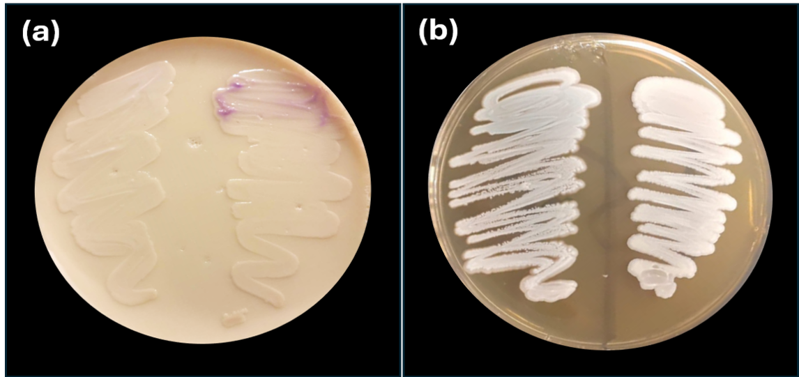
Phenotypic characteristics of Candida auris. (a) Colony appearance on chromogenic media, color ranging from pale or white to lavender. (b) Robust growth at 42°C, demonstrating C. auris thermotolerance.
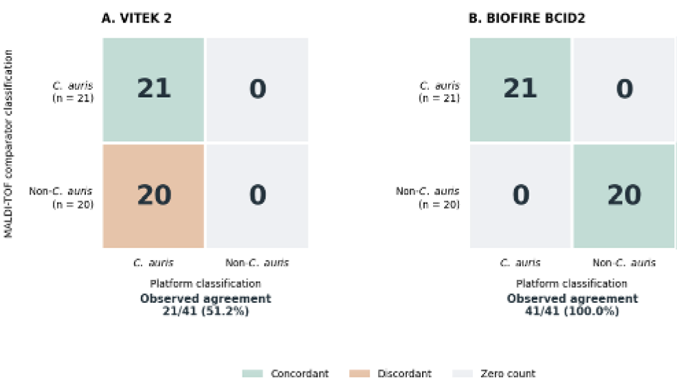
Agreement of VITEK 2 and BIOFIRE FilmArray BCID2 with MALDI-TOF MS for Candida auris classification. Matrices represent the shared subset of 41 cultures, comprising 21 C. auris-positive cultures and 20 non-C. auris controls. Rows indicate MALDI-TOF comparator classifications and columns display classifications by (A) VITEK 2 and (B) BIOFIRE BCID2. Cell values represent culture counts. Observed agreement was calculated as the sum of diagonal counts divided by 41. The four C. famata controls were assessed for C. auris non-detection, not species-level identification, because C. famata was not a BCID2 target.
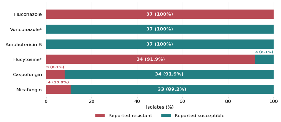
Antifungal susceptibility profiles of 37 Candida auris isolates. Stacked bars show the number and percentage of isolates in each reported susceptibility category. ᵃVoriconazole categories represent non–wild-type/wild-type under the stated derivatization method-based epidemiological cutoff values (dECOFF) criterion. ᵇFlucytosine classification uses the stated breakpoint extrapolated from the other Candida species.
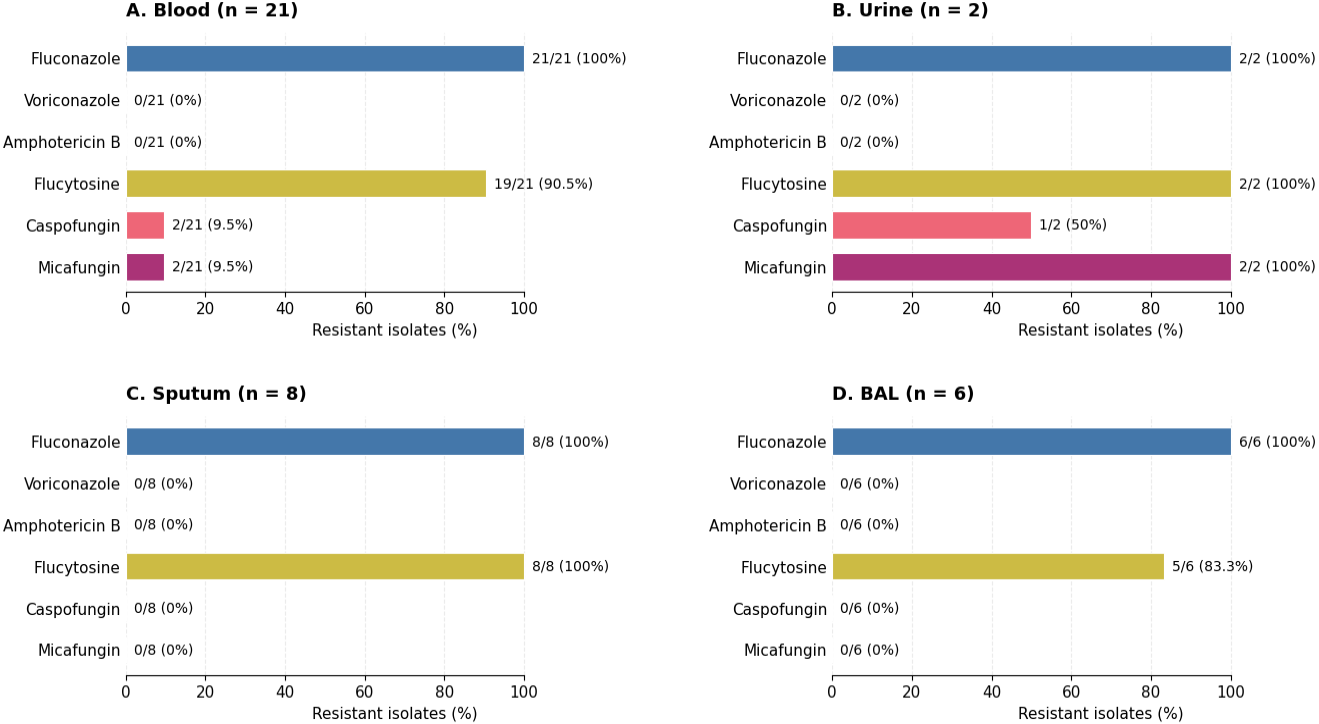
Antifungal resistance of Candida auris isolates by source. Panels represent (A) blood, (B) urine, (C) sputum, and (D) bronchoalveolar lavage (BAL). Bars indicate resistance percentages within each source, while labels show the resistant/total isolates (%).
References
- Abdolrasouli, A. and Fraser, M.A., 2022. Candida auris identification and profiling by MALDI-ToF mass spectrometry. Methods Mol. Biol., 2517: 1-18. https://doi.org/10.1007/978-1-0716-2417-3_2[CrossRef]
- Ahmad, S. and Alfouzan, W., 2021. Candida auris: Epidemiology, diagnosis, pathogenesis, antifungal susceptibility, and infection control measures to combat the spread of infections in healthcare facilities. Microorganisms, 9(4): 807. https://doi.org/10.3390/microorganisms9040807[CrossRef]
- Ahmad, S. and Asadzadeh, M., 2023. Strategies to prevent transmission of Candida auris in healthcare settings. Curr. Fungal Infect. Rep., 17(1): 36-48. https://doi.org/10.1007/s12281-023-00451-7[CrossRef]
- Ahmed, F., El-Kholy, I.M., Abdou, D.A.M., El-Mehalawy, A.A. and Elkady, N.A., 2025. Epidemiological and microbiological characterization of Candidozyma auris (Candida auris) Isolates from a Tertiary Hospital in Cairo, Egypt: An 18-Month Study. Sci. Rep., 15(1): 32427. https://doi.org/10.1038/s41598-025-15302-3[CrossRef]
- Al-Balushi, A., Khamis, F., Klaassen, C.H.W., Gangneux, J.P., Van Hellemond, J.J. and Petersen, E., 2018. Double infection with Leishmania tropica and L. major in an HIV patient controlled with high doses of amphotericin B. Open Forum Infect. Dis., 5(12): ofy323. https://doi.org/10.1093/ofid/ofy323[CrossRef]
- Altun, O., Almuhayawi, M., Ullberg, M. and Ozenci, V., 2013. Clinical evaluation of the FilmArray blood culture identification panel in identification of bacteria and yeasts from positive blood culture bottles. J. Clin. Microbiol., 51(12): 4130-4136. https://doi.org/10.1128/JCM.01835-13[CrossRef]
- Ambaraghassi, G., Dufresne, P.J., Dufresne, S.F., Vallières, É., Muñoz, J.F., Cuomo, C.A., Berkow, E.L., Lockhart, S.R. and Luong, M.L., 2019. Identification of Candida auris by use of the updated Vitek 2 yeast identification system, version 8.01: A multilaboratory evaluation study. J. Clin. Microbiol., 57(11): e00884-19. https://doi.org/10.1128/JCM.00884-19[CrossRef]
- Arendrup, M.C., Prakash, A., Meletiadis, J., Sharma, C. and Chowdhary, A., 2017. Comparison of EUCAST and CLSI reference microdilution mics of eight antifungal compounds for Candida auris and associated tentative epidemiological cutoff values. Antimicrob. Agents Chemother., 61(6): e00485-17. https://doi.org/10.1128/AAC.00485-17[CrossRef]
- Aygar, İ.S. and Hoşbul, T., 2025. Diagnostic accuracy and clinical impact of FilmArray multiplex PCR system in bloodstream infections: A comparative study with conventional methods in a tertiary health care setting. Medicine, 104(29): e43263. https://doi.org/10.1097/MD.0000000000043263[CrossRef]
- Balla, N., Kovács, F., Balázs, B., Borman, A.M., Bozó, A., Jakab, Á., Tóth, Z., Kobaissi, O., Majoros, L. and Kovács, R., 2022. Synergistic interaction of caspofungin combined with posaconazole against FKS wild-type and mutant Candida auris planktonic cells and biofilms. Antibiotics, 11(11): 1601. https://doi.org/10.3390/antibiotics11111601[CrossRef]
- Bassetti, M., Vena, A., Meroi, M., Cardozo, C., Cuervo, G., Giacobbe, D. R., Salavert, M., Merino, P., Gioia, F., Fernández-Ruiz, M., López-Cortés, L. E., Almirante, B., Escolà-Vergé, L., Montejo, M., Aguilar-Guisado, M., Puerta-Alcalde, P., Tasias, M., Ruiz-Gaitán, A., González, F., Puig-Asensio, M. and Muñoz, P. 2020. Factors associated with the development of septic shock in patients with candidemia: A post hoc analysis from two prospective cohorts. Crit. Care, 24: 117. https://doi.org/10.1186/s13054-020-2793-y[CrossRef]
- Bergamasco, M.D., Pereira, C.A.P., Arrais-Rodrigues, C., Ferreira, D.B., Baiocchi, O., Kerbauy, F., Nucci, M. and Colombo, A.L., 2021. Epidemiology of invasive fungal diseases in patients with hematologic malignancies and hematopoietic cell transplantation recipients managed with an antifungal diagnostic driven approach. J. Fungi, 7(8): 588. https://doi.org/10.3390/jof7080588[CrossRef]
- Bhargava, A., Klamer, K., Sharma, M., Ortiz, D. and Saravolatz, L., 2025. Candida auris: A continuing threat. Microorganisms, 13(3): 652. https://doi.org/10.3390/microorganisms13030652[CrossRef]
- Biagi, M.J., Wiederhold, N.P., Gibas, C., Wickes, B.L., Lozano, V., Bleasdale, S.C. and Danziger, L., 2019. Development of high-level echinocandin resistance in a patient with recurrent Candida auris Candidemia secondary to chronic candiduria. Open Forum Infect. Dis., 6(7): ofz262. https://doi.org/10.1093/ofid/ofz262[CrossRef]
- Bing, J., Huang, Y., Du, H., Guo, P., Cao, J., Kang, M., Nobile, C.J. and Huang, G., 2025. Rapid spread of Candida auris in China after COVID-19. J. Infect., 90(4): 106476. https://doi.org/10.1016/j.jinf.2025.106476[CrossRef]
- Bing, J., Li, S., Ji, L., Du, H., Shamoon, N.M., Nobile, C.J. and Huang, G., 2026. Global emergence and rapid spread of Candidozyma auris (syn. Candida auris): Epidemiology, biology, and antifungal resistance. Clin. Microbiol. Rev., 39: e00394-25. https://doi.org/10.1128/cmr.00394-25[CrossRef]
- Bohner, F., Papp, C., Takacs, T., Varga, M., Szekeres, A., Nosanchuk, J.D., Vágvölgyi, C., Tóth, R. and Gacser, A., 2023. Acquired triazole resistance alters pathogenicity-associated features in Candida auris in an isolate-dependent manner. J. Fungi, 9(12): 1148. https://doi.org/10.3390/jof9121148[CrossRef]
- Borman, A.M., Szekely, A. and Johnson, E.M., 2016. Comparative pathogenicity of United Kingdom isolates of the emerging pathogen Candida auris and other key pathogenic Candida Species. mSphere, 1(4): e00189-16. https://doi.org/10.1128/mSphere.00189-16[CrossRef]
- Bruins, M.J., Bloembergen, P., Ruijs, G.J. and Wolfhagen, M.J., 2004. Identification and susceptibility testing of enterobacteriaceae and Pseudomonas aeruginosa by direct inoculation from positive BACTEC blood culture bottles into Vitek 2. J. Clin. Microbiol., 42(1): 7-11. https://doi.org/10.1128/JCM.42.1.7-11.2004[CrossRef]
- Burrack, L.S., Todd, R.T., Soisangwan, N., Wiederhold, N.P. and Selmecki, A., 2022. Genomic diversity across Candida auris clinical isolates shapes rapid development of antifungal resistance in vitro and in vivo. mBio, 13(3): e00842-22. https://doi.org/10.1128/mbio.00842-22[CrossRef]
- Caceres, D.H., Forsberg, K., Welsh, R.M., Sexton, D.J., Lockhart, S.R., Jackson, B.R. and Chiller, T., 2019. Candida auris: A review of recommendations for detection and control in healthcare settings. J. Fungi, 5(4): 111. https://doi.org/10.3390/jof5040111[CrossRef]
- Caméléna, F., Péan de Ponfilly, G., Pailhoriès, H., Bonzon, L., Alanio, A., Poncin, T., Lafaurie, M., Dépret, F., Cambau, E., Godreuil, S., Chenouard, R., Le Monnier, A., Jacquier, H. and Berçot, B., 2023. Multicenter evaluation of the FilmArray blood culture identification 2 panel for pathogen detection in bloodstream infections. Microbiol. Spectr., 11(1): e02547-22. https://doi.org/10.1128/spectrum.02547-22[CrossRef]
- Casadevall, A., Kontoyiannis, D.P. and Robert, V., 2019. On the emergence of Candida auris: Climate change, Azoles, Swamps, and Birds. mBio, 10(4): e01397-19. https://doi.org/10.1128/mBio.01397-19[CrossRef]
- Centers for Disease Control and Prevention (CDC), 2024a. Antifungal susceptibility testing for Candida auris. Available at: https://www.cdc.gov/candida-auris/hcp/laboratories/antifungal-susceptibility-testing.html.
- Centers for Disease Control and Prevention (CDC), 2024b. Clinical treatment of Candida auris infections. Available at: https://www.cdc.gov/candida-auris/hcp/clinical-care/index.html
- Centers for Disease Control and Prevention (CDC), 2024c. Detection of colonization. Available at: https://www.cdc.gov/candida-auris/hcp/laboratories/detection-colonization.html
- Centers for Disease Control and Prevention (CDC), 2026. Identification of Candida auris. Available at: https://www.cdc.gov/candida-auris/hcp/laboratories/identification-of-c-auris.html.
- Chen, S.C., Slavin, M.A. and Sorrell, T.C., 2011. Echinocandin antifungal drugs in fungal infections: A comparison. Drugs, 71(1): 11–41. https://doi.org/10.2165/11585270-000000000-00000[CrossRef]
- Chowdhary, A., Lionakis, M.S. and Chauhan, N., 2026. Candida auris: Host interactions, antifungal drug resistance, and diagnostics. Microbiol. Mol. Biol. Rev., 90(1): e00187-22. https://doi.org/10.1128/mmbr.00187-22[CrossRef]
- Chowdhary, A., Prakash, A., Sharma, C., Kordalewska, M., Kumar, A., Sarma, S., Tarai, B., Singh, A., Upadhyaya, G., Upadhyay, S., Yadav, P., Singh, P.K., Khillan, V., Sachdeva, N., Perlin, D.S. and Meis, J.F., 2018. A multicentre study of antifungal susceptibility patterns among 350 Candida auris isolates (2009-17) in India: Role of the ERG11 and FKS1 genes in azole and echinocandin resistance. J. Antimicrob. Chemother., 73(4): 891–899. https://doi.org/10.1093/jac/dkx480[CrossRef]
- Chowdhary, A., Tarai, B., Sharma, C. and Kumar, D., 2020. Multidrug-resistant Candida auris infections in critically Ill coronavirus disease patients, India, April-July 2020. Emerg. Infect. Dis., 26(11): 2694-2696. https://doi.org/10.3201/eid2611.203504[CrossRef]
- Clinical and Laboratory Standards Institute (CLSI), 2017. M27: Reference method for broth dilution antifungal susceptibility testing of yeasts. Clinical and Laboratory Standards Institute. Available at: https://clsi.org/shop/standards/m27/.
- Cortegiani, A., Misseri, G., Fasciana, T., Giammanco, A., Giarratano, A. and Chowdhary, A., 2018. Epidemiology, clinical characteristics, resistance, and treatment of infections by Candida auris. J. Intens. Care, 6: 69. https://doi.org/10.1186/s40560-018-0342-4[CrossRef]
- Deshkar, S., Patil, N., Amberkar, S., Lad, A., Siddiqui, F. and Sharan, S., 2022. Identification and antifungal drug susceptibility pattern of Candida auris in India. J. Glob. Infect. Dis., 14(4): 131–135. https://doi.org/10.4103/jgid.jgid_44_22[CrossRef]
- Di Luca, M., Koliszak, A., Karbysheva, S., Chowdhary, A., Meis, J.F. and Trampuz, A., 2019. Thermogenic characterization and antifungal susceptibility of Candida auris by microcalorimetry. J. Fungi, 5(4): 103. https://doi.org/10.3390/jof5040103[CrossRef]
- Ding, C.H., Situ, S.F., Steven, A. and Razak, M.F.A., 2019. The pitfall of utilizing a commercial biochemical yeast identification kit to detect Candida auris. Ann. Clin. Lab. Sci., 49(4): 546–549.
- El-Sherif, H.M., Elsayed, M., El-Ansary, M.R., Aboshanab, K.M., El-Borhamy, M.I. and Elsayed, K.M., 2022. BioFire FilmArray BCID2 versus VITEK-2 system in determining microbial etiology and antibiotic-resistant genes of pathogens recovered from central line-associated bloodstream infections. Biology, 11(11): 1573. https://doi.org/10.3390/biology11111573[CrossRef]
- El-Zakhem, A., Mahmoud, O., Zakhour, J., Nahhal, S.B., El Ghawi, N., Omran, N., El-Sheikh, W.G., Tamim, H. and Kanj, S.S., 2023. The impact of COVID-19 on the epidemiology and outcomes of candidemia: A retrospective study from a tertiary care center in Lebanon. J. Fungi., 9(7): 769. https://doi.org/10.3390/jof9070769[CrossRef]
- Erturk Sengel, B., Ekren, B.Y., Sayin, E., Tukenmez Tigen, E., Seydaliyeva, A., Cerikcioglu, N., Cinel, I., Korten, V., Sezerman, U. and Odabasi, Z., 2023. Nosocomial infection of C. auris in COVID-19 intensive care unit in Türkiye and phylogenetic analysis of isolates. Mycopathologia, 188(5): 765-773. https://doi.org/10.1007/s11046-023-00782-6[CrossRef]
- Eyre, D.W., Sheppard, A.E., Madder, H., Moir, I., Moroney, R., Quan, T.P. and Jeffery, K.J., 2018. A Candida auris outbreak and its control in an intensive care setting. N. Engl. J. Med., 379(14): 1322-1331. https://doi.org/10.1056/NEJMoa1714373[CrossRef]
- Gamal, D.M., Ibrahim, R.A. and Samaan, S.F., 2022. Post COVID-19 syndrome in a prospective cohort study of Egyptian patients. Egypt. Rheumatol. Rehabil., 49: 12. https://doi.org/10.1186/s43166-021-00104-y[CrossRef]
- Garcia-Bustos, V., Cabanero-Navalon, M.D., Ruiz-Saurí, A., Ruiz-Gaitán, A.C., Salavert, M., Tormo, M.Á. and Pemán, J., 2021. What do we know about Candida auris? State of the art, knowledge gaps, and future directions. Microorganisms, 9(10): 2177. https://doi.org/10.3390/microorganisms9102177[CrossRef]
- Ghannoum, M.A. and Kuhn, D.M., 2002. Voriconazole better chances for patients with invasive mycoses. Eur. J. Med. Res., 7(5): 242–256.
- Goravey, W., Ali, G.A., Ali, M., Ibrahim, E.B., Al-Maslamani, M. and Abdel Hadi, H., 2021. Ominous combination: COVID-19 disease and Candida auris Fungemia case report and review of the literature. Clin. Case Rep., 9(9): e04827. https://doi.org/10.1002/ccr3.4827[CrossRef]
- Ham, Y.Y., Lewis, J.S. and Thompson, G.R., 2021. Rezafungin: A novel antifungal for the treatment of invasive candidiasis. Fut. Microbiol., 16(1): 27-36. https://doi.org/10.2217/fmb-2020-0217[CrossRef]
- Hsu, C. and Yassin, M., 2025. Diagnostic approaches for Candida auris: A comprehensive review of screening, identification, and susceptibility testing. Microorganisms, 13(7): 1461. https://doi.org/10.3390/microorganisms13071461[CrossRef]
- Huma, Z.E., Saleem, S., Imran, M., Jabeen, K., Arshad, F. and Amar, A., 2023. Screening of C. auris among Candida isolates from various tertiary care institutions in Lahore by VITEK 2 and real time PCR based molecular technique. PLoS One, 18(10): e0293390. https://doi.org/10.1371/journal.pone.0293390[CrossRef]
- Ismail, H., Perovic, O., Mpembe, R., Lowman, W., Govind, C., Ekermans, P., Jassat, W., Welch, R. and Govender, N.P., 2026. Accelerated increase in Candida auris bloodstream infections during COVID-19 Pandemic, South Africa. Emerg. Infect. Dis., 32(4): 563-572. https://doi.org/10.3201/eid3204.251407[CrossRef]
- Ivanova, Z., Philipova, I., Boyanova, L. and Kantardjiev, T., 2023. Retrospective re-identification of Candida strains by MALDI-TOF in Seek of Candida auris. Probl. Infect. Parasit. Dis., 50(2): 8-12. https://doi.org/10.58395/pipd.v50i2.90[CrossRef]
- Jackson, B.R., Chow, N., Forsberg, K., Litvintseva, A.P., Lockhart, S.R., Welsh, R., Vallabhaneni, S. and Chiller, T., 2019. On the origins of a species: What might explain the rise of Candida auris? J. Fungi, 5(3): 58. https://doi.org/10.3390/jof5030058[CrossRef]
- Jamalian, A., Freeke, J., Chowdhary, A., de Hoog, G.S., Stielow, J.B. and Meis, J.F., 2023. Fast and accurate identification of Candida auris by high resolution mass spectrometry. J. Fungi, 9(2): 267. https://doi.org/10.3390/jof9020267[CrossRef]
- Jenks, J.D., Tobin, E.H. and Zahra, F., 2025. Candida auris. In: StatPearls Treasure Island (FL): StatPearls Publishing. Available at: https://www.ncbi.nlm.nih.gov/sites/books/NBK563297/.
- John, L.L.H., Thomson, D.D., Bicanic, T., Hoenigl, M., Brown, A.J.P., Harrison, T.S. and Bignell, E.M., 2023. Heightened efficacy of anidulafungin when used in combination with manogepix or 5-Flucytosine against Candida auris in vitro. Antimicrob. Agents Chemother., 67(6): e01645-22. https://doi.org/10.1128/aac.01645-22[CrossRef]
- Jordan, V., Perera, M., Unsworth, A., Basile, K., Halliday, C.L. and Chen, S.C., 2026. The challenges of Candidozyma auris: Updates on the laboratory investigation, infection prevention and management. Future Microbiol., 21(2): 217-230. https://doi.org/10.1080/17460913.2026.2634539[CrossRef]
- Kang, C.M., Chen, X.J., Chih, C.C., Hsu, C.C., Chen, P.H., Lee, T.F., Teng, L.J. and Hsueh, P.R., 2020. Rapid identification of bloodstream bacterial and fungal pathogens and their antibiotic resistance determinants from positively flagged blood cultures using the BioFire FilmArray blood culture identification panel. J. Microbiol. Immunol. Infect., 53(6): 882-891. https://doi.org/10.1016/j.jmii.2020.03.018[CrossRef]
- Kazancioglu, S., Bodur, H., Mumcuoglu, I., Bastug, A., Ozbay, B.O., Aydos, O. and Dinc, B., 2024. Candidemia in Critically Ill COVID-19 patients: Risk factors and impact on mortality. Heliyon, 10(6): e28033. https://doi.org/10.1016/j.heliyon.2024.e28033[CrossRef]
- Khairat, S.M., Anany, M.G., Ashmawy, M.M. and Hussein, A.F.A., 2021. Setting a protocol for identification and detecting the prevalence of Candida auris in Tertiary Egyptian Hospitals using the CDC steps. Open Access Maced. J. Med. Sci., 9(A): 397-402. https://doi.org/10.3889/oamjms.2021.6095[CrossRef]
- Kilburn, S., Innes, G., Quinn, M., Southwick, K., Ostrowsky, B., Greenko, J.A., Magleby, R., Chaturvedi, V. and Chaturvedi, S., 2022. Antifungal resistance trends of Candida auris clinical isolates in New York and New Jersey from 2016 to 2020. Antimicrob. Agents Chemother., 66(8): e02242-21. https://doi.org/10.1128/aac.02242-21[CrossRef]
- Kim, H.Y., Nguyen, T.A., Kidd, S., Chambers, J., Alastruey-Izquierdo, A., Shin, J.H., Dao, A., Forastiero, A., Wahyuningsih, R., Chakrabarti, A., Beyer, P., Gigante, V., Beardsley, J., Sati, H., Morrissey, C.O. and Alffenaar, J.W., 2024. Candida auris a systematic review to inform the world health organization fungal priority pathogens list. Med. Mycol., 62(6): myae042. https://doi.org/10.1093/mmy/myae042[CrossRef]
- Kwon, Y.J., Shin, J.H., Byun, S.A., Choi, M.J., Won, E.J., Lee, D., Lee, S.Y., Chun, S., Lee, J.H., Choi, H.J., Kee, S.J., Kim, S.H. and Shin, M.G., 2019. Candida auris clinical isolates from South Korea: Identification, antifungal susceptibility, and genotyping. J. Clin. Microbiol., 57(4): e01624-18. https://doi.org/10.1128/JCM.01624-18[CrossRef]
- Larkin, E., Hager, C., Chandra, J., Mukherjee, P.K., Retuerto, M., Salem, I., Long, L., Isham, N., Kovanda, L., Borroto-Esoda, K., Wring, S., Angulo, D. and Ghannoum, M., 2017. The emerging pathogen Candida auris: Growth phenotype, virulence factors, activity of antifungals, and effect of SCY-078, a novel glucan synthesis inhibitor, on growth morphology and biofilm formation. Antimicrob. Agents Chemother., 61(5): e02396-16. https://doi.org/10.1128/AAC.02396-16[CrossRef]
- Lepak, A.J., Zhao, M., Berkow, E.L., Lockhart, S.R. and Andes, D.R., 2017. Pharmacodynamic optimization for treatment of invasive Candida auris infection. Antimicrob. Agents Chemother., 61(8): e00791-17. https://doi.org/10.1128/AAC.00791-17[CrossRef]
- Li, H., Miao, M.X., Jia, C.L., Cao, Y.B., Yan, T.H., Jiang, Y.Y. and Yang, F., 2022. Interactions between Candida albicans and the resident microbiota. Front. Microbiol., 13: 930495. https://doi.org/10.3389/fmicb.2022.930495[CrossRef]
- Lockhart, S.R., 2019. Candida auris and multidrug resistance: Defining the new normal. Fungal Genet. Biol., 131: 103243. https://doi.org/10.1016/j.fgb.2019.103243[CrossRef]
- Lockhart, S.R., Etienne, K.A., Vallabhaneni, S., Farooqi, J., Chowdhary, A., Govender, N.P., Colombo, A.L., Calvo, B., Cuomo, C.A., Desjardins, C.A., Berkow, E.L., Castanheira, M., Magobo, R.E., Jabeen, K., Asghar, R.J., Meis, J.F., Jackson, B., Chiller, T. and Litvintseva, A.P., 2017. Simultaneous emergence of multidrug-resistant Candida auris on 3 continents confirmed by whole-genome sequencing and epidemiological analyses. Clin. Infect. Dis., 64(2): 134-140. https://doi.org/10.1093/cid/ciw691[CrossRef]
- Lockhart, S.R., Lyman, M.M. and Sexton, D.J., 2022. Tools for detecting a “Superbug”: Updates on Candida auris testing. J. Clin. Microbiol., 60(5): e00808-21. https://doi.org/10.1128/jcm.00808-21[CrossRef]
- Maphanga, T.G., Naicker, S.D., Kwenda, S., Munoz, J.F., van Schalkwyk, E., Wadula, J., Nana, T., Ismail, A., Coetzee, J., Govind, C., Mtshali, P.S., Mpembe, R.S. and Govender, N.P., 2021. In-vitro antifungal resistance of Candida auris isolates from bloodstream infections, South Africa. Antimicrob. Agents Chemother., 65(5): e00517-21. https://doi.org/10.1128/AAC.00517-21[CrossRef]
- Melinte, V., Tudor, A.D., Bujoi, A.G., Radu, M.-A., Văcăriou, M.C., Cismaru, I.M., Holban, T.S., Mîrzan, C.L., Popescu, R., Ciupan, R.C., Baciu, A., Moraru, O.E., Popa-Cherecheanu, M. and Gheorghiță, V., 2024. Candida auris outbreak in a multidisciplinary hospital in romania during the post-pandemic era: Potential solutions and challenges in surveillance and epidemiological control. Antibiotics, 13(4): 325. https://doi.org/10.3390/antibiotics13040325[CrossRef]
- Mina, S., Yaakoub, H., Annweiler, C., Dubée, V. and Papon, N., 2022. COVID-19 and fungal infections: A double debacle. Microbes Infect., 24(8): 105039. https://doi.org/10.1016/j.micinf.2022.105039[CrossRef]
- Mirzaei, R., Goodarzi, P., Asadi, M., Soltani, A., Aljanabi, H.A., Jeda, A.S., Dashtbin, S., Jalalifar, S., Mohammadzadeh, R., Teimoori, A., Tari, K., Salari, M., Ghiasvand, S., Kazemi, S., Yousefimashouf, R., Keyvani, H. and Karampoor, S., 2020. Bacterial co-infections with SARS-CoV-2. IUBMB Life, 72(10): 2097-2111. https://doi.org/10.1002/iub.2356[CrossRef]
- Ninan, M.M., Sahni, R.D., Chacko, B., Balaji, V. and Michael, J.S., 2020. Candida auris: Clinical profile, diagnostic challenge and susceptibility pattern experience from a tertiary-care centre in South India. J. Glob. Antimicrob. Resist., 21: 181-185. https://doi.org/10.1016/j.jgar.2019.10.018[CrossRef]
- Nucci, M., Barreiros, G., Guimarães, L.F., Deriquehem, V.A., Castiñeiras, A.C. and Nouér, S.A., 2021. Increased incidence of candidemia in a tertiary care hospital with the covid-19 pandemic. Mycoses, 64(2): 152-156. https://doi.org/10.1111/myc.13225[CrossRef]
- O’Brien, B., Chaturvedi, S. and Chaturvedi, V., 2020a. In vitro evaluation of antifungal drug combinations against multidrug-resistant Candida auris isolates from New York outbreak. Antimicrob. Agents Chemother., 64(4): e02195-19. https://doi.org/10.1128/AAC.02195-19[CrossRef]
- O’Brien, B., Liang, J., Chaturvedi, S., Jacobs, J.L. and Chaturvedi, V., 2020b. Pan-resistant Candida auris: New York Subcluster susceptible to antifungal combinations. Lancet Microbe, 1(5): e193-e194. https://doi.org/10.1016/S2666-5247(20)30090-2[CrossRef]
- Osaigbovo, I.I., Ekeng, B.E., Davies, A.A., Ebeigbe E., Bongomin, F., Kanyua, A., Revathi G., Oladele, R.O., 2024. Candida auris: A systematic review of a globally emerging fungal pathogen in Africa. Open Forum Infect. Dis., 11(6): ofad681. https://doi.org/10.1093/ofid/ofad681[CrossRef]
- Osman, M., Al-Bikai, A. and Rafei, R., 2020. Update on invasive fungal infections in the Middle Eastern and North African region. Braz. J. Microbiol., 51: 1771–1789. https://doi.org/10.1007/s42770-020-00325-x[CrossRef]
- Pappas, P.G., Kauffman, C.A., Andes, D.R., Clancy, C.J., Marr, K.A., Ostrosky-Zeichner, L., Reboli, A.C., Schuster, M.G., Vazquez, J.A., Walsh, T.J., Zaoutis, T.E. and Sobel, J.D., 2016. Clinical practice guideline for the management of candidiasis: 2016 update by the infectious diseases society of America. Clin. Infect. Dis., 62(4): e1-e50. https://doi.org/10.1093/cid/civ933[CrossRef]
- Park, B., Oh, E.H., Won, E.J., Kang, J., Jin, D., Yoo, C., Park, J., Sung, H. and Kim, M.N., 2025. Detection of clinically relevant Candida species from positive blood cultures using a novel sample-to-answer molecular assay. Sci. Rep., 15: 5417. https://doi.org/10.1038/s41598-025-90090-4[CrossRef]
- Parnell, L.A., Ribeiro Dos Santos, A., Forsberg, K., Lyman, M., Misas, E., Gade, L., Sexton, D.J., Litvintseva, A.P. and Chow, N.A., 2026. Updated genomic epidemiologic description of Candida (Candidozyma) auris, United States. Emerg. Infect. Dis., 32(5): 728-739. https://doi.org/10.3201/eid3205.250760[CrossRef]
- Patwardhan, S.A., Prayag, P.S., Soman, R.N., Purandare, B.D., Ramya, S., Dawra, R., Joshi, R. and Prayag, A.P., 2024. Candida auris comparison of sensititre yeast one and Vitek 2 AST systems for antifungal susceptibility testing a single centre experience. Indian J. Med. Microbiol., 50: 100618. https://doi.org/10.1016/j.ijmmb.2024.100618[CrossRef]
- Perlin D.S., 2011. Current perspectives on echinocandin class drugs. Future Microbiol., 6(4): 441–457. https://doi.org/10.2217/fmb.11.19[CrossRef]
- Posteraro, B., Menchinelli, G., Ivagnes, V., Cortazzo, V., Liotti, F.M., Falasca, B., Fiori, B., D’Inzeo, T., Spanu, T., De Angelis, G. and Sanguinetti, M., 2023. Efficient recovery of Candida auris and five other medically important Candida species from blood cultures containing clinically relevant concentrations of antifungal agents. Microbiol. Spectr., 11(2): e04104-22. https://doi.org/10.1128/spectrum.04104-22[CrossRef]
- Prestel, C., Anderson, E., Forsberg, K., Lyman, M., de Perio, M.A., Kuhar, D., Edwards, K., Rivera, M., Shugart, A., Walters, M. and Dotson, N.Q., 2021. Candida auris outbreak in a COVID-19 specialty care unit Florida. MMWR., 70(2): 56–57. https://doi.org/10.15585/mmwr.mm7002e3[CrossRef]
- Puumala, E., Adler, N.H., Deml, S.M. and Wengenack, N.L., 2025. A retrospective evaluation of the BIOFIRE FilmArray blood culture identification 2 panel for the detection of pathogenic yeasts. Microbiol. Spectr., 13(12): e02258-25. https://doi.org/10.1128/spectrum.02258-25[CrossRef]
- Rahman, M.A., Victoros, E., Shanjana, Y., Thomas, M.R. and Islam, M.R., 2024. The Candida auris infection after the COVID-19 pandemic seems to be an urgent public health emergency: A call to attention. Health Sci. Rep., 7(11): e70160. https://doi.org/10.1002/hsr2.70160[CrossRef]
- Ravichandran, S., Shanmugam, P. and Thayikkannu, A.B., 2022. Phenotypic and genotypic characterization of Candida auris, an emerging pathogen isolated from blood. J. Commun. Dis., 54(4): 54-61. https://doi.org/10.24321/0019.5138.2022103[CrossRef]
- Rhoads, D.D., Pournaras, S., Leber, A., Balada-Llasat, J.-M., Harrington, A., Sambri, V., She, R., Berry, G.J., Daly, J., Good, C., Tarpatzi, A., Everhart, K., Henry, T., McKinley, K., Zannoli, S., Pak, P., Zhang, F., Barr, R., Holmberg, K., Kensinger, B. and Lu, D.Y., 2023. Multicenter evaluation of the BIOFIRE blood culture identification 2 panel for detection of bacteria, yeasts, and antimicrobial resistance genes in positive blood culture samples. J. Clin. Microbiol., 61(6): e01891-22. https://doi.org/10.1128/jcm.01891-22[CrossRef]
- Rossato, L. and Colombo, A.L., 2018. Candida auris: What have we learned about its mechanisms of pathogenicity? Front. Microbiol., 9: 3081. https://doi.org/10.3389/fmicb.2018.03081[CrossRef]
- Rybak, J.M., Fortwendel, J.R. and Rogers, P.D., 2019. Emerging threat of triazole-resistant Aspergillus fumigatus. J. Antimicrob. Chemother., 74(4): 835–842. https://doi.org/10.1093/jac/dky517[CrossRef]
- Salehi, M., Ahmadikia, K., Mahmoudi, S., Kalantari, S., Jamalimoghadamsiahkali, S., Izadi, A., Kord, M., Dehghan, M.S.A., Seifi, A., Ghiasvand, F., Khajavirad, N., Ebrahimi, S., Koohfar, A., Boekhout, T. and Khodavaisy, S., 2020. Oropharyngeal candidiasis in hospitalised COVID-19 patients from Iran: Species identification and antifungal susceptibility pattern. Mycoses, 63(8): 771–778. https://doi.org/10.1111/myc.13137[CrossRef]
- Salimnia, H., Fairfax, M.R., Lephart, P.R., Schreckenberger, P., DesJarlais, S.M., Johnson, J.K., Robinson, G., Carroll, K.C., Greer, A., Morgan, M., Chan, R., Loeffelholz, M., Valencia-Shelton, F., Jenkins, S., Schuetz, A.N., Daly, J.A., Barney, T., Hemmert, A. and Kanack, K.J., 2016. Evaluation of the FilmArray blood culture identification panel: Results of a multicenter controlled trial. J. Clin. Microbiol., 54(3): 687-698. https://doi.org/10.1128/JCM.01679-15[CrossRef]
- Salmanton-García, J., Sprute, R., Stemler, J., Bartoletti, M., Dupont, D., Valerio, M., Garcia-Vidal, C., Falces-Romero, I., Maisel, C., Jacob, A., Reinhold, I., Meintker, L., Prenzel, F., Stecher, M. and Cornely, O.A., 2021. COVID-19-associated pulmonary aspergillosis. Emerg. Infect. Dis., 27(4): 1077-1086. https://doi.org/10.3201/eid2704.204895[CrossRef]
- Schaefer, S., Walits, E., Thaler, K. and Patel, G., 2024. Impact of the COVID-19 Pandemic on Candida auris infections: A retrospective analysis in an academic medical center in New York City. Open Forum Infect. Dis., 11(6): 148. https://doi.org/10.1093/ofid/ofae148[CrossRef]
- Semenya, M.D., Aladejana, A.E. and Ndlovu, S.I., 2025. Characterization of susceptibility patterns and adaptability of the newly emerged Candida auris. Int. Microbiol., 28(3): 575-587. https://doi.org/10.1007/s10123-024-00563-1[CrossRef]
- Senok, A., Dabal, L.A., Alfaresi, M., Habous, M., Celiloglu, H., Bashiri, S., Almaazmi, N., Ahmed, H., Mohmed, A.A., Bahaaldin, O., Elimam, M.A.E., Rizvi, I.H., Olowoyeye, V., Powell, M. and Salama, B., 2023. Clinical impact of the BIOFIRE blood culture identification 2 panel in adult patients with bloodstream infection: A multicentre observational study in the United Arab Emirates. Diagnostics, 13(14): 2433. https://doi.org/10.3390/diagnostics13142433[CrossRef]
- Spruijtenburg, B., Melchior do Prado, C., Kempen, M.V., Shawky, S.M., Meis, J.F., Aparecida Vicente, V., Queiroz-Telles, F., Groot, T., El-Kholy, M.A. and Meijer, E.F.J., 2025. Genetic epidemiology and resistance investigations of clinical yeasts in Alexandria, Egypt. Pathogens, 14(5): 486. https://doi.org/10.3390/pathogens14050486[CrossRef]
- Tan, Y.E., Teo, J.Q., Rahman, N.B.A., Ng, O.T., Kalisvar, M., Tan, A.L., Koh, T.H. and Ong, R.T.H., 2019. Candida auris in Singapore: Genomic epidemiology, antifungal drug resistance, and identification using the updated 8.01 VITEK®2 system. Int. J. Antimicrob. Agents, 54(6): 709-715. https://doi.org/10.1016/j.ijantimicag.2019.09.016[CrossRef]
- Timsit, J.F., Ruppé, E., Barbier, F., Tabah, A. and Bassetti, M., 2020. Bloodstream infections in critically ill patients: An expert statement. ICM., 46(2): 266-284. https://doi.org/10.1007/s00134-020-05950-6[CrossRef]
- Tóth, Z., Forgács, L., Locke, J.B., Kardos, G., Nagy, F., Kovács, R., Szekely, A., Borman, A.M. and Majoros, L., 2019. In vitro activity of rezafungin against common and rare Candida species and Saccharomyces cerevisiae. J. Antimicrob. Chemother., 74(12): 3505-3510. https://doi.org/10.1093/jac/dkz390[CrossRef]
- Ulrich, P.S., Bastian, I.N. and Chen, D.J., 2022. Clinical significance of BD Bactec FX blood culture incubation beyond 96 hours (4 days). J. Clin. Microbiol., 60(7): e00549-22. https://doi.org/10.1128/jcm.00549-22[CrossRef]
- Vazquez, J.A., Pappas, P.G., Boffard, K., Paruk, F., Bien, P.A., Tawadrous, M., Ople, E., Wedel, P., Oborska, I. and Hodges, M.R., 2023. Clinical efficacy and safety of a novel antifungal, fosmanogepix, in patients with candidemia caused by Candida auris: Results from a Phase 2 Trial. Antimicrob. Agents Chemother., 67(5): e01419-22. https://doi.org/10.1128/aac.01419-22[CrossRef]
- Wang, S., Pan, J., Gu, L., Wang, W., Wei, B., Zhang, H., Chen, J. and Wang, H., 2024. Review of treatment options for a multidrug-resistant fungus: Candida auris. Med. Mycol., 62(1): myad127. https://doi.org/10.1093/mmy/myad127[CrossRef]
- Watkins, R.R., Gowen, R., Lionakis, M.S. and Ghannoum, M., 2022. Update on the pathogenesis, virulence, and treatment of Candida auris. Pathog. Immun., 7(2): 46-65. https://doi.org/10.20411/pai.v7i2.535[CrossRef]
- Widyasari, K., Lee, S., Cho, O.H., Hong, S.I., Ryu, B.H. and Kim, S., 2023. The significance of FilmArray blood culture identification panel (FA-BCID) for managing patients with positive blood cultures. Diagnostics, 13(21): 3335. https://doi.org/10.3390/diagnostics13213335[CrossRef]
- Yadav, A., Singh, A., Wang, Y., van Haren, M.H., Singh, A., de Groot, T., Meis, J. F., Xu, J. and Chowdhary, A., 2021. Colonisation and transmission dynamics of Candida auris among chronic respiratory diseases patients hospitalised in a Chest Hospital, Delhi, India: A comparative analysis of whole genome sequencing and microsatellite typing. J. Fungi, 7(2): 81. https://doi.org/10.3390/jof7020081[CrossRef]
- Yassin, H. and Abo Elyazeed, E.R., 2023. Prediction of the morbidity and mortality rates of COVID-19 in Egypt using non-extensive statistics. Sci. Rep., 13(1): 10056. https://doi.org/10.1038/s41598-023-36959-8[CrossRef]
- Zhang, S.X., Carroll, K.C., Lewis, S., Totten, M., Mead, P., Samuel, L., Steed, L.L., Nolte, F.S., Thornberg, A., Reid, J.L., Whitfield, N.N. and Babady, N.E., 2020. Multicenter evaluation of a PCR-based digital microfluidics and electrochemical detection system for the rapid identification of 15 fungal pathogens directly from positive blood cultures. J. Clin. Microbiol., 58(5): e02096-19. https://doi.org/10.1128/JCM.02096-19[CrossRef]
Services
Article usage statistics
Loading…