The results of the liver function tests revealed variations in serum liver enzyme values between treatment groups.
The results of this study approved the activities of Sc-RE to inhibit in vivo-induced hepatic and kidney cancer in mice.
Therefore, Saussurea costus may be a promising agent for inhibiting induced cancer growth due to various carcinogenic substances.
The authors suggest a future study investigating the pathway of Saussurea costus-Root extract effects as an anticancer and determining the anticancer bioactive components.
Selected from the article's own text.
Abstract
The Saussurea lappa (Sc), traditionally known as Costus, is a perennial effective root extensively used to treat various ailments. This study intends to evaluate the anticancer effect of Sc- Root Extract (Sc-RE) against cancer induced by intraperitoneal injection of Polyvinyl pyrrolidone K-30 (PVP) in mice. 72 mice divided randomly into eight groups (G1, control group; G2, Sc-RE group; G3, chemotherapy alone; G4 cancer- group without treatment, G5, cancer-induced + chemotherapy; G6, cancer induced+ Sc-RE/ low dose; G7, cancer induced+ Sc-RE/ moderate dose; G8, cancer induced+ Sc-RE/ high dose). Liver and kidney samples were collected from all mice during different experiment periods, kept in 10% neutral buffered formalin, and sent for histological analysis. No abnormal histological features were seen in liver and kidney sections from the control and other mice groups before various treatments. The various treatment groups revealed diverse histopathological changes compared to the control groups. Various histological features were also seen in low, moderate, and high-dose Sc-RE treatment groups, accompanied by regeneration related to the treatment dose. Severe damage comprising necrosis of the hepatic cells, renal tubules, congestion, and infiltration of inflammatory cells was seen in the chemotherapy-treated group and mice suffering from cancer without treatment. This study also revealed variations in serum liver enzyme values between treatment groups. The values of ALT, AST, and ALP (U/L) evaluation in control groups were ˃ 29.01±1.8, 87.55±2.9 and ˃ 98.12±8.8, respectively. In conclusion, this study investigated that the ethanolic extract of Sc-RE has an anticancer effect that reduces the proliferation of cancer cells caused by the carcinogenic substance.
Cancer is a general name for many diseases that can affect any body part . Four characteristics distinguish cancer cells from normal cells: uncontrolled proliferation, a lack of capacity to differentiate, which leads to loss of function, invasiveness, and the ability to metastasis . Cancer is a worldwide public health problem that is likely to affect almost every person on the planet, with the estimation that one in three people will develop cancer at some point during their lifetime . It is second only to heart disease as a leading cause of death in developed nations and is among the three leading causes of death for adults in developing countries .
Medicinal plants have contributed richly to the health of human beings. The vast potential of plant-based medicines in treating numerous diseases has always contributed to the value of the plant community as a major area of research and development. Medicinal plants are a storehouse of numerous bioactive compounds with various therapeutic properties. The therapeutic potential of plants has been well explored over a very long period . The vast array of therapeutic effects associated with medicinal plants includes anti-inflammatory, antiviral, antitumor, antimalarial, and analgesic .
The Saussurea costus (Family Asteraceae) is a perennial or biennial herb naturally found in sub-Himalayan regions . Sc is well-known in Islamic medicine and the Indian systems of medicine, and it is used either as a single drug or in combination with other drugs . Sc roots are used mainly as an antispasmodic in asthma and cough and also in the treatment of cholera, chronic skin diseases, rheumatism , cold, quartan malaria, leprosy, persistent hiccups, hair wash, stomachache, toothache, and typhoid fever . It is an important medicine for gout, erysipelas and promotes spermatogenesis, chronic inflammation of the lungs, and chest congestion . Several previous studies have suggested that Sc has anticancer effects in neuroblastoma , lung cancer , hepatocellular carcinoma , gastric cancer , and prostate cancer . The hexane extract of Sc induced apoptosis in androgen-insensitive human prostate cells , and MgO nanoparticles (MgONPs) were synthesized using biomasses of Sc roots against breast cancer cells . A literature review revealed scarce publications on the activities of Sc-RE for treating epithelial cell cancer. Consequently, this study intends to detect the anticancer activities of Sc-RE against experimentally induced epithelial cell cancer in white mice in vivo.
Materials and Methods
The Sc-Roots were purchased from an Iraqi herbal market and were free from defects and contaminants. The roots were dried at 40°C. The dried pieces were then ground into powder, aseptically dispensed into sterile plastic bags, and stored at −40°C. Extraction was performed using 80% ethanol at 25°C for 72 hours, as optimized in the previous study . The solvent was evaporated using a rotary vacuum evaporator (EYELA A-1000S, USA) at 40°C, and the subsequent extract was freeze-dried and stored in dark bottles at 4°C.
Seventy-two Albino Swiss mice (aged 8-10 weeks, with an average body weight of 28±1.5 g) obtained from (the Iraqi Center for Cancer and Medical Genetics Research/ Baghdad) were used in the present study. This study was approved by the Ethical and research committee/College of Science/ Al-Muthanna University (No. 404 Date 10-12- 2021). The mice were allowed to acclimate to the standard laboratory conditions for 2 weeks and had free access to tap water and a chow diet for one week. Later on, the mice were randomly divided into eight groups (nine each) as follows: (G1, control group (Received distilled water only); G2, Sc-RE group (Received Sc-80% ethyl extract/ 600 mg/Kg) as a single oral dose only.; G3, chemotherapy alone/Mice received Ebetrex (2.5mg/Kg) chemotherapy alone.; G4, cancer- group without treatment; G5, cancer-induced + chemotherapy; G6, cancer induced+ Sc-RE / low dose; G7, cancer induced+ Sc-RE/moderate dose; G8, cancer induced+ Sc-RE / high dose (Table 1).
Cancer was induced by intraperitoneal injection of PVP-K30 at a 15 mg/ kg body mice dose. After 2 weeks, all mice were fed their diets, and mild, moderate, and high doses of Sc-RE were administered for G6, G7, and G8 through gastric intubation for four weeks. After the end of each treatment period, mice were euthanized.
Table 1: Shows the eight experimental groups including 5 control groups and 3 Sc-RE treated groups.
Groups
Control groups
Type of treatment
Treated group
Type of treatment
G1
Received distilled water only.
G6
Received PVP (IPI) as a carcinogenic agent before two weeks from treatment with low dose (400 mg/Kg) of Sc 80% ethyl extract
G2
Received Sc-RE 80% ethyl extract (600 mg/Kg) as a single oral dose only.
G3
Mice received Ebetrex (2.5mg/Kg)
chemotherapy alone.
G7
Received PVP (IPI) as a carcinogenic agent before two weeks from treatment with moderate dose (600 mg/Kg) of Sc 80% ethyl extract
G4
Received PVP as a carcinogenic agent / intraperitoneal injection
G8
Received PVP (IPI) as a carcinogenic agent before two weeks from treatment with high dose (800 mg/Kg) of Sc 80% ethyl extract.
G5
Received PVP (IPI) as a carcinogenic agent before two weeks from treatment with Ebetrex (2.5mg/Kg) as chemotherapy
Blood samples were collected by cardiac puncture, kept in test tubes without anticoagulant, and centrifuged at 3000 rpm at room temperature for 10 minutes. Later on, serum was collected and stored at -40 °C until further analysis. Liver and kidney specimens were collected in 10% neutral buffered formalin.
After fixation of the specimens for 48 hours, the specimens were dehydrated using ascending grades of ethyl alcohol (50-100%), then cleared using xylene (2 changes), then embedded in melted paraffin wax (59c), then blocked and sectioned with an ordinary microtome (3-6 micrometer thickness), stained by Hematoxylin and Eosin stain and examined under a light microscope. All sections were examined in magnification (X4, X10, X20, X40). Furthermore, images were acquired by a Leica image analyzer . Serum was used to analyze the activities of liver aminotransferases (AST and ALT) and alkaline phosphatase (ALP) using Cobas C111 automated Chemistry analyzer Roche Company, Germany.
Statistical analysis
SPSS was used to examine the collected data. One-way analysis of variance (ANOVA) was used to compare the samples and controls statistically, and Tukey’s post-test analysis was used to obtain statistical differences between the samples and the controls. Results were expressed as mean ± standard deviation (SD). A difference in the mean values with p < 0.05 was considered statistically significant .
Results and Discussion
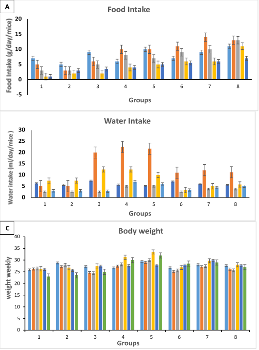
The study’s results revealed no significant differences in food and water intake among the mice (Figure 1A, B and C). Moreover, the cumulative tumor incidence was, on average and revealed 42% lower in the calorie-restricted groups. Multivariate regression analyses revealed that, regardless of the level of dietary fat, tumor incidence increased with increased caloric intake and body weight over a wide range of intakes, including moderate caloric restriction (i.e., 7-20%).
The results of the liver function tests revealed variations in serum liver enzyme values between treatment groups. The values of ALT (U/L) evaluation were ˃ 29.01±1.8, > 37±1.1, > 360±8.7, > 175±7.5, > 313±5.4, > 53±1.1, > 46±1.5 and > 52±0.87 for G1, G2, G3, G4, G5, G6, G7 and G8, respectively. The results of AST (U/L) measurements were > 87.55±2.9, > 98.98±3.7, > 255±8.8, > 167±5.2, > 474±7.9, > 106±8.4, > 97±8.9, and > 99.4±7.6 for G1, G2, G3, G4, G5, G6, G7 and G8, respectively. While the results of ALP U/L determination were > 98.12±8.8, > 99±6.5, > 278±5.87, > 260±8.9, > 440±8.8, > 135.3±7.4, > 110.5±6.7 and > 125.2±7.6 for G1, G2, G3, G4, G5, G6, G7 and G8, respectively Table 2.
Table 2: Serum level of AST, ALT, and ALP in mice.
Groups
ALT UI/L
AST UI/L
ALP UI/L
1/ control group
29.01±1.8a
87.55±2.9a
98.12±8.8d
2/ Sc-RE group
37±1.1b
98.98±3.7b
99±6.5e
3/chemotherapy alone
360±8.7e
255±8.8f
278±5.87c
4/ cancer- group without treatment
175±7.5b
167±5.2c
260±8.9b
5/cancer-induced + chemotherapy
313±5.4f
474±7.9f
440±8.8c
6/cancer induced+
Sc-RE/ low dose
53±1.1d
106±8.4e
135.3±7.4a
7/ cancer induced+
Sc-RE/moderate dose
46±1.5c
97±8.9c
110.5±6.7b
8/cancer induced+
Sc-RE/ high dose
52±0.87c
99.4±7.6d
125.2±7.6c
Different letters indicate a significance difference (p < 0.05) between groups. Values are mean ± SD.
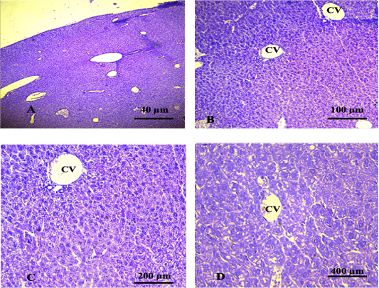
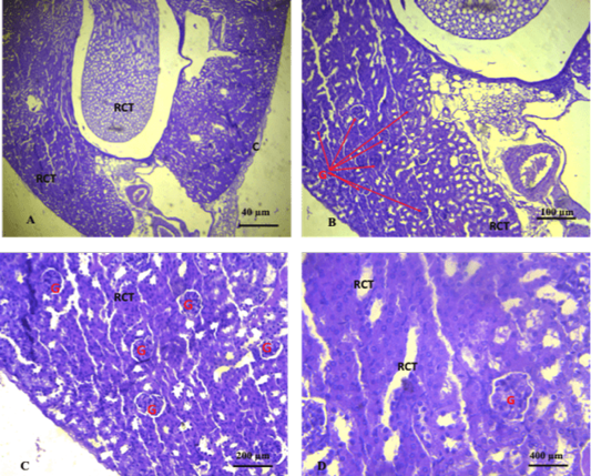
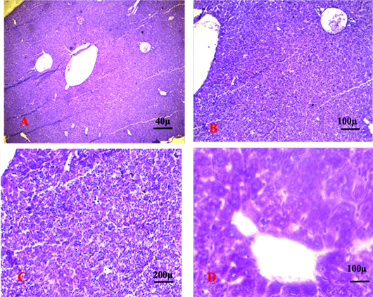
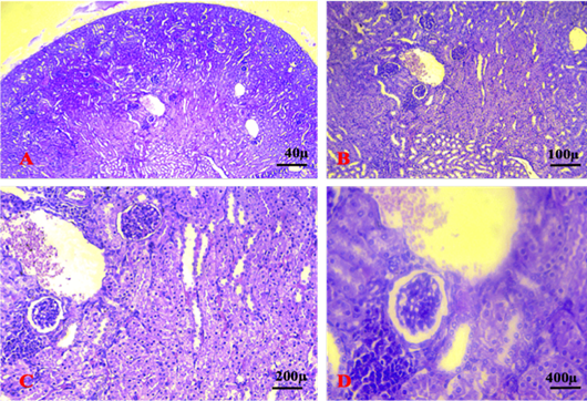
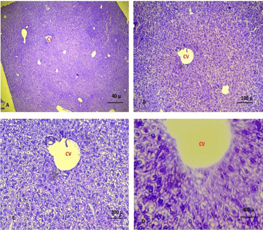
No abnormal histopathological changes were seen in the liver and kidney sections of mice in G1/ the control group mice without treatment along the experimental period (Figures 2A, B, C, D and 3A, B, C, D).
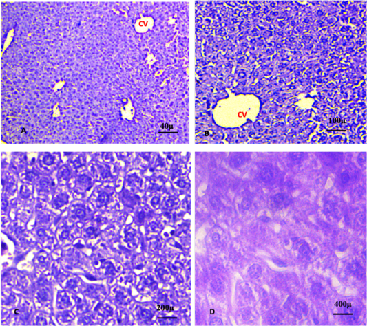
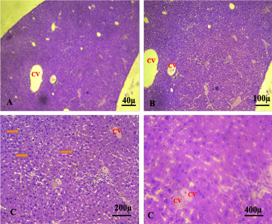
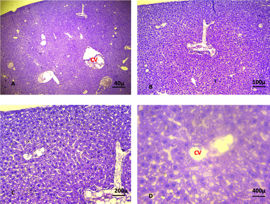
The histological features of the kidney and liver from G2 (mice received Sc-RE only) revealed normal histological features similar to those seen in control group 1 (Figures 4A, B, C, D and 5A, B, C, D).
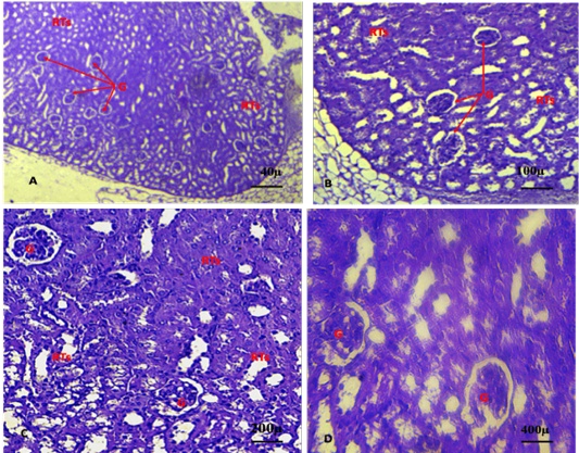
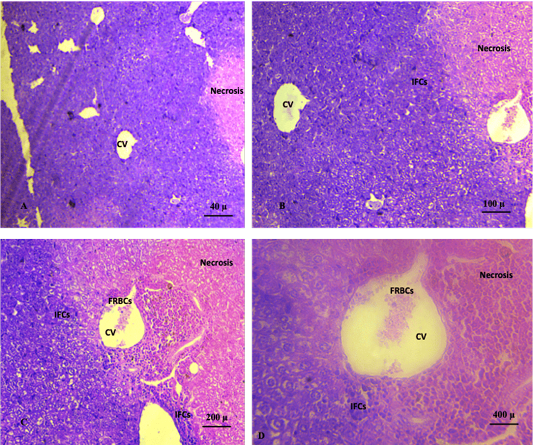
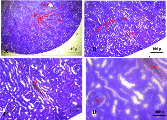
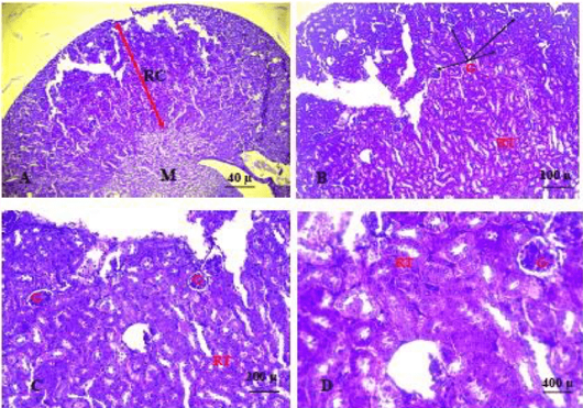
Liver and kidney sections of mice in control group 3/ chemotherapy alone (2.5mg/Kg Ebetrex) revealed various severe histopathological changes at different treatment periods. The liver sections revealed severe necrotic areas and heavy infiltration of inflammatory cells around the central vein. Moreover, various degenerative changes were seen in the hepatic cells comprising cytoplasmic condensation. Congestion and free RBCs were also seen in central veins and the hepatic parenchyma (Figure 6A, B, C, D). The kidney sections in the 5th week revealed severe degenerative and necrosis of glomeruli and renal tubules with obvious congestion and free red blood cells. Infiltration of inflammatory cells was also observed (Figure 7A, B, C, D).
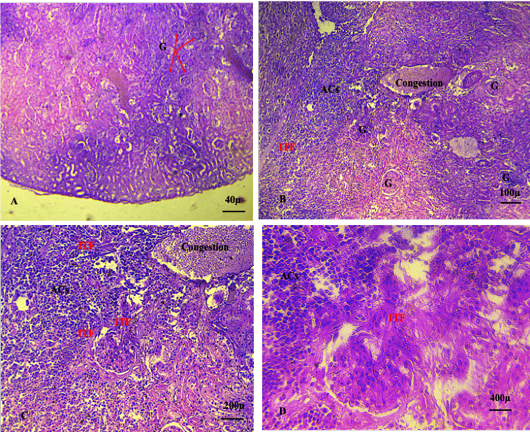
Various histopathological features were seen in mice’s liver and kidney sections in group 4/ mice treated with carcinogenic PVP-K30 only. Mice in this group showed advanced stages of cancer in kidney and liver sections in the 5th week. The liver sections revealed hepatic cell degeneration associated with nuclear pyknosis (orange arrow) and multiple aggregations of cancer cells around the portal area, accompanied by the infiltration of inflammatory cells and congestion. There was severe damage of renal parenchyma, including degeneration of convoluted renal tubules and invading cancer cells with Intra-tumoral fibrosis (ITF) related to poor prognosis of this cancer (Figures 8A, B, C, D and 9A, B, C, D).
Sections of the liver and kidney of mice in group 5/ cancer treated with chemotherapy (2.5mg/Kg Ebetrex) revealed various severe histopathological changes at different treatment periods. The liver sections revealed advanced stages of cancer, severe degeneration, and necrosis of the hepatic cells accompanied by congestion and infiltration of inflammatory cells. Besides, the liver sections in the 5th week revealed loss of the central vein and portal area’s structural organization and extensive hepatic parenchyma necrosis (Figure 10A, B, C, D). Moreover, kidney sections showed severe degeneration and necrosis of glomeruli accompanied by a widening of the glomerular wall and damaged renal tubules. Aggregations of immature cancer cells heavily invaded the renal parenchyma, leading to the loss of normal histological features. Intra-tumoral fibrous tissue was also seen that indicating the advanced stages of renal cancer and an unfavorable prognosis. The kidney sections also showed severe infiltration of the inflammatory cells and congestion (Figure 11A, B, C, D).
The liver and kidney sections of mice in treatment group 6 (mice received PVP as a carcinogenic agent two weeks after treatment with a low dose (400 mg/Kg) of Sc-RE) revealed various histopathological changes at different periods of treatment. Liver sections in 5th week revealed aggregation of immature cancer cells surrounding the central veins and portal areas.
Degeneration and necrosis of the hepatic cells and necrosis of hepatic parenchyma were also noticeable. Liver sections revealed structural organization loss accompanied by congestion and infiltration of inflammatory cells (Figure 12A, B, C, D). At the same time, kidney sections in the 5th week revealed severe degeneration and necrosis of the renal cortex involving the renal tubules. Degeneration of glomeruli and widening of its wall was also observed. Small aggregations of immature cancer cells were seen in the renal parenchyma. The kidney sections also showed infiltration of the inflammatory cells and congestion (Figure 13A, B, C, D).
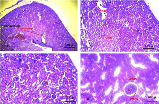
The liver and kidney sections of mice in treatment group 7 (mice received PVP as a carcinogenic agent two weeks from treatment with a moderate dose of 600 mg/Kg of Sc-RE) revealed diverse histopathological changes at different periods of treatment. Liver sections revealed slight degeneration and a few infiltrations of inflammatory cells. Additionally, normal structural organization of the central vein and portal area was seen, and no apparent immature cancer cell was seen (Figure 14A, B, C, D). Section of the kidney at 5th week revealed degeneration and necrosis of the renal cortex involving the renal tubules. There was an obvious degeneration of the glomeruli and extension of its wall. The kidney sections also showed infiltration of the inflammatory cells and congestion, but no apparent aggregations of immature cancer cells were seen in the renal parenchyma (Figure 15A, B, C, D).
The liver and kidney sections of mice in treatment group 8 (mice received PVP as a carcinogenic agent two weeks from treatment with a high dose of 800 mg/Kg of Sc-RE) revealed various histopathological changes at different periods of treatment.
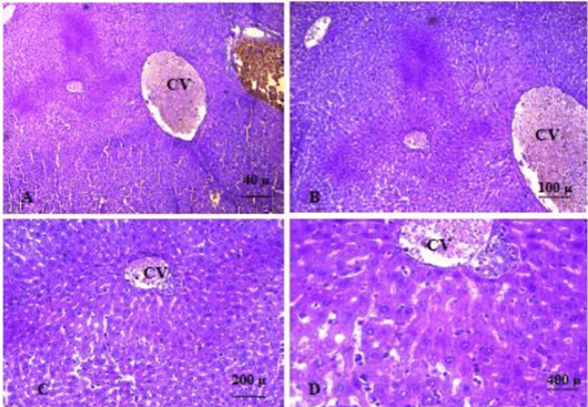
The liver section revealed slight degeneration and a few infiltrations of inflammatory cells. Additionally, severe congestion of the central vein and portal area was seen. No prominent immature cancer cell was seen (Figure 16A, B, C, D).
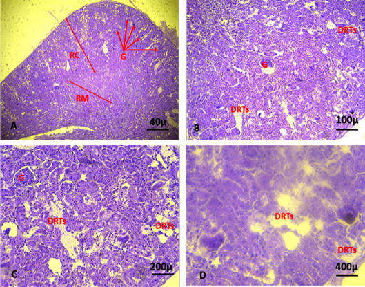
Section of the kidney in 5th week revealed slight degeneration of the renal cortex and medulla that involved the renal tubules. Additionally, there were infiltration of the inflammatory cells, an obvious degeneration of glomeruli, and extending of its wall. The kidney sections also showed congestion, but no apparent aggregations of immature cancer cells were seen in the renal parenchyma (Figure 17A, B, C, D).
Polyvinyl pyrrolidone K-30 (PVP-K-30) is a macromolecule with 35.000-40.000 molecular weight used in the pharmaceutical industry . Repeated subcutaneous Polyvinyl pyrrolidone injections was induced local sarcoma in vivo in mice, rats, and rabbits. Additionally, its single or several intraperitoneal implantations revealed tumor development at distant sites comprising the reticuloendothelial system . However, the inhalation of Polyvinyl pyrrolidone to Sprague-Dawley rats for 24 months revealed adenomas and adenocarcinoma of the nasal cavity accompanied by hepatocellular carcinomas significantly related to the dose. Larynx squamous carcinoma was also observed in the high-dose group . The results of the current study approved the development of liver and kidney cancer in experimental groups (G4, G5, G6, G7, G8) due to intraperitoneal injections of mice with a high dose of PVP-K-30.
Moreover, various advanced histopathological features comprising cancer cells were seen in the last weeks of the experiment in group 4 (mice treated with carcinogenic substance PVP-K30 only). These results are compatible with previously reported studies that approved the development of local and distant cancer in various experimental animals (mice, rats, rabbits) due to repeated administration of Polyvinyl pyrrolidone by several routes using materials of different molecular weights. The reported cancers were adenoma and adenocarcinoma of the nasal cavity, hepatocellular carcinoma, and larynx squamous carcinoma ; . These studies approved the development of tumors due to injection or implantation of PVP that was documented by various macroscopical and microscopical changes in the liver and kidney of experimental animals. Likewise, liver biopsy collected from patients injected intravenously with PVP revealed mild inflammation accompanied by basophilic globular deposits in Kupffer cells. Additionally, a foam-cell storage phenomenon was seen due to the accumulation of PVP in reticulated nuclei. Likewise, PVP can be excreted via the kidneys, and autopsy investigation approved its accumulation in the kidneys, liver, spleen, and lymph nodes .
The results of the current study revealed no significant differences in water and food intake between experimental groups, with 42% lower in the calorie-restricted groups and average cumulative tumor incidence. Nonetheless, dietary fat and tumor incidence increased with increased caloric intake and body weight over a wide range of intakes, including moderate caloric restriction (i.e., 7-20%). These data indicate that total caloric intake is an important determinant of tumorigenesis in mice. Body weight may be a more sensitive indicator for this effect than caloric intake alone . Previous studies approved that the food intake increased significantly in cachectic tumor-bearing mice (TB), synchronously with losing body weight . However, another study revealed variation in fed mice housed at 22 ˚C consumed 30% more calories than ad libitum–fed mice at 27 ˚C, although there was no difference between groups in body composition or cancer progression .
Liver enzyme tests consider obvious prognostic factors of survival time in advanced cancer patients and are useful for patients with hepatobiliary cancer or liver metastasis. The current study revealed variations in the liver enzyme evaluation between experimental groups compared to the values of the control group. The serum levels of liver enzymes ALT, AST, and ALP (U/L) in mice fed with the Sc-RE for 4 weeks were significant (P < 0.05) compared to that of mice in the control group. Nonetheless, significant increases were seen in G3, G4, and G5. Moreover, slight increases were observed in G6, G7, and G8, compared to the control group, but minor variations were seen within Sc-RE low, moderate, and high treatment doses. These results are compatible with previous studies that approved an increase in the values of liver enzymes in treatment groups indicating damage to the hepatocytes . A previous study also revealed that two times higher AST and ALT levels, prolonged PT, and hypoalbuminemia are likely prognostic factors of poor survival in advanced cancer patients. These parameters provide new insights into prognostication in advanced cancer .
The primary importance of measuring alkaline phosphatase (ALP) is to check the possibility of bone diseases or liver disease. The transaminase enzymes (ALT and AST) are important in producing various amino acids. Measuring the concentrations of various transaminases is important in diagnosing and tracking many diseases . The liver is crucial in the metabolism (i.e., inactivation or activation) of many commonly used anticancer agents cytotoxic or new biological agents. Therefore, the assessment of liver function is a fundamental part of the initial work-up and management of patients with cancer .
Histopathological examination of liver and kidney sections of mice in different experimental groups is considered a diagnostic tool to evaluate cancer and malignancy’s progress and treatment efficacy in different tissues. The results of the current study showed normal liver and kidney histological features of the control group 1, in accordance with reference histological features described in the textbook . Additionally, normal histological features similar to those in the control group were also seen in the kidney and liver sections of G2 (mice received Sc-RE only).
These results are compatible with the results of the previously reported researcher , who approved the notable protective effect of Sc-ethanolic extract via its anti-apoptotic, anti-inflammatory, and antioxidant capability. Consequently, they considered Sc, the best nominee, a natural antioxidant that faces the harmful effects of glucocorticoids.
In the current study, liver and kidney sections of mice in Group 5/ cancer treated with chemotherapy (2.5mg/Kg Ebetrex) revealed diverse severe histopathological sections at different treatment periods, which became more severe at 5th week, indicating the advanced stages of cancer. These results agree with previously reported studies ; ; . These studies approved the hepatotoxicity, kidney damage, and administration disorders of Ebetrex as anticancer. Methotrexate is the active component of Ebetrex. It is a chemotherapy agent and immunosuppressive. Ebetrex treats breast, leukemia, lung, lymphoma, gestational trophoblastic diseases, and osteosarcoma. Moreover, it is also used as an antimetabolite and antirheumatic (https://web.archive.org/web/20161008130258/; https:/www.drugs.com/monograph/methotrexate.html). Though, Methotrexate has adverse effects on the treated patients. The previous studies approved the adverse effects, including hepatotoxicity, blood abnormalities (leukopenia, anemia, and thrombocytopenia), stomatitis, increased risk of infection, hair loss, nausea, fatigue, acute pneumonitis, renal impairment and mucositis ; ; . Consequently, the severe histopathological features in this group might be related to the adverse effects of Methotrexate used as chemotherapy to treat the induced cancer in experimental mice. Nonetheless, these observations were evident in the histopathological examination of the liver and kidney sections of control group 3, where the animal received only chemotherapy (2.5mg/Kg Ebetrex). In 5th week, both liver and kidney sections revealed severe degenerative and necrosis changes accompanied by heavy infiltration of inflammatory cells. These results are compatible with the previous study that approved Methotrexate administration induced an acute reaction, tissue edema, marked vascular congestion, and leucocyte infiltration. Other studies found that Methotrexate induces oxidative and nitrative stresses accompanied by activation of nuclear factor-KB (NF- KB) and p38 pathways. Additionally, numerous degenerative changes, including karyorrhexis nuclei, cellular vacuolization that displays the smooth endoplasmic reticulum’s dilatation, and tissue architecture disruption ; .
In groups 6, 7, and 8, mice received PVP (IPI) as a carcinogenic agent; two weeks later, mice received Sc-RE treatment at 400 mg/kg, 600 mg/kg, and 800mg/kg for G6, G7, and G8, respectively. The sections of the liver and kidney from these mice revealed numerous diverse histopathological features related to various treatment groups. In 5th week, the liver sections of G6 revealed aggregation of immature cancer cells. The kidney sections revealed severe degeneration and necrosis of the renal cortex with small aggregations of immature cancer cells in the renal parenchyma. Additionally, the liver sections of G7 revealed slight degeneration, few infiltrations of inflammatory cells, nearly typical structural organization of the hepatic central vein and portal area, and no apparent immature cancer cells. At the same time, kidney sections showed degeneration and necrosis of the renal cortex. There was an obvious degeneration of glomeruli but no apparent aggregations of immature cancer cells in renal parenchyma. Nonetheless, the liver sections of G8 revealed slight degeneration and few infiltrations of inflammatory cells accompanied by severe congestion of the central vein and portal area with no apparent immature cancer cells. While kidney sections revealed slight degeneration of the renal cortex and medulla, accompanied by infiltration of the inflammatory cells. However, no apparent aggregations of immature cancer cells were seen in the renal parenchyma. These results are compatible with previously reported studies ; ; ; ; ; ; ; .
These in vitro studies approved the presence of the active anticancer substance in the roots of Saussurea lappa. They found a new sesquiterpene lactone, which induced apoptosis in cancer cells mediated via ROS generation and mitochondrial dysfunction. Korean, Japanese, and China traditional medicine uses SC-dried root for the treatment of different diseases and conditions, such as abdominal pain. A previous study investigated two major components of Sesquiterpene lactones in the root of Sc.
The costunolide and dehyrocostus lactones own numerous biological activities, comprising antitumor activity . At the same time, another researcher determined the bioactive compound a costunolide in the Sc (S. lappa) root. This substance has been explored for its cause of inhibition of human leukemia cells and its hypothetical action pathway. The costunolide is a potent apoptosis inducer enabling its action by forming reactive oxygen species, mediating mitochondrial permeability change, and liberating cytochrome C . The root extract of S. lappa is approved to treat gastric cancer cells via G2- growth arrest. It can produce growth inhibition of gastric cancer cells and is also effective on leukemia and intestinal carcinoma cells in vitro.
Nonetheless, approved the cure with S. lappa that drastically diminished the viabilities of cells in a dose and time-dependent manner. In vitro, cytotoxicity was determined by S. lappa bioactive Costunolide, dehydrocostus lactone, and lappa dilactone. Likewise, S. lappa root cynaropicrin was approved to have potential against some leukocyte cancer cells like leukemia or lymphoma .
Consequently, the evaluation of the histopathological features for all treatment groups found that a low dose of 400 mg\ kg Sc-RE was less effective in inhibiting the cancer cells in the liver and kidney. In contrast, moderate 600 mg/ kg and high 800 mg/ kg treatment doses inhibited the growth of cancer cells in the liver and kidney. These results are compatible with previously reported studies ; ; ; , they approved that Saussurea lappa ethanolic extract can reduce the pulmonary and splenic tissue damage caused by triamcinolone acetonide through modulation of the apoptosis, inflammation, and oxidative stress. However, severe damages were seen in the histological structure of the kidney in the last weeks of treatment due to the excretion and accumulation of the Sc-RE in the renal tubules. These observations must be addressed by adjusting the treatment dose to prevent these adverse effects on the kidney.
Conclusions and Recommendations
The results of this study approved the activities of Sc-RE to inhibit in vivo-induced hepatic and kidney cancer in mice. Therefore, Saussurea costus may be a promising agent for inhibiting induced cancer growth due to various carcinogenic substances. The authors suggest a future study investigating the pathway of Saussurea costus-Root extract effects as an anticancer and determining the anticancer bioactive components.
Acknowledgment
The authors would like to thank the technician in the Graduated student Biology department laboratory /Al Muthanna University for help and support.
Novelty statement
For the authors knowledge, this is the first study approved the activities of Saussurea costus to inhibit in vivo induced hepatic and kidney cancer in mice.
Author’s Contribution
The study was carried out by ZAA, HKS, and KAA, and all authors contributed equally. In addition, KAA and ZAA made essential contributions to the experimental experiment. HKS organization and analysis of the data for this work. All authors above consented to be held responsible for all parts of the work and participated in its preparation, drafting, and revision. They also provided final permission for the version that was published.
Ethical consideration
The authors have no any ethical issues such as plagiarism, information fabrication, misconduct and/or falsification, permission to publish, duplicate publication and/or submission, and redundancy.
Conflict of interest
The authors have declared no conflict of interest.
Figures
Figure 1:.
Shows the effects of date extract on (A) weekly food intake, (B) weekly water intake, and (C) body weights of the tested mice groups.
Figure 2:.
Shows liver of the control group showing normal histological features. The sections revealed normal organisation of the hepatic cells surrounded the central vein. (CV): central vein. (A. X 4, B. X10, C.X20, D. X40) (H and E).
Figure 3:.
Shows section of the kidney of the control group. The sections revealed normal glomeruli (G) with normal urinary space (US) and renal convoluted tubules (RCT) (A. X 4, B. X10, C.X20, D. X40) (H and E).
Figure 4:.
A, B, C, D shows liver of the control group 3 (plant extract alone). The sections revealed normal organisation of the hepatic cells surrounded the central vein. (CV): central vein. (A. X 4, B. X10, C.X20, D. X40). (H and E).
Figure 5:.
A, B, C, D shows the kidneys of the control group 2 (plant extract alone). Sections showing normal histological features of the kidney. The sections revealed normal glomeruli (G) with normal urinary space (US) and renal convoluted tubules (RCT) (A. X 4, B. X10, C.X20, D. X40). (H and E).
Figure 6:.
5th weeks: Shows liver section for Group 3/ chemotherapy alone (2.5mg/Kg Ebetrex) revealing large necrosis hepatic area and heavy infiltration of inflammatory cells around central vein. A congestion and free RBCs were also seen in the section. (A. X 4, B. X10, C.X20, D. X40). (H and E).
Figure 7:.
3rd and 4th and 5th weeks: Shows kidney sections with various degenerative changes of the glomeruli and renal tubules. Congestion was also seen. (G: Glomeruli, RC: Renal Congestion, DRP: Damage renal parenchyma). (A. X 4, B. X10, C.X20, D. X40). (H and E).
Figure 8:.
A, B, C, D shows the liver control group 3 (Cancer alone without treatment) at 5th week of intraperitoneal injection of Polyvinyl pyrrolidone K-30. Hepatic cell degeneration associated with nuclear pyknosis (orange arrow) and cancer cell aggregation (ACs) around the portal area, accompanied by the infiltration of inflammatory cell and congestion (CV: central vein). (A. X 4, B. X10, C.X20, D. X40) (H and E).
Figure 9:.
A, B, C, D shows advanced stages of renal cancer characterised by Intra-tumoral fibrosis (ITF), severe damage of renal parenchyma, including degeneration of renal convoluted tubules and congestion. (ACs: Abnormal cell aggregation, G: glomeruli). (A. X 4, B. X10, C.X20, D. X40) (H and E).
Figure 10:.
A, B, C, D shows liver sections with sever degeneration and necrosis of the hepatic cells accompanied by congestion and infiltration of inflammatory cells. (A. X 4, B. X10, C.X20, D. X40). (H and E).
Figure 11:.
A, B, C & D: Shows the kidney section in group 5 at 5th week revealing severe degeneration and necrosis of glomeruli accompanied by widening of the glomerular wall and damaged renal tubules. Aggregations of immature cancer cell and heavily invaded the renal parenchyma lead to loss of normal histological features. (A. X 4, B. X10, C.X20, D. X40). (H&E).
Figure 12:.
A, B, C, D shows sections of the liver at the 5th week of the experiment revealed slight degeneration of the hepatic cells, congestion, fatty degeneration, necrosis of hepatic cells around the central vein and absence of immature cancer cells (A. X 4, B. X10, C.X20, D. X40). (H and E).
Figure 13:.
A, B, C, D shows section of kidney at the 5th week of the experiment. The kidney section revealed severe degeneration and necrosis of renal parenchyma, damage of renal tubules, infiltration of the inflammatory cells and severe congestion and absence of immature cancer cell aggregation (Damage of renal tubules: DRTs; renal cortex: RC; Renal medulla: RM; Glomeruli: G). (A. X 4, B. X10, C.X20, D. X40). (H and E).
Figure 14:.
A, B, C, D shows liver sections of mice in group 7 at 4th week of experiment. Liver sections revealed slight degeneration and few infiltrations of inflammatory cells. Additionally, normal structural organization of the central vein and portal area was seen and no obvious immature cancer cell was seen (A. X 4, B. X10, C.X20, D. X40). (H and E).
Figure 15:.
A, B, C, D shows section of kidney of group 7 at the 5th week of the experiment. The kidney sections revealed severe congestion, degeneration and necrosis of renal parenchyma and renal tubules, infiltration of the inflammatory cells, absence of immature cancer cell aggregation (Damaged renal tubules: DRTs; Necrosis of renal tubules: NRTs; Glomeruli: G; Widening of glomerular wall: WGW). (A. X 4, B. X10, C.X20, D. X40). (H and E).
Figure 16:.
A, B, C, D shows liver of mice in group 8 at 5th week of the experiments, the liver section showed slight degeneration and few infiltrations of inflammatory cells. Additionally, severe congestion of the central vein and portal area was seen. No obvious immature cancer cell was seen. (A. X 4, B. X10, C.X20, D. X40). (H and E).
Figure 17:.
A, B, C, D: Shows kidney sections of mice in group 8 at 5th week of the experiments, the kidney section revealed slightly degeneration of renal cortex and medulla, infiltration of the inflammatory cells, degeneration of glomeruli, congestion, and no obvious aggregations of immature cancer cell A. X 4, B. X10, C.X20, D. X40). (H&E).
References
Ahmed A, Ahmad S, Soni K, Lapa B, Afzal M, Sharma K, and Kumar G (2016). Suitable solvent and drying condition to enhance phenolics and extractive value of Saussurea costus. J. Ayurvedic Herb. Med., 2(5): 165-170. https://doi.org/10.31254/jahm.2016.2504[CrossRef]
Alaa K, Hamad, Mariam A, Kadhem (2020). Prophylactic effect of ethanolic extract of Saussurea Costus roots against hepato- renal toxicity induced by diazinon in chickens. Al-Anbar J. Vet. Sci., 13(2): 34-51. https://doi.org/10.37940/AJVS.2020.13.2.4[CrossRef]
Albanes D (1987). Total calories, body weight, and tumor incidence in mice. Cancer Res., 47(8): 1987-1992.
Alotaibi AA, Bepari A, Assiri RA, Niazi SK, Nayaka S, Rudrappa M, Nagaraja SK, Bhat MP (2021). Saussurea lappa exhibits anti-oncogenic effect in hepatocellular carcinoma, HepG2 cancer cell line by Bcl-2 mediated apoptotic pathway and mitochondrial cytochrome C release. Curr. Issues Mol. Biol., 43(2): 1114-1132. https://doi.org/10.3390/cimb43020079[CrossRef]
Amina M, Al Musayeib NM, Alarfaj NA, El-Tohamy MF, Oraby HF, Al-Hamoud GA, Moubayed NM (2020). Biogenic green synthesis of MgO nanoparticles using Saussurea costus biomasses for a comprehensive detection of their antimicrobial, cytotoxicity against MCF-7 breast cancer cells and photocatalysis potentials. PLoS One, 15(8): e0237567. https://doi.org/10.1371/journal.pone.0237567[CrossRef]
Azevedo T, Ferreira T, Barros L, Ferreira ICFR, Pinto MdL, Oliveira PA (2022). Histopathological evaluation of mice’s liver and kidney after exposure to an elderberry extract. Biol. Life Sci. Forum, 18: 6. https://doi.org/10.3390/Foods2022-13031[CrossRef]
Bancroft JD, Gamble M (2008). Theory and practice of histological techniques. 6th Edition, Churchill Livingstone, Elsevier, China.
Choudhary M, Kumar V, Malhotra H, Singh S (2015). Medicinal plants with potential anti-arthritic activity. J. Intercult. Ethnopharmacol., 4(2): 147-179. https://doi.org/10.5455/jice.20150313021918[CrossRef]
Dar KK, Ali S, Ejaz M, Nasreen S, Ashraf N, Gillani SF, Shafi N, Safeer S, Khan MA, Andleeb S, Mughal TA (2019). In vivo induction of hepatocellular carcinoma by diethylnitrosoamine and pharmacological intervention in Balb C mice using Bergenia ciliata extracts. Braz. J. Biol., 79(4): 629-638. https://doi.org/10.1590/1519-6984.186565[CrossRef]
Deabes MM, Fatah AE, Sally I, Salem SHE, Naguib KM (2021). Antimicrobial activity of bioactive compounds extracts from Saussurea costus against food spoilage microorganisms. Egypt. J. Chem., 64(6): 2833-2843. https://doi.org/10.21608/ejchem.2021.69572.3528[CrossRef]
Doha S, Mohamed E, Nor-Eldin K (2018). Histological and immunohistochemical study of the possible protective effect of folic acid on the methotrexate- induced cardiac muscle toxicity in male albino rats. Egypt. J. Histol., 41(1): 73-82. https://doi.org/10.21608/EJH.2017.7523[CrossRef]
Dwarkasing JT, van Dijk M, Dijk FJ, Boekschoten MV, Faber J, Argilès JM, van Norren K (2014). Hypothalamic food intake regulation in a cancer-cachectic mouse model. J. Cachexia, Sarcopenia Muscle 5(2): 159-169. https://doi.org/10.1007/s13539-013-0121-y[CrossRef]
Elgharabawy RM, El-Tantawy, El-Sayed I, Abd-Allah RN, Tousson E (2021). Therapeutic impact of Costus (Saussurea lappa) against ehrlich solid tumor-induced cardiac toxicity and DNA damage in female mice. Front. Pharmacol., 28(12): 708785. https://doi.org/10.3389/fphar.2021.708785[CrossRef]
Elsebai MF, Mocan A, Atanasov AG (2016). Cynaropicrin: A comprehensive research review and therapeutic potential as an anti-hepatitis C virus agent. Front. Pharmacol., 8(7): 472. https://doi.org/10.3389/fphar.2016.00472[CrossRef]
Ferlay J, Ervik M, Lam F, Colombet M, Mery L, Piñeros M, Znaor A, Soerjomataram I, Bray F (2020). Global cancer observatory: Cancer today. Lyon: International Agency for Research on Cancer. (https://gco.iarc.fr/today, accessed February 2023).
Ghada IAE-R, Amany B, Nora ME, Gaber E-SB, Wael NH, Dina MK, Yasmina MA-E (2020). Saussurea lappa ethanolic extract attenuates triamcinolone acetonide-induced pulmonary and splenic tissue damage in rats via modulation of oxidative stress, inflammation, and apoptosis. Antioxidants, 9: 396. https://doi.org/10.3390/antiox9050396[CrossRef]
Hanahan D, Weinberg RA (2011). Hallmarks of cancer: The next generation. Cell, 144(5): 646-674. https://doi.org/10.1016/j.cell.2011.02.013[CrossRef]
Hersh EM, Wong VG, Henderson ES, Freireich EJ (1966). Hepatotoxic effects of methotrexate. Cancer, 19(4): 600–606. https://doi.org/10.1002/1097-0142(196604)19:4<600::AID-CNCR2820190420>3.0.CO;2-3[CrossRef]
Hsu YL, Wu LY, Kuo PL (2009). Dehydrocostuslactone, a medicinal plant-derived sesquiterpene lactone, induces apoptosis coupled to endoplasmic reticulum stress in liver cancer cells. J. Pharmacol. Exp. Therapeut., 329(2): 808-819. https://doi.org/10.1124/jpet.108.148395[CrossRef]
The American Society of Health-System Pharmacists. Methotrexate. https://web.archive.org/web/20161008130258/https:/www.drugs.com/monograph/methotrexate.html.
Hua W, Lihua Y, Weihong F, Guoxu M, Yong P, Zhimin W (2014). Research progress on active ingredients and pharmacologic properties of saussurealappa. Curr. Opin. Complement. Altern. Med., 1(1): 1- 7.
Huffman DM, Johnson MS, Watts A, Elgavish A, Eltoum IA, Nagy TR (2007). Cancer progression in the transgenic adenocarcinoma of mouse prostate mouse is related to energy balance, body mass, and body composition, but not food intake. Cancer Res., 67(1): 417-424. https://doi.org/10.1158/0008-5472.CAN-06-1244[CrossRef]
Hung JY, Hsu YL, Ni WC, Tsai YM, Yang CJ, Kuo PL, Huang MS (2010). Oxidative and endoplasmic reticulum stress signaling are involved in dehydrocostuslactone-mediated apoptosis in human non-small cell lung cancer cells. Lung Cancer, 68(3): 355-365. https://doi.org/10.1016/j.lungcan.2009.07.017[CrossRef]
IARC Monographs on the Evaluation of Carcinogenic Risks to Humans, (1999). Re-evaluation of Some Organic Chemicals, Hydrazine and Hydrogen Peroxide Volume 71. World Health Organization International Agency for Research on Cancer. Lyon France.
IARC, 1999. Working Group on the Evaluation of Carcinogenic Risks to Humans. Lyon (FR): International Agency for Research on Cancer; 1999.
Kamen BA, Nylen PA, Camitta BM, Bertino JR (1981). Methotrexate accumulation and folate depletion in cells as a possible mechanism of chronic toxicity to the drug. Br. J. Haematol., 49(3): 355–360. https://doi.org/10.1111/j.1365-2141.1981.tb07237.x[CrossRef]
Kim YJ, Song M, Ryu JC (2009). Inflammation in methotrexate-induced pulmonary toxicity occurs via the p38 MAPK pathway. Toxicology, 256(3): 183–190. https://doi.org/10.1016/j.tox.2008.11.016[CrossRef]
Klimisch HJ, Deckardt K, Gembardt C, Hildebrand B, Küttler K, Roe FJC (1997b). Subchronic inhalation and oral toxicity of N-vinylpyrrolidone-2. Studies in rodents. Food Chem. Toxicol., 35: 1061–1074. https://doi.org/10.1016/S0278-6915(97)87274-2[CrossRef]
Ko SG, Kim HP, Jin DH, Bae HS, Kim SH, Park CH, Lee JW (2005). Saussurea lappa induces G2-growth arrest and apoptosis in AGS gastric cancer cells. Cancer Lett., 220: 11–19. https://doi.org/10.1016/j.canlet.2004.06.026[CrossRef]
Ko SG, Koh SH, Jun CY, Nam CG, Bae HS, Shin MK (2004). Induction of apoptosis by Saussurea lappa and Pharbitis nil on AGS gastric cancer cells. Biol. Pharma. Bull., 27(10): 1604-1610. https://doi.org/10.1248/bpb.27.1604[CrossRef]
Kulsoom Z, Shaista T, Sidra S, Muhammad A, Rahmatullah Q, Muhammad SA, Sunbal KC (2014). A review of therapeutic potential of Saussurea lappa-An endangered plant from Himalaya. Asian Pac. J. Trop. Med. 7(Suppl 1): S60-S69. https://doi.org/10.1016/S1995-7645(14)60204-2[CrossRef]
Lala V, Zubair M, Minter DA (2023). Liver function tests. In: Stat Pearls [Internet]. Treasure Island (FL): StatPearls Publishing.
Lee MG, Lee KT, Chi SG, Park JH (2001). Costunolide induces apoptosis by ROS-mediated mitochondrial permeability and cytochrome C release. Biol. Pharma. Bull., 24: 303–306. https://doi.org/10.1248/bpb.24.303[CrossRef]
Leopold M, Marguerite H, Leonard W, John S, Samuel G (1954). Some physical-chemical properties of polyvinylpyrrolidone. 35(2): 191-196.
Liu CY, Chang HS, Chen IS, Chen CJ, Hsu ML, Fu SL, Chen YJ (2011). Costunolide causes mitotic arrest and enhances radiosensitivity in human hepatocellular carcinoma cells. Radiat. Oncol., 6: 56. https://doi.org/10.1186/1748-717X-6-56[CrossRef]
Mishra P, Singh U, Pandey CM, Mishra P, Pandey G (2019). Application of student’s t-test, analysis of variance, and covariance. Ann. Card Anaesth., 22(4): 407-411. https://doi.org/10.4103/aca.ACA_94_19[CrossRef]
Mukherjee S, Ghosh S, Choudhury S Adhikary A, Manna K, Dey S, Sa G, Das T, Chattopadhyay S (2013). Pomegranate reverses methotrexate-induced oxidative stress and apoptosis in hepatocytes by modulating Nrf2-NF-κB pathways. J. Nutr. Biochem., 24(12): 2040–2050. https://doi.org/10.1016/j.jnutbio.2013.07.005[CrossRef]
Nageswara R, Satyanarayana R, Suresh B, Vadaparthi R (2013). HPLC determination of costunolide as a marker of saussurealappa and its herbal formulations. Int. J. Res. Pharm. Chem., 3(1): 99-107.
Nguyen J, Kitzmiller P, Khungar P, Dabbade N, Nguyen KT, Flynn EL (2017). Phytochemical, hematologic and anti-tumor activity evaluations of Carica papaya leaf extract. J. Transl. Sci., 3(3): 1–7. https://doi.org/10.15761/JTS.1000185[CrossRef]
Odiegwu CN, Chianella I, Azubike NC, Odiegwu UO, and Ogbuowelu OS (2021). Liver function tests values in albino wistar rats administered with isolated Nigeria Achatina achatina Snail Lectin. GSC Biol. Pharm. Sci., 15(2): 92-102. https://doi.org/10.30574/gscbps.2021.15.2.0115[CrossRef]
Oliff A, Bleyer WA, Poplack DG (1979). Methotrexate-induced oral mucositis and salivary methotrexate concentrations. Cancer Chemother. Pharmacol., 2(3): 225–226. https://doi.org/10.1007/BF00258300[CrossRef]
Pandey MM, Rastogi S, Rawat AKS (2007). Saussurea costus: Botanical, chemical and pharmacological review of an ayurvedic medicinal plant. J. Ethnopharmacol., 110(3): 379-390. https://doi.org/10.1016/j.jep.2006.12.033[CrossRef]
Patel AA, Amanullah M, Eissa M, Elsaid FG, Soliman T (2020). A study on anti-cancer properties of Saussurea lappa (Asteraceae) against breast and colonic cancer cell lines. Clin. Oncol., 5: 1702.
Qassadi FI, Zhu Z, Monaghan TM (2023). Plant-derived products with therapeutic potential against gastrointestinal bacteria. Pathogens, 12(2): 333. https://doi.org/10.3390/pathogens12020333[CrossRef]
Raina H, Soni G, Jauhari N, Sharma N, Bharadvaja N (2014). Phytochemical importance of medicinal plants as potential sources of anticancer agents. Turk. J. Bot., 38(6): 1027-1035. https://doi.org/10.3906/bot-1405-93[CrossRef]
Rasul A, Bao R, Malhi M, Zhao B, Tsuji I, Li J, Li X (2013). Induction of apoptosis by costunolide in bladder cancer cells is mediated through ROS generation and mitochondrial dysfunction. Molecules, 18(2): 1418-1433. https://doi.org/10.3390/molecules18021418[CrossRef]
Robinson A, Kumar TV, Sreedhar E, Naidu VG, Krishna SR, Babu KS, Srinivas PV, Rao JM (2008). Anew sesquiterpene lactone from the roots of Saussurealappa: Structure- anticancer activity study. Bioorg. Med. Chem. Lett., 18(14): 4015-4017. https://doi.org/10.1016/j.bmcl.2008.06.008[CrossRef]
Rossi S (2013). Australian Medicines Handbook (2013 ed.). Adelaide: The Australian Medicines Handbook Unit Trust. ISBN 978-0-9805790-9-3
Siegel RL, Miller KD, Wagle NS, Jemal A (2023). Cancer statistics, 2023. CA Cancer J Clin. 73(1): 17-48. https://doi.org/10.3322/caac.21763[CrossRef]
Sun CM, Syu WJ, Don MJ, Lu JJ, Lee GH (2003). Cytotoxic sesquiterpene lactones from the root of Saussurealappa. J. Nat. Prod., 66(9): 1175-1180. https://doi.org/10.1021/np030147e[CrossRef]
Tabata K, Nishimura Y, Takeda T, Kurita M, Uchiyama T, Suzuki T (2015). Sesquiterpene lactones derived from Saussurea lappa induce apoptosis and inhibit invasion and migration in neuroblastoma cells. J. Pharm. Sci., 127(4): 397-403. https://doi.org/10.1016/j.jphs.2015.01.002[CrossRef]
Tian X, Song HS, Cho YM, Park B, Song YJ, Jang S, Kang SC (2017). Anticancer effect of Saussurea lappa extract via dual control of apoptosis and autophagy in prostate cancer cells. Medicine, 96: 30. https://doi.org/10.1097/MD.0000000000007606[CrossRef]
Tobias H, Auerbach R (1973). Hepatotoxicity of long-term methotrexate therapy for psoriasis. Arch. Intern. Med., 132(3): 391–396. https://doi.org/10.1001/archinte.132.3.391[CrossRef]
Tsai HJ, Hsieh MY, Tsai YC, Liu ZY, Hsieh HY, Lee CM, Huang CT (2014). Liver function tests may be useful tools for advanced cancer patient care: A preliminary single-center result. Kaohsiung J. Med. Sci., 30(3): 146-152. https://doi.org/10.1016/j.kjms.2013.09.005[CrossRef]
Evaluation of the Anticancer Effect of Saussurea costus Root Extract Against Induced Hepatic and Renal Cancer in White Mice: A Histopathological Study · ResearchersLinks