Research Article · Volume 12, Issue 8, 27 June 2024 · Open Access
Functional and Histological Signs of Early Detection of Liver and Kidney Stress Under the Influence of Tramadol Oral Administration in Male Rats
· ·
Affiliations & Notes ⌄
1College of Dentistry, University of Misan, Iraq
2Department of Anatomy and Histology, College of Veterinary Medicine, University of Basrah, Iraq. *Correspondence | Hawraa F.H. Alowaid, College of Dentistry, University of Misan, Iraq
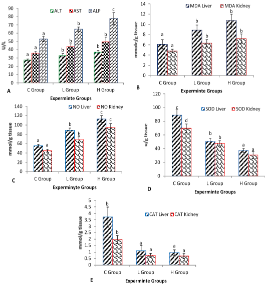
While we detected a significant increase (P≤ 0.05) in MDA from sampled liver and kidney tissues in L and H groups compare to the C group, the higher values were 10.77 and 7.19mmol/g tissue respectively detected in animals from the H group (Figure 1B).
NO concentration showed a Significance difference (P≤ 0.05) among all groups, the higher NO values 112.98 and 94.87mmol/g tissue respectively were detected in the H group (Figure 1C).
All tramadol groups showed a lower mean of SOD activity (p≤0.05) in liver and kidney tissue compare to the control group, Absolutely the lowest value in the H group was 37.33 and 30.71u/g at liver and kidney tissue respectively (Figure 1D).
Administration of the tramadol to the treatment animals group led to inhibition in CAT activity (p≤0.05) in liver and kidney tissue, and the lower values 0.97 and 0.71mmol/g tissue were shown in the H group respectively (Figure 1E).
Selected from the article's own text.
Abstract
The opioid painkiller tramadol (TML) is frequently used to treat moderate to severe pain. On the other side, TML is frequently misused in many nations due to its accessibility and low price. High doses or prolonged administration of TML are linked to hepatic and renal damage. There is currently no known precise mechanism for how TML damages the liver and kidneys. The goal of the current investigation was to assess the potential contribution of oxidative stress dysfunction to the etiology of renal and hepatic injury brought on by TML. For this purpose, male adult rats were treated with TML (15 and 30 mg/kg/day oral administration) 30 days in a row. A significant decrease in liver and kidney tissue SOD, CAT, NO, and MAD was detected in TML groups, while, hepatic and Renal histopathological alterations included inflammation, necrosis, hemorrhage, and congestion, in TML-treated animals, generally, the complementary results of the histological study, by evaluating the degree of histopathological changes, the distribution of lesions by scores (Sc) and an important factor (Fi), and the injury severity index in both kidney and liver tissue, revealed the severity of the pathological changes in the highest dose group compared with the rest of the groups. On the other hand, TML (30 ​mg/kg/day) caused more dysfunction in the studied biomarkers compared to the other groups. Prolonged tramadol use carries a risk of greater hepatic and renal damage. As such, tramadol’s harmful effects must be taken into account during ongoing treatment.
All around the world, Addiction to drugs is a social and health problem, one of the most widely abused medicines is analgesics, particularly opioids . Tramadol is a synthetic centrally-acting analgesic that has similar effects to codeine but is ten times less expensive than morphine . Tramadol has several applications, however, it is most commonly employed in the treatment of moderate to severe acute or chronic pain . Repeated TR treatment may cause toxic metabolites to build up in the body and reduce TR clearance, raising the risk of TR poisoning . Especially in high doses, the harmful consequences of TR are anticipated during long-term therapy . The oral bioavailability of tramadol is high, ranging from 70 to 80%. After an oral dose, peak blood levels are attained in around 2 hours . The cytochrome p450 enzyme in the liver breaks it down, and the kidneys are used to eliminate the byproducts. O-desmethyl-tramadol, which is bio-transformed into it in the liver and is 2 to 4 times more effective than tramadol, is an active chemical . The liver and kidney are in charge of controlling tramadol metabolism and excretion . As a result, during its metabolism, it may cause hepatotoxicity and nephrotoxicity . Generally, due to the liver’s crucial involvement in drug metabolism, almost every drug has been linked to hepatotoxicity . Drugs’ metabolites, which are excreted by the kidneys, may also induce cellular damage that results in renal failure and may be more active or dangerous than the original drug . The current study aimed to assess the negative effect of chronic use of tramadol on the liver and kidney, and its relationship with oxidative stress and functional impairment in a rat model. Since tramadol is a commonly used analgesic and can cause addiction when misused, so our information results might be used to create new preventive and therapeutic approaches to combat this problem.
Materials and Methods
Standard solution and single-administration toxicity
Tramadol tablets that each included100 mg of tramadol hydrochloride were provided by Dimidics Company, (Kingdom of Saudi Arabia), A standard solution of tramadol was prepared by dissolving in 100mg/5ml of normal saline to create a solution containing 20 milligrams per 1ml and given group L and H 1.5 and 0.75 ml/kg/day as a single dose by oral gavage, which represents 1/10 and 1/20 of LD50 .
Experimental design
The study protocols were approved by the Scientific Research Ethics Committee of the Faculty of Veterinary Medicine at Basrah University after the committee took into account the guidelines set forth by the Institutional Animal Care and Use Committees (IACUC) Center for Animal Care. A total of male rats (n= 15) weighing 225±25g, were kept in a controlled environment with a 12-hour cycle of darkness and light at 25±1°C, 48% relative humidity. Rats had unlimited access to tap water and were fed a typical rodent pellet chow diet. Rats were randomly divided according to Essam et al. (2015) into three experimental groups (n= 5): (C) group as control: (2.5ml/kg/day of normal saline), L group (15mg/kg/day of TML), and H group (30mg/kg/day of TML). On day 30, the animals were anesthetized and blood, liver, and kidney samples were collected.
Clinical signs
During the course of the experiment, no deaths were reported using the tramadol medication concentrations employed in this investigation. Comparing drug-treated rats to control rats, the former displayed reduced activity and a decline in feed initiation rates.
Serum enzymes and preparation of tissue homogenate
The biochemical characteristics for biological liver function indicators of male rats serum, Alanine aminotransferase (ALT), Aspartate aminotransferase (AST), and alkaline phosphatase (ALP) enzymes activities were evaluated in all groups by collecting blood into coagulant tubes and centrifuging at 3500 rpm /10 min. The serum was isolated (stored at -20 degrees Celsius for analysis). The enzyme activity of (ALT) and (AST) was determined in IU/l using the IFCC method . While, (ALP) enzyme is evaluated in mg/100 ml using the pNPP Kinetic technique . While, the histochemical measurements in the liver and kidney tissues, it was treated as follows: To create 10% homogenates, liver and kidney tissues were cut, weighed, and minced into minute pieces before being homogenized in 9 ml of ice-cold 0.05 mM potassium phosphate buffer (pH 7.4) with a glass homogenizer. The homogenates were centrifuged for 15 minutes at 4°C at 6000 rpm, and the supernatant was used to test malondialdehyde (MDA), nitric oxide (NO), superoxide dismutase (SOD), and catalase (CAT). Furthermore, the levels of MDA, NO, CAT, and SOD in liver and kidney tissues were determined using the procedures described by ; ; ; , respectively.
Histological examination
The liver and kidneys organs of all animals euthanized at the end of the experiment were removed and preserved in a 10% neutral buffered formalin solution. After 72 hours of fixation, the samples were washed under running water for two hours. They were then dehydrated by gradually increasing the alcohol concentration from 70% to 100% over a two-hour period for each step. Xylene was used to clean the samples, and then paraffin wax was applied. Finally, the samples were sliced with a microtome to a thickness of 5 um for each tissue. The stains utilized were hematoxylin and eosin (H and E) . Histological alterations were evaluated using an unpretentious scoring system and classified as (-) none, (+) low modification, (++) moderate modification, and (+++) important modification .
Histopathological evaluation of lesion scores
Histological examinations were used to evaluate the degree of the alterations and the distribution of lesions inside the studied organ, depending on the analysis method . the distribution of the lesions was determined by scores (Sc) and an important factor (Fi). Sc was represented distributed the total tissue under analysis as (0= absence - 10% of lesions) of, 1= (11-25% of lesions), 2= (26-50% of lesions) 3 = (51-75% of lesions), and 4= (76-100% of lesions). While Fi specifies how such a change will effect organ function, the following values are assigned to it: 1: denotes a lesion that is quickly reversible and has little histological significance. 2: reversible lesions of moderate to neutral histopathological significance; and 3: lesions with high histolopathological significance. The lesion index was determined as follows for each lesion (I Lesion) as the following: I Lesion= (Sc x Fi) and the lesion organ index (I LOrg) is denoted as I LOrgan equals Σ I Lesion.
Data analysis
presented as mean SE, and statistical significance was determined using one-way ANOVA in the (SPSS), version 18. the program was followed by the least significant difference (LSD). When P0.05, values were considered statistically significant.
Results and Discussion
Administration of the tramadol 15 and 30mg/kg/day (L and H groups) at 30 days led to a significant elevation (P≤ 0.05) of AST, ALT, and ALP serum enzymes concentration compare to the control group (Figure 1A). While we detected a significant increase (P≤ 0.05) in MDA from sampled liver and kidney tissues in L and H groups compare to the C group, the higher values were 10.77 and 7.19mmol/g tissue respectively detected in animals from the H group (Figure 1B). NO concentration showed a Significance difference (P≤ 0.05) among all groups, the higher NO values 112.98 and 94.87mmol/g tissue respectively were detected in the H group (Figure 1C). All tramadol groups showed a lower mean of SOD activity (p≤0.05) in liver and kidney tissue compare to the control group, Absolutely the lowest value in the H group was 37.33 and 30.71u/g at liver and kidney tissue respectively (Figure 1D). Administration of the tramadol to the treatment animals group led to inhibition in CAT activity (p≤0.05) in liver and kidney tissue, and the lower values 0.97 and 0.71mmol/g tissue were shown in the H group respectively (Figure 1E).
Histological data
The light microscopic examination of the hematoxylin and eosin-stained paraffin 5 um sections revealed different types of histological lesions of the liver and kidney, which are perfectly in relation with our proven results about the irregular levels of enzymatic and metabolite biomarkers in blood and tissue.
Liver
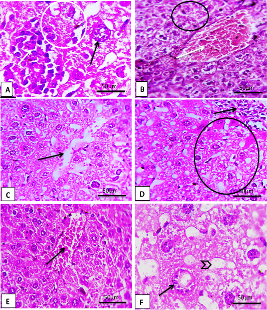
Liver sections revealed in L and H groups, loss of cord-like parenchymal architecture, acute Inflammation infiltration, necrosis and apoptosis state, also showed a hemorrhage, vein congestion, as well as degeneration and vaculation cytoplasmic, excessive sinusoidal dilatation. In general, these histopathological signs were more severe, and clear in the H group of experimental animals (Figure 2).
Kidney
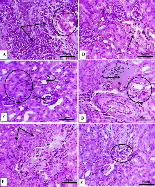
histopathological sections of L and H groups kidneys were a sensitive tool to detect the toxic effect of an overdose of tramadol (15 and 30 mg/kg/day), respectively. The histological alterations revealed slight to severe in each of the following, degeneration of cytoplasm, necrosis of renal tubular cells, necrosis of renal glomeruli, wider urinary spaces, edema of renal tubular cells, hemorrhage, and vessel congestion, generally the H group reversed severe histological alterations.
Histological changes feature of the liver
The values of total histological alteration scores in the control, L, and H groups are shown in Tables 1 and 2. Significant changes in (I Lesion) occurred in rat liver in conjunction with tramadol overdose. L and H groups revealed Subcellular disarrangements included loss of parenchymal cord-like architecture, degeneration and necrosis of hepatocytes, as well as hemorrhage, Vein congestion, Excessive sinusoidal dilatation, and Infiltration of inflammatory cells, in general, these histopathological signs were more severe and clear in the H group of experimental animals, while represented control group, typical appearance of the hepatocytes and parenchymal architecture.
Histological changes feature of the kidney
The values of total histological alteration scores in the control, L, and H groups are shown in Tables 3 and 4. Significant changes in (I Lesion) occurred in rat kidney in conjunction with tramadol overdose. L and H groups revealed the histological alterations, discrepancies between slight to severe histopathological alteration, and as blogged below, degeneration of cytoplasm, necrosis of renal tubular cells, atrophied glomeruli, wider Bowman’s spaces, edema, hemorrhage, and vessel congestion, while the control group’s kidney histopathology examination showed the typical appearance and normal of the glomerulus and renal tubular epithelium in all rats (Figure 3).
Table 1: Distribution of the histological damage features in the liver of the experimental groups.
Histological signs
Experimental group
C
L
H
loss of parenchymal cord-like architecture
-
+
++
Degeneration of hepatocytes
-
+
++
necrosis of hepatocytes
-
+
++
hemorrhage
-
+
+++
Vein congestion
+
+++
+++
inflammatory cells
-
+
++
sinusoidal dilatation
+
++
+++
(-) nil or mild histological changes; (+) moderate histological changes; (++) severe histological changes; and (+++) very severe histological changes in the liver architecture.
Table 2: Liver alteration index (I Lesion%) of rats groups, scores (Sc) and an important factor (Fi) in the study groups.
Histological signs
Liver lesion index (I Lesion%)
C
L
H
Fi
Sc
Fi
Sc
Fi
Sc
loss of parenchymal cord-like architecture
0
0
2
55.5
3
73.6
Degeneration of hepatocytes
1
5
3
60.5
3
80.9
necrosis of hepatocytes
1
5
2
50.8
3
78.9
hemorrhage
0
0
2
33.7
3
63.8
Vein congestion
1
5
3
60.6
3
90.5
inflammatory cells
0
0
3
20.8
3
52.9
I LOrgan= Σ I Lesion
1
15%
3
281.9%
3
440.6%
I Lesion= (Sc x Fi) and the lesion organ index (I LOrg is denoted by I LOrgan = Σ I Lesion. (Sc): Distributed and Frequently of lesions. (Fi): reversible lesions, reversible lesions, and irreversible.
Table 3: Distribution of the histological damage features in the kidney of the experimental groups.
Histological signs
Experimental group
C
L
H
degeneration of cytoplasm
+
++
+++
necrosis of renal tubular cells
-
++
+++
atrophied glomeruli
-
+
++
edema
-
+
++
hemorrhage
-
++
++
vessel congestion
+
++
+++
inflammatory cells
-
+
++
(-) nil or mild histological changes; (+) moderate histological changes; (++) severe histological changes; and (+++) very severe histological changes in the kidney architecture.
Table 4: Kidney alteration index (I Lesion %) of rats groups, scores (Sc) and an important factor (Fi).
Histological signs
Kidney lesion index (I Lesion%)
C
L
H
Fi
Sc
Fi
Sc
Fi
Sc
Degeneration of cytoplasm
0
0
2
55.5
3
73.6
Necrosis of renal tubular cells
Atrophied glomeruli
1
5
3
60.5
3
80.9
Edema
1
5
2
50.8
3
78.9
Hemorrhage
0
0
2
33.7
3
63.8
Vessel congestion
1
5
3
60.6
3
90.5
Inflammatory cells
0
0
3
20.8
3
52.9
I LOrgan= Σ I Lesion
1
15%
3
281.9%
3
440.6%
I Lesion= (Sc x Fi) and the lesion organ index (I LOrg is denoted by I LOrgan= ΣI Lesion. (Sc): Distributed and Frequently of lesions. (Fi): reversible lesions, reversible lesions, and irreversible lesions.
To better comprehend the intricate mechanics of histology and its potentially harmful biochemical effects, experimental models of opioid addiction in laboratory rats that simulate a human context are a crucial tool, as well as for developing therapeutic strategies for addictive diseases. Over the last ten years, there has been a considerable increase in the abuse of opioid analgesics and anxiolytics around the world, according to the European Medicines Agency’s Committee for Medicinal Products in 2009 stated that there was a lack of information regarding long-term overdose side effects . All antipsychotic and morphine drugs derivatives and in acute doses with prolonged periods of abuse for addicted persons cause hepatotoxicity and nephrotoxicity during its metabolism . The marker of oxidative stress (MDA and NO) significantly increased in the current investigation, dysfunction markers in the liver and kidney (ALT, AST, ALP enzymes), and (SOD and CAT enzymes), and histopathological markers, were evident in the overdose oral administration of TML-treated, on liver and kidneys. These data indicate that oxidative stress and dysfunction, play a crucial role in histopathogenesis. tramadol is classified as a drug category C by the American Food and Drug Administration (AFDA), and it is not recommended because of its effects on kidney, liver, or stomach disorders, mental illness, or depression . Our findings suggest that tramadol causes oxidative stress in the tissues of the liver and the kidney, it is consistent with and . When the liver breaks down different substances, it is known to produce certain metabolites; the toxic metabolites are subsequently packaged for elimination. our results were outcomes resemble those reported by . Who discovered that the rats in the treatment group had significantly greater blood AST and ALT activity after receiving oral tramadol for 8 weeks. Our results are also came in accordance with the recorded data . Who observed that using tramadol for 63 days caused a noticeable rise in MAD and NO levels in brain tissue, . Which demonstrated a significant reduction in the SOD, CAT, and GSH activities in the liver tissue as compared to control rats. Additionally, . demonstrated that abuse of tramadol for a month caused a considerable rise in MDA and a fall in antioxidant (CAT) activity. Ahmed and Kurkar (2014) also had similar findings in testicular tissue, reporting that tramadol enhanced testicular levels of No while dramatically decreasing enzymatic antioxidant activity. ROS renders all antioxidant enzymes inactive, generally, Nitric oxide (NO) and malondialdehyde (MDA) levels are frequently utilized as a biomarker of free radical-mediated lipid peroxidation because they show how much cellular harm that free radicals have indirectly caused . Our histopathological results of overdose tramadol toxicity was confirmed in liver and kidney tissue, This may be explained by the fact that the liver is in charge of tramadol’s metabolism and excretion . While the kidneys play a role in the secretion of numerous toxins, they are also prone to release large amounts of free radicals, which in turn contribute to high levels of oxidative stress and kidney damage . Our findings supported the findings of , which claimed that tramadol usage over an extended period of time causes localized vessel congestion, necrosis hepatocyte, and cytoplasmic degeneration in rats. Because the kidneys are involved in the release of numerous toxins, they are prone to producing significant numbers of free radicals, which enhance oxidative stress and cause kidney disease . Most of the histopathological findings obtained in our study were similar to what was reported . Which confirmed that given that tramadol and its metabolites are eliminated through the kidneys, there is a chance that they could cause nephrotoxicity and histological changes. Overall, the current study’s findings point to oxidative stress and free radicals as processes implicated in the histopathological of TML-induced hepatocytes and renal cell impairment, so more research is needed to create preventive/ therapeutic measures based on these findings.
Acknowledgement
Data gathering, experimental setup, and data visualization were carried out by Majdy Faisal Majeed and Alaa Hussein Sadoon. Dr. Majdy Faisal Majeed worked on data visualization, formal analysis, and research topics. The creation and modification of the paper draft involved both writers. The final version of the manuscript, as well as the draft(s), were all read and approved by all authors.
Author’s Contribution
HFHA: Writing and analyzing the physiological results of the research, AAH analyzing and interpreting biochemical results. MFM design experiments and analysis of data statistically and interpret histopathological results.
Ethical clearance
This experiment followed the animal ethics committee’s set rules, which were based on internationally accepted principles for laboratory animal use and care.
Additional information
There is no extra information for this paper.
Abbreviations
Tramadol (TML), High dose (H), Low dose (L), superoxide dismutase (SOD), catalase (CAT), Malondialdehyde (MDA), Nitric oxide (NO), Scores (Sc), important factor (Fi), Alanine aminotransferase enzymes (ALT), Aspartate aminotransferase enzymes (AST), Alkaline phosphatase enzymes (ALP), Institutional Animal Care and Use Committees (IACUC).
Conflict of interest
The authors state that they have no known competing financial or personal interests that could appear to have impacted the work presented in this paper.
Figures
Figure 1:.
Semi quantified data of biochemical alteration as biomarker to overdose stress of TML (15 and 30md/kg/day) for up 30 days, (A) serum enzymatic concentration, AST, ALT, and ALP, (B and C) The concentration of MAD and NO metabolites, (D and E) tissue enzymatic activity, SOD and CAT. Mean ± SD, n= 5 per sample, P≤0.05.
Figure 2:.
Paraffin sections of experiment groups liver, (A 1000 X) H group liver note severe Inflammation infiltration (white arrow), necrosis (circle), and apoptosis state (black arrow), as well as (B 400X) shown a hemorrhage (white arrow), severe degeneration and vaculation of cytoplasm (circle), while (C 400X) note dilated sinusoids (black arrow). L group notes (D 400X) mild Inflammation infiltration (black arrow), and necrosis (circle), while (E 400X) shown a hemorrhage (black arrow), (F 100X) note apoptosis state (black arrow) and dilated sinusoids (arrow head). (H and E staining).
Figure 3:.
Paraffin sections of experiment groups kidney, (A, B and C) H group kidney note: showing (A 400X) severe inflammation infiltration (black arrow), and severe necrosis (circle), while (B 400X) hemorrhage (black arrow), (C 400X) necrosis of renal glomerulus (black arrow), degeneration of renal tubular cells (arrow head), and hypertrophy of renal epithelial (circle), while (D 400X) artery congestion (white arrow), Accumulation of edema fluid (black arrow), and acute degeneration of renal tubules cells (circle). L group notes (E 400X) necrosis of renal tubules cells (white arrow), and degeneration (black arrow), while (F 400X), note mild inflammation infiltration (circle) and hemorrhage (black arrow). (H and E staining).
References
Abdelraouf A, Elmanama AA, Abu Tayyem NE, Essawaf HN, Hmaid IM (2015). Tramadol-induced liver and kidney toxicity among abusers in Gaza Strip, Palestine. Jordan J. Biol. Sci., 8(2): 133–137. https://doi.org/10.12816/0027559[CrossRef]
Ahmed MA, Kurkar A (2014). Effects of opioid (tramadol) treatment on testicular functions in adult male rats: The role of nitric oxide and oxidative stress. Clin. Exp. Pharmacol. Physiol., 41: 317-323. https://doi.org/10.1111/1440-1681.12213[CrossRef]
Alexander M, John RA (2021). Drug therapies affecting renal function: An overview. Cureus, 13(11): 2-11.
Al-Mashhadane FA, Ismail HK, Al-Saidya AM (2019). Histopathological effects of chronic use of tramadol on liver and kidney in sheep. Model J. Pharm. Sci. Res., 11(6): 2208-2212.
Atici, S., Cinel, I., Cinel, L., Doruk, N., Eskandari, G., & Oral, U. (2005). Liver and kidney toxicity in chronic use of opioids: an experimental long term treatment model. J. Biosci., 30:245-252.
Bernet D, Schmidt H, Meier W, Burkhardt-Holm P, Wahli T (1999). Histopathology in fish: Proposal for a protocol to assess aquatic pollution. J. Fish Dis., 22: 25-34. https://doi.org/10.1046/j.1365-2761.1999.00134.x[CrossRef]
Bowers GN, McCommb RB (1972). Recommendation of the German society for clinical chemistry. Z. Clin. Chem. Biol., 10: 182.
Costa PF, Nunes N, Belmonte EA, Moro JV, Lopes PC (2013). Hematologic changes in propofol-anesthetized dogs with or without tramadol administration. Arq. Bras. Med. Vet. Zootec., 65: 1306-1312. https://doi.org/10.1590/S0102-09352013000500007[CrossRef]
Daniel C (2022). Histological staining and its method. J. Interdiscip. Histopath., 10(9): 1-2.
Elwy AM, Tabl G (2014). Effects of chronic usage of tramadol, acetaminophen and tramacet on some biochemical and immunological changes in male rats. J. Drug Res. Energy, 35(1): 63-71.
Essam HM, Sahar IY, Safaa ARM (2015). Parenchymatous toxicity of tramadol: Histopathological and biochemical study. J. Alcohol Drug Depend., 3(5): 2-6.
Ghosh JJ, Das P, Manna, Sil PC (2010). Acetaminophen induced renal injury via oxidative stress and TNF-alpha production: Therapeutic potential of arjunolic acid. Toxicology, 268: 8-18. https://doi.org/10.1016/j.tox.2009.11.011[CrossRef]
Halling J, Weihe P, Brosen K (2008). CYP2D6 polymorphism in relation to tramadol metabolism: A study of Faroese patients. Therap. Drug. Monitor., 30: 271-275. https://doi.org/10.1097/FTD.0b013e3181666b2f[CrossRef]
İsa A, Muhammet EG, Mustafa A, Yavuz Y (2019). Murat ozturk4 histopathological findings in patients with lipoid proteinosis. Turk. J. Dermatol., 13(3): 99-102. https://doi.org/10.4103/TJD.TJD_8_19[CrossRef]
Jasim NH, Mohammed YJ, Alahmed JAS, Majeed MF (2023). Methylphenidate hydrochloride overdose induces liver damage in rats: The evaluation of histopathology and some antioxidant enzymes biomarkers. Adv. Anim. Vet. Sci., 11(6): 933-938. https://doi.org/10.17582/journal.aavs/2023/11.6.933.938[Full text][CrossRef]
Kakkar P, Das B, Viswanathan PN (1984). A modified spectrophotometric assay of superoxide dismutase. Indian J. Biochem. Biophys., 21: 130-132.
Kallen MR (2015). Use of tramadol in early pregnancy and congenital malformation. Risk Reprod. Toxicol., 58: 246–251. https://doi.org/10.1016/j.reprotox.2015.10.007[CrossRef]
Kavita G, Mohd RR, Nishant R, Arunabha R (2018). Hepatotoxicity: Its mechanisms, experimental evaluation and protective strategies. Am. J. Pharmacol., 1(1): 2-9.
Luck H (1974). Estimation of catalase. In: Methods in enzymatic analysis. 2nd ed, New York. Bergmeyer. Academic Press, pp. 885-890.
Marquardt KA, Alsop JA, Albertson TE (2005). Tramadol exposures reported to statewide poison control system. Ann. Pharmacother., 39(6): 1039–1044. https://doi.org/10.1345/aph.1E577[CrossRef]
Matthiesen T, Wöhrmann T, Coogan T, Uragg PH (1998). The experimental toxicology of tramadol: An overview. Toxicol. Lett., 95(1): 63-71. https://doi.org/10.1016/S0378-4274(98)00023-X[CrossRef]
Mesbah L, Soraya B, Narimane S, Jean PF (2004). Protective effect of flavonides against the toxicity of vinblastine cyclophosphamide and paracetamol by inhibition of lipid peroxydation and increase of liver glutathione. Haematology, 7(1): 59-67.
Mustafa SG, Dhuha AK, Majdy FM (2021). Histopathological changes in the liver and kidney in rabbits exposed to overdose of Olanzapine drug. Biochem. Cell. Arch., 21: 1853-1857.
Nafea OE, El-Khishin IA, Awad OA, Mohamed DA (2016). A study of the neurotoxic effects of tramadol and cannabis in adolescent male albino rats. Int. J. Sci. Rep., 2(7): 143-154. https://doi.org/10.18203/issn.2454-2156.IntJSciRep20162164[CrossRef]
Nematollah A, Mahmoud B, Arash K, Mahmoud R (2017). The impact of oxidative stress on testicular function and the role of antioxidants in improving it: A review. J. Clin. Diagn. Res., 11(5): 1-5.
Nna VU, Akpan UP, Okon VE, Atangwho IJ (2015). Hepatotoxicity following separate administration of two phosphodiesterase-5 inhibitors (sildenafil and tadalafil) and opioid (tramadol); evaluation of possible reversal following their withdrawal. J. Appl. Pharm. Sci., 5(8): 105-113. https://doi.org/10.7324/JAPS.2015.50817[CrossRef]
Nossaman VE, Ramadhyani U, Kadowitz PJ, Nossaman BD (2010). Advances in perioperative pain management: Use of medications with dual analgesic mechanisms, tramadol and tapentadol. Anesthesiol. Clin., 28: 647–666. https://doi.org/10.1016/j.anclin.2010.08.009[CrossRef]
Samy AH, Samir ALA, Hany KI (2017). Neurodegeneration and oxidative stress induced by tramadol administration in male rats: The effect of its withdrawal. Benha Vet. Med. J., 33(2): 149-159. https://doi.org/10.21608/bvmj.2017.30017[CrossRef]
Shah NH, Thomas E, Jose R, Peedicayil J (2013). Tramadol inhibits the contractility of isolated human myometrium. Auton. Autacoid. Pharmacol., 33: 1-5. https://doi.org/10.1111/aap.12003[CrossRef]
Stefan G, Armin S (2004). Clinical pharmacology of tramadol. Clin. Pharmacol. Kinet., 43(13): 879-923. https://doi.org/10.2165/00003088-200443130-00004[CrossRef]
Stephen FB, Ryan AB, Theresa AC, Taryn MD, Simon HB (2011). Abuse risks and routes of administration of different prescription opioid compounds and formulations. Harm. Redu. J., 8 (29): 2-17. https://doi.org/10.1186/1477-7517-8-29[CrossRef]
Suvarna M, Anuradha C, Kumar KK, Sekhar PC, Chandra KLP, and Reddy BR (2012). Cytomorphometric analysis of exfoliative buccal cells in type II diabetic patients. J. Dr. NTR Univ. Health Sci.1(1): 33-37.
Tjäderborn M, Jönsson AK, Hägg S, Ahlner J (2007). Fatal unintentional intoxication with tramadol during 1995-2005. Forensic Sci. Int., 173: 107-111. https://doi.org/10.1016/j.forsciint.2007.02.007[CrossRef]
Vassault A (1986). Protocol de validation de techniques. Ann. Biol. Clin., 44: 686.
Vodovotz Y (1996). Modified microassay for serum nitrite and nitrate. Biol. Tech., 20: 390-394. https://doi.org/10.2144/19962003390[CrossRef]
Watson W, Litovitz TL, Klein-Schwartz W, Rodgers GC, Youniss J, Reid N, Borys D 2004. Toxic exposure surveillance system. Annual report of the American Association of Poison Control Centers (Ultram). Am. J. Emerg. Med., 22: 335–404. https://doi.org/10.1016/j.ajem.2004.06.001[CrossRef]
Functional and Histological Signs of Early Detection of Liver and Kidney Stress Under the Influence of Tramadol Oral Administration in Male Rats · ResearchersLinks