Research Article · Volume 14, Issue 7, 6 July 2026 · Open Access
Effects of FUT1 and MUC4 Polymorphisms on Intestinal Gene Expression in Yorkshire Pigs under Tropical Conditions
· · · ·
Affiliations & Notes ⌄
1Department of Animal Breeding and Genetics, Faculty of Animal Science, Vietnam National University of Agriculture, Hanoi 10000, Vietnam
2Department of Animal Science, Faculty of Agriculture - Forestry - Fishery, Hong Duc University, Thanh Hoa 40132, Vietnam
3Sunjin Vina Co., Ltd – Ha Nam Branch, Ha Nam 400000, Vietnam. *Correspondence | Luc Duc Do, Department of Animal Breeding and Genetics, Faculty of Animal Science, Vietnam National University of Agriculture, Trau Quy, Gia Lam, Hanoi 12406, Email: ddluc@vnua.edu.vn
The results of this study show that FUT1 gene expression did not differ among genotypes in any examined tissues.
In addition, male pigs showed higher FUT1 expression in the colon, jejunum, and overall tissues, while higher MUC4 expression in male pigs was observed only in the colon.
Selected from the article's own text.
Abstract
This study was conducted at a nucleus breeding pig farm in northern Vietnam from August to September 2018 to evaluate the effects of mutation c.307G>A in alpha (1,2)-fucosyltransferase (FUT1) and g.243A>G in mucin 4 (MUC4) on gene expression in intestinal tissues of Yorkshire pigs. The polymorphisms of FUT1 and MUC4 were identified from ear tissue samples of 28 21-day-old pigs using the PCR-RFLP method. From the genotype information, a total of 12 clinically healthy piglets (6 females and 6 uncastrated males) with average age 32.5 days were selected for analyzing gene expression. Colon, duodenum and jejunum tissues were collected and determined the expression levels of FUT1 and MUC4 gene using q-PCR technique. The genotypes of interested genes (FUT1 and MUC4), tissue (colon, duodenum and jejunum) and gender (male and female) were included in the statistical model to estimate the effects of factors on the gene expression. The results showed that FUT1 gene expression did not differ among genotypes in any examined tissues. In contrast, MUC4 expression was significantly higher in pigs with the AG genotype than in those with the GG genotype in the colon and across the overall tissues. FUT1 expression was similar among intestinal tissues, whereas MUC4 expression was significantly higher in the colon than in the duodenum and jejunum. In addition, male pigs showed higher FUT1 expression in the colon, jejunum, and overall tissues, while higher MUC4 expression in male pigs was observed only in the colon.
In the pig farms, enterotoxigenic Escherichia coli (ETEC) has been considered as one of the main causes of morbidity and mortality due to diarrhea in neonatal and just weaned piglets, and resulting in huge economic losses in swine husbandry ; ; . Antibiotic administration, a popular strategy for controlling diarrhea for a long time, has been reconsidered due to the increased risk of antibiotic resistance in pathogens . Another strategy is vaccination the sows, that could protect piglets but only during lactation, not for post-weaning diarrhea ; . Oral vaccination has been suggested to reduce the incidence of post-weaning diarrhea and fecal shedding in pigs infected with ETEC from 18 days of age, however, its cost was 3-fold of antibiotic treatment . On the other hand, the effects of nutritional supplements on post-weaning diarrhea in pigs were high variables . Recently, research has focused on preventing and controlling diseases with minimal veterinary intervention or non-chemotherapeutic approaches. The potential for discovering genetic resistibility through the selection of genotypes resistant to diseases has been interested .
There are two types of important factors that allow ETEC to cause diarrhea, namely surface adhesions and enterotoxins. For the former, in the gastrointestinal tract of the animals, ETEC attach with their fimbriae or pili to specific receptors on the epithelium of the host small intestine or in the mucus, which coat the small intestine. For the latter, the bacteria proliferate rapidly, colonize the small intestine, and release enterotoxins and lipopolysaccharide, that trigger diarrhea ; . Of the five well described types of fimbriae in ETEC, F4 and F18 are the most prevalent. F4 fimbriae are related to diarrhea in both neonatal and post-weaning piglets, while F18 fimbriae are associated with post-weaning piglets . To cause diarrhea, F4 or F18 fimbriae of ETEC necessarily bind to the specific receptor in the host brush border, which do not express at the same level in all pigs. The literature studies have suggested that, alpha (1,2)-fucosyltransferase (FUT1) gene located on chromosome 6 was candidate gene for genetical modulation of F18 receptor expression ; . An association between a mutation at position c.307G>A in FUT1 gene and the susceptibility to ETEC F18 was reported, in which genotype AA was considered as resistant, whereas GG or AG as sensitive genotypes ; . On the other hand, MUC4 g.243A>G mutation in intron 17 was demonstrated the association with susceptibility to ETEC F4ab/ac infection, in which GG related to resistant genotype, while AA and AG were sensitive .
In addition, the associations between FUT1, MUC4 polymorphisms and the economically important traits in pigs were investigated. A study by Liu et al. (2015b) demonstrated the positive effect of MUC4GG genotype on age reached 100kg body weight of pigs compared with the genotypes AA and AG. Inversely, Geraci et al. (2019) reported no association between the polymorphisms of MUC4 g.243A>G with average daily gain and backfat thickness of Italian Large White pigs. For the reproductive traits, the effect of MUC4 genotypes on total number of piglets born or number born alive were not detected ; b). Similarly, polymorphism of MUC4 did not have negative effects on the growth performance and sperm quality traits of Landrace and Yorkshire pigs. Previous studies have confirmed the positive effect of genotype FUT1AA on the productive traits of pigs, including body length, body height, back width , survival rate of piglets . Whereas, no significant effects of FUT1 polymorphisms on productive traits of pigs were observed ; .
Despite extensive investigations on the associations between FUT1, MUC4 polymorphisms with ETEC susceptibility, the biological mechanisms underlying these associations remain poorly understood. One possible mechanism is that disease-resistant genotypes may influence gene expression patterns in intestinal tissues during the critical post-weaning period, thereby affecting host resistance to ETEC infection . Previous studies have demonstrated that the expression of MUC4 and FUT1 genes can be influenced by several factors, including age, breed, and dietary components such as zinc oxide, laminarin, resistant starch, and inulin ; ; Liu Y et al., 2015; ; ; ; ; . While previous studies have examined these relationships in other breeds or under temperate conditions ; , investigations specifically focusing on post-weaned Yorkshire pigs raised under tropical conditions in Vietnam remain limited. The objective of this study was to investigate the effects of mutations c.307G>A in FUT1 and g.243A>G in MUC4 on their expression in different intestinal tissues of post-weaning Yorkshire pigs.
MATERIALS AND METHODS
The study protocol was approved by the Faculty Council, Faculty of Animal Science, Vietnam National University of Agriculture under approval number FAS-VNUA-2018/602.
Time and location
The study was conducted at Dabaco nucleus breeding pig farm in northern Vietnam from August to September 2018. FUT1 and MUC4 genotyping, and gene expression were conducted at the Genetic Laboratory, Faculty of Animal Science, Vietnam National University of Agriculture.
Animals and sample collection
The ear tissue samples of 28 Yorkshire piglets at 21 days of age were collected to identify FUT1 and MUC4 genotypes. Among the 28 individuals, the numbers of animals with FUT1 genotypes AA, AG, and GG were 3, 10, and 15, respectively, corresponding to genotype frequencies of 0.11, 0.36, and 0.54. The numbers of individuals carrying MUC4 genotypes AA, AG, and GG were 11, 15, and 2, respectively, corresponding to genotype frequencies of 0.39, 0.54, and 0.07. Based on this genotype information, 12 clinically healthy piglets at 32-33 days old of age (average 32.5 days, 6 females and 6 uncastrated males) were chosen from above 28 piglets for evaluating gene expression. The animals were slaughtered at Dabaco food processing company according to TCVN 8402:2010 (Ministry of Science and Technology, 2010). The tissue samples were collected from 3 regions of the intestine, including colon, duodenum, and jejunum. At each region, all tissues were taken in duplicates and stored individually in nuclease-free collection tubes with NucleoProtect® RNA for stabilization, protection and storage tissues (followed the User manual - NucleoProtect® RNA (MACHEREY-NAGEL GmbH & Co. KG). After complete permeation of samples with NucleoProtect® RNA solution (≥ 12 h at 4 °C – 25 °C), the samples were frozen at -30 °C and stored until RNA extraction.
Fut1 and muc4 genotypes identification
DNA extraction was followed method of with slight modifications for suitable of laboratory condition. The determination of concentration, quality and purity of DNA was tested by agarose gel electrophoresis and measured using NanoDrop 2000 Spectrophotometer device (Thermo Fisher Scientific, USA). Then DNA working solution (concentration of 50 ng/µl) was prepared by dilution of DNA stock solution with nuclease-free water for PCR reaction. The genotypes of FUT1 G307A and MUC4 G243A were identified by identified using the PCR-RFLP technique based on the methods described in study of Meijerink et al. (1997) and Liu et al. (2015a), respectively. The information on primer sequences, PCR product size, restriction enzymes and allele sizes are shown in Table 1.
The components of a PCR reaction were a total 25 μL mixture containing 2 μL (50 ng/ μL) of template DNA, 0.5 μL (10 μM) of each primer, 2.5 μL Dream TaqTM buffer (including MgCl2), 0.5 μL dNTP, 0.2 μL Dream TaqTM and 18.8 μL ddH2O. The PCR thermal cycling condition for FUT1 and MUC4 were presented in Table 2.
Fut1 and muc4 genes expression
The tissue samples (50-100 mg) from each intestinal region (colon, duodenum, or jejunum) were homogenized and a total RNA extraction was conducted by using NucleoSpin® RNA Set for NucleoZOL kit (Mecherey Nagel, Germany) according to the manufacturer instructions. The RNA quality and integrity were controlled by NanoDrop 2000 Spectrophotometer device (Thermo Fisher Scientific, USA).
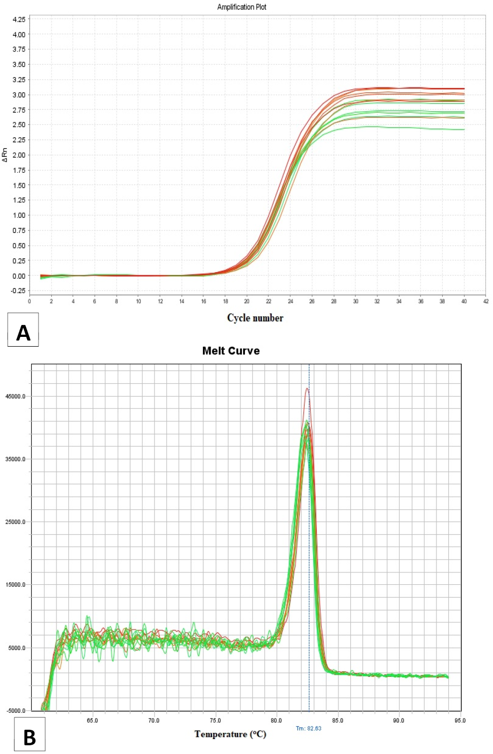
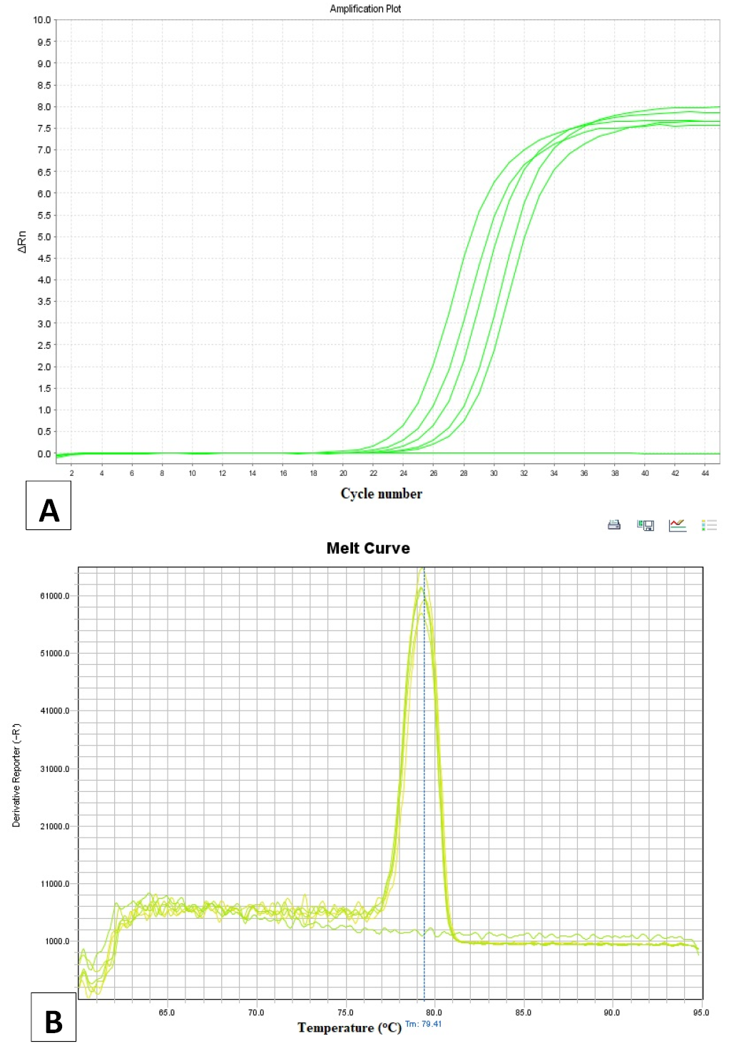
Reaction of q-PCR was performed on an ABI Step one Plus system (Applied Biosystems, USA) using the Universal One-Step RT-qPCR kit with SYBR Green dye. FUT1 and MUC4 primers for q-PCR were followed to study of Xia et al. (2016) and Dong WenHua et al. (2015), respectively. Three commonly reference genes including GAPDH (Glyceraldehyde 3-phosphate dehydrogenase), transferrin-binding protein1 (TBP1) and Actin beta (ACTB) was evaluated under laboratory conditions across the examined tissues. Based on these assessments, TBP1 was selected as housekeeping gene in this study. The information on primer sequences, PCR product size of FUT1, MUC4 and housekeeping genes are presented in Table 3. A q-PCR reaction conditions were initial step (15s at 95°C) and denaturation step (5s at 95°C and 34s at 62°C) for 40 cycles. Analysis of the melt curve was performed after the amplification. The peak values Tm (82.63 for FUT1 and 79.41 for MUC4) of the melt curve were used to evaluate the specificity of the amplification reactions. The Tm value of each sample at each region was an average of 3 replicates from the q-PCR reaction. The 2-ΔΔCt method was used to quantification for gene expression.
Statistical analysis
The effects of genotype (FUT1 or MUC4), tissue and gender on gene expression were assessed using the statistical mixed model using REML method as below:
Figure 1.
Where: yijkl = values of gene expression, μ = overall mean, Gi = fixed effect of genotype i (AA, AG and GG), Oj = fixed effect of tissue j (colon, duodenum and jejunum), Sk = fixed effect of gender k (male and female) IDl = random effect of animal l, and + εijklm = residual error. Additionally, the effect of genotype and gender on gene expression at each tissue was also estimated using the above statistical model without random (animal) and tissue effects.
Table 1: Primer sequences, PCR products, restriction enzymes, allele sizes of FUT1 and MUC4 and referenced methods.
Gene
SNP
Primer sequence
PCR product size (bp)
Restriction enzyme
Allele size (bp)
Reference
FUT1
G307A
F - CTTCAGCCAGGGCTCCTTTAAG
R - CTGCCTGAACGTCTATCAAGACC
421
Hin6I
A: 328, 93
G: 241, 93, 87
Meijerink et al. (1997)
MUC4
G243A
F – CAGGATGCCCAATGGCTCTAC
R - CCCCGAAGTTGTGAAAGGAAG
538
HhaI
A: 538
G: 295, 243
Peng et al. (2007)
Note: R: Reverse, F: Forward.
Table 2: Thermo cycling conditions of PCR reactions according to temperature (T, °C) and time (Time, sec).
Steps
FUT1
MUC4
T (°C)
Time (sec)
Number of cycles
T (°C)
Time (sec)
Number of cycles
Initial
94
180
1
95
180
1
Denaturation
94
45
35
95
45
30
Annealing
58
30
62.5
45
Extension
72
45
72
45
Final Extension
72
300
1
72
300
1
Table 3: Primer sequences, PCR product size of FUT1, MUC4 and housekeeping gene TBP1.
Gene
SNP
Primer sequence
PCR product size (bp)
Reference
FUT1
G307A
F - TTTTAAGCCCCCAAACTGCC
R - TAAATCGACCCCATCAGCCTC
126
MUC4
G243A
F - GGCCCACCTTAAGATTCCCA
R - GCTTCTCCTTAGCATGCCCAG
131
TBP1
F: AACAGTTCAGTAGTTATGAGC
R: AGATGTTCTCAAACGCTTCG
153
Note: R: Reverse, F: Forward.
Normality and homogeneity of variances were checked. These assumptions were met for FUT1 data but not with MUC4. Therefore, data transformation was applied for MUC4 using log10 of the initial value plus 9. All values for MUC4 are displayed in the log10 scale.
The statistical parameters were observation (n), Least Square Mean (LSM) and standard error (SE). The Tukey test was used for pairwise comparison between LSM. All data were performed using SAS® OnDemand for Academics.
RESULTS AND DISCUSSION
This study was conducted at a pig farm in northern Vietnam, a region climatically characterized by high ambient temperatures (approximately 28–35 °C), high relative humidity (often exceeding 80%), and frequent rainfall, reflecting a hot and humid monsoon season. These environmental factors may influence physiological status and gene expression in livestock. For example, heat and humidity can induce thermal stress, which may alter metabolic activity, immune responses, and intestinal function. Such changes could, in turn, affect the expression of genes such as FUT1 and MUC4, particularly in tissues associated with digestion and host–microbe interactions.
Figures 1 and 2 show the melting curves for the FUT1 and MUC4 qPCR products. We performed simultaneous amplification of multiple samples, which resulted in multiple peaks. However, the critical point is that these peaks cluster around nearly the same melting temperature (Tm), thereby confirming the specificity of the qPCR assay and suggesting the accurate quantification of the target genes.
Table 4: Gene expression of FUT1 and MUC4 according to genotype and tissue.
Tissue
Genotype
FUT1
MUC4
n
LSM
SE
n
LSM
SE
Overall
AA
9
0.785
0.305
12
0.999ab
0.009
AG
12
0.990
0.261
18
1.020a
0.008
GG
15
1.363
0.235
6
0.975b
0.014
P-value
0.314
0.0352
Colon
AA
3
0.688
0.337
4
1.021ab
0.019
AG
4
0.972
0.288
6
1.078a
0.016
GG
5
1.514
0.259
2
0.976b
0.029
P-value
0.189
0.0334
Duodenum
AA
3
0.899
0.416
4
0.999
0.015
AG
4
1.064
0.355
6
0.992
0.013
GG
5
1.236
0.321
2
0.977
0.023
P-value
0.818
0.7286
Jejunum
AA
3
0.768
0.284
4
0.976
0.006
AG
4
0.934
0.242
6
0.989
0.005
GG
5
1.339
0.219
2
0.973
0.010
P-value
0.290
0.2694
Within each gene, means with different superscript letters are significantly different (P<0.05).
The gene expression according to the genotypes of FUT1 and MUC4 genes is presented in Table 4. Three expected genotypes (AA, AG and GG) of each gene and their expressions were detected at all the selected tissues. The expression levels of FUT1 did not differ significantly among pigs carrying the AA, AG, and GG genotypes, either across all examined tissues or within each of the three intestinal tissues individually. These findings were
consistent with the previous study in Sutai piglets, which reported FUT1 expressions at 11 different tissues of 35-day-old pigs with the highly expression levels at lung, liver, stomach and duodenum . Similarly, the effect of FUT1 polymorphisms (AA, AG and GG) on gene expressions in each of four highly expressing tissues were not significant . The non-significant effect of FUT1 polymorphisms on gene expression in various tissues of pigs suggest that the expression of this gene may be regulated by other loci or factors.
On the other hand, MUC4 expression in the colon was
significantly higher in pigs with the AG genotype than associated with differences in the physiological status of the pigs, particularly during the post-weaning period. In Vietnam, piglets are typically weaned at 25–28 days of age; therefore, the period from 30 to 35 days is considered the post-weaning stage. This is a period during which piglets are highly susceptible to diarrhea; therefore, we hypothesize that genes associated with resistance to diarrhea may exhibit elevated expression during this stage. In the current study, the pigs were approximately 32.5 days old, corresponding to the early post-weaning stage, which is characterized by substantial intestinal stress, abrupt dietary transition, and marked alterations in gut microbiota composition. These physiological changes may strongly influence the transcriptional regulation of the MUC4 gene and consequently modify the relationship between genotype and gene expression. Supporting this hypothesis, Comelli et al. (2008) demonstrated that intestinal microbiota can regulate the mucus layer at the transcriptional level and that variations in bacterial composition significantly affect this process. Therefore, the higher MUC4 expression observed in pigs carrying the AG genotype compared with the GG genotype in the present study may reflect genotype-specific responses to weaning-associated intestinal stress and microbiota alterations.
In addition, previous studies have shown that MUC4 expression is influenced by age and developmental stage. Wenhua Dong et al. (2015) reported significantly higher MUC4 expression levels in pigs at 30 and 35 days of age compared with those at 8 and 18 days of age. This supports the view that the post-weaning period is a critical stage for MUC4 transcriptional activity. The discrepancy between studies may therefore be explained by differences in intestinal physiological conditions at the time of sampling, especially factors related to weaning stress and microbiota composition, rather than by genotype alone. Nevertheless, other factors such as genetic background, environmental conditions, diet, and methodological differences in tissue sampling, RNA extraction, normalization procedures, or statistical analyses may also contribute to the variation among studies. Moreover, only two individuals carrying the MUC4GG genotype were identified in the present study. Therefore, the limited sample size may have affected the statistical power of the analysis. Further studies with larger sample sizes are needed to draw more reliable conclusions regarding the effects of MUC4 genotypes on gene expression in tissues.
The gene expression of FUT1 and MUC4 in different tissues is presented in Table 5. FUT1 expression level was similar in all tested tissues (P=0.887) while the expression level of MUC4 in colon was significantly higher than which in duodenum and jejunum (P<0.001). The result of FUT1 expression in this study was consistent with previous articles, which reported that the expression of FUT1 gene was relatively high in lung, stomach, liver and duodenum, jejunum among 11 tissues ; a; . In addition, there was not any significant difference at FUT1 expression level in duodenal and jejunal tissues of experimental pigs in mentioned studies. According to previous study, post-weaning diarrhea in piglets normally associated with ETEC F18, which attach their F18 fimbriae to the specific receptors in the intestinal of the piglets . The piglets in this study were 32.5 day-old, in the period of post-weaning. Therefore, the relatively high expression of FUT1 in the intestinal tissues such as colon, duodenum and jejunum, where suggests a role for FUT1 in regulating the expression or structure of the ETEC F18 receptor.
Table 5: Gene expression of FUT1 and MUC4 according to tissues
Tissue
FUT1
MUC4
n
LSM
SE
n
LSM
SE
Colon
12
1.079
0.172
12
1.035a
0.009
Duodenum
12
1.046
0.172
12
0.984b
0.009
Jejunum
12
1.013
0.172
12
0.975b
0.009
P-value
0.887
0.0002
Within each gene, the LSMs with different superscript letters are significantly different (P<0.05).
In this study, MUC4 expressed in colon with significantly higher level (1.035) than those in duodenum (0.984) and jejunum (0.975). The result was consistent with the finding by Jacobsen et al. (2011), which observed the MUC4 expression in the colon was markedly higher than those in the jejunum and duodenum of crossbred pigs, respectively. A study on Sutai pigs reported highly expression of MUC4 gene in liver, spleen, stomach and intestinal tissues and the expression level were similar between duodenum and jejunum . It is widely known that mucin was generally expressed in the epithelial line of secretory systems including the digestive system, respiratory system and reproductive system and played an important role in lubrication and protection of the mucosal epithelium . Therefore, the relatively high expression of MUC4 observed in gastrointestinal tissues in the present study is likely related to its physiological role in maintaining mucosal integrity and epithelial protection. In this study, in colon, the expression levels of MUC4 gene in the pigs with the GG genotype (known as resistant genotype) were lower than in those with the AG genotype. It is hypothesized that, if the disease-resistant genotype exhibits higher gene expression during critical susceptible periods, it may contribute to enhancing resistance to pathogenic microorganisms in weaned piglets and reducing the incidence of post-weaning diarrhea. . Neverthless, resistance phenotypes were not evaluated, as no pathogen challenge experiment was performed. Therefore, the current data do not support a direct link between MUC4 expression and disease resistance in pigs. Any potential role of MUC4 in resistance to pathogens should be considered hypothetical and requires further investigation through studies integrating gene expression with phenotypic resistance data.
FUT1 gene expression levels in the colon, jejunum, and overall tissues were significantly higher in male pigs than in female pigs (P<0.05). However, no significant difference between sexes was observed in the duodenum (Table 6). For the MUC4 gene, expression levels in the colon were significantly higher in male pigs than in female pigs (P = 0.048). Inversely, no significant sex-related differences were observed in the duodenum, jejunum, or across the overall tissues. The physiological reason for this sex difference in pre-pubertal piglets is unclear. While hormonal influences are possible, they are less likely at this young age. Further research is needed to explore potential sex-linked genetic or epigenetic regulatory mechanisms.
Table 6: Gene expression of FUT1 and MUC4 according to gender and tissue
Tissue
Gender
FUT1
MUC4
n
LSM
SE
n
LSM
SE
Overall
Female
18
0.673b
0.213
18
0.987
0.009
Male
18
1.149a
0.213
18
1.009
0.007
P-value
0.024
0.0763
Colon
Female
6
0.610b
0.245
6
0.995b
0.019
Male
6
1.506a
0.235
6
1.055a
0.015
P-value
0.030
0.0480
Duodenum
Female
6
0.765
0.303
6
0.984
0.015
Male
6
1.367
0.290
6
0.994
0.012
P-value
0.189
0.6253
Jejunum
Female
6
0.645b
0.207
6
0.981
0.006
Male
6
1.382a
0.198
6
0.978
0.005
P-value
0.033
0.7020
Within the gene and each tissue, the LSMs with different superscript letters are significantly different (P<0.05).
In this study, all pigs were of the same age and maintained under identical dietary conditions; therefore, the effects of age and diet were not evaluated. Although previous studies have reported influences of these factors on gene expression and the expression of MUC4 in the intestinal tissues and of pigs were reported ; ; ; . On the other hand, the effects of age and breed a; , they are beyond the scope of the present data. Further studies are needed to clarify the potential role factors in regulating FUT1 and MUC4 expression in the intestinal tissues.
In presented study, given the limited sample size, the findings should be considered preliminary. Further studies with larger sample sizes and more robust experimental designs are required to validate these observations and clarify their biological and practical significance.
CONCLUSIONS
The results of this study show that FUT1 gene expression did not differ among genotypes in any examined tissues. In contrast, MUC4 expression was significantly higher in pigs with the AG genotype than in those with the GG genotype in the colon. However, given the very small number (n=2) of pigs with the GG genotype, this finding should be considered preliminary and requires validation in a larger cohort. FUT1 expression was similar among intestinal tissues, whereas MUC4 expression was significantly higher in the colon than in the duodenum and jejunum. In addition, male pigs showed higher FUT1 expression in the colon, jejunum, and overall tissues, while higher MUC4 expression in male pigs was observed only in the colon.
ACKNOWLEDGMENTS
This study was carried out in the framework of the project No: 11/FIRST/1.a/VNUA3 funded by FIRST (Fostering Innovation through Research, Science and Technology, Vietnam).
NOVELTY STATEMENT
This study provides novel evidence on the relationship between polymorphisms of the FUT1 (c.307G>A) and MUC4 (g.243A>G) genes and their expression levels in intestinal tissues of Yorkshire pigs under Vietnamese production conditions. Unlike previous studies focusing mainly on genotypephenotype associations, this work is among the first to investigate how these specific mutations influence gene expression in different intestinal tissues (colon, duodenum, and jejunum) in young pigs. In addition, the study highlights tissue-specific and sex-dependent differences in MUC4 expression, contributing new insights into the biological regulation of genes associated with disease resistance in pigs.
AUTHORS CONTRIBUTIONS
Do Duc Luc, Vu Dinh Ton, Nguyen Hoang Thinh; Conceptualization and design the experiment, supervision, editing and finalization; Do Thi Phuong; data collection, laboratory manipulation, data analysis, manuscript preparation; Phan Thi Tuoi; data analysis, manuscript preparation.
Generative AI and AI assisted technology statement
The authors declare that no generative AI and AI assisted technology was used in the creation of this manuscript.
Conflict of interest
The authors declare that there is no conflict of interests regarding the publication of this article.
Figures
Figure 1:.
Amplification curve (A) and dissociation curve (B) for the FUT1 gene.
Figure 2:.
Amplification curve (A) and dissociation curve (B) for the MUC4 gene.
References
Balcells I, Castelló A, Mercadé, A, Noguera JL, Fernández-Rodríguez A, Sànchez A, Tomàs A (2011). Analysis of porcine MUC4 gene as a candidate gene for prolificacy QTL on SSC13 in an Iberian× Meishan F2 population. BMC Genet., 12(93): 1-6. https://doi.org/10.1186/1471-2156-12-93[CrossRef]
Bao WB, Ye L, Pan ZY, Zhu J, Du ZD, Zhu GQ, Huang XG, Wu SL (2012a). The effect of mutation at M307 in FUT1 gene on susceptibility of Escherichia coli F18 and gene expression in Sutai piglets. Mol. Biol. Rep., 39(3): 3131-3136. https://doi.org/10.1007/s11033-011-1078-6[CrossRef]
Bao WB, Ye L, Pan ZY, Zhu J, Zhu GQ, Huang XG, Wu SL (2011). Beneficial genotype of swine FUT1 gene governing resistance to E. coli F18 is associated with important economic traits. J. Genet., 90(2): 315. https://doi.org/10.1007/s12041-011-0059-9[CrossRef]
Bao WB, Ye L, Zi C, Su XM, Pan ZY, Zhu J, Zhu GQ, Huang XG, Wu SL (2012b). Study on the age-dependent tissue expression of FUT1 gene in porcine and its relationship to E. coli F18 receptor. Gene, 497(2): 336-339. https://doi.org/10.1016/j.gene.2012.01.035[CrossRef]
Canibe N, Højberg O, Kongsted H, Vodolazska D, Lauridsen C, Nielsen TS, Schönherz AA (2022). Review on preventive measures to reduce post-weaning diarrhoea in piglets. Anim. (Basel), 12(19): 2585. https://doi.org/10.3390/ani12192585[CrossRef]
Comelli EM, Faure RM, Donnicola D, Mansourian R, Rochat F, Corthesy-Theulaz I, Cherbut C (2008). Multifaceted transcriptional regulation of the murine intestinal mucus layer by endogenous microbiota. Genomics, 91(1):70-7. https://doi.org/10.1016/j.ygeno.2007.09.006[CrossRef]
Dong WenHua HX, Xia RiWei, Sen W, Bao WenBin, Wu ShengLong (2016). Polymorphism of MUC4 gene and its effect on mRNA expression and cytokines level in large white pigs. Chin. J. Anim. Sci., 3(2): 1-6.
Dong WenHua ZQ, Gan Lina, Qin Weiyun, Zhu Guoqiang, Bao Wenbin, Wu Shenglong (2015). Age-dependent expression of MUC4 and MUC13 gene in Sutai (Duroc x Meishan) piglets. J. Sci., 5(11): 1024-1028.
Emam M, Livernois A, Paibomesai M, Atalla H, Mallard B (2019). Genetic and epigenetic regulation of immune response and resistance to infectious diseases in domestic ruminants. Vet. Clin. North Anim. Food Anim. Pract., 35(3): 405-429. https://doi.org/10.1016/j.cvfa.2019.07.002[CrossRef]
Fairbrother JM, Nadeau É, Gyles CL (2005). Escherichia coli in postweaning diarrhea in pigs: an update on bacterial types, pathogenesis, and prevention strategies. Anim. Health Res. Rev., 6(1): 17-39. https://doi.org/10.1079/AHR2005105[CrossRef]
Fratto A, Torricelli M, Sebastiani C, Ciullo M, Felici A, Biagetti M (2024). Survey on resistance occurrence for F4+ and F18+ enterotoxigenic Escherichia coli (ETEC) among pigs reared in Central Italy regions. Vet. Res. Commun., 48(2): 1279-1284 https://doi.org/10.1007/s11259-023-10287-8.[CrossRef]
Geraci C, Varzandi AR, Schiavo G, Bovo S, Ribani A, Utzeri VJ, Galimberti G, Buttazzoni L, Ovilo C, Gallo M (2019). Genetic markers associated with resistance to infectious diseases have no effects on production traits and haematological parameters in Italian Large White pigs. Livest. Sci., 223: 32-38. https://doi.org/10.1016/j.livsci.2019.03.003[CrossRef]
Jacobsen M, Cirera S, Joller D, Esteso G, Kracht SS, Edfors I, Bendixen C, Archibald AL, Vogeli P, Neuenschwander S (2011). Characterisation of five candidate genes within the ETEC F4ab/ac candidate region in pigs. BMC Res. Notes, 30(4): 1-8. https://doi.org/10.1186/1756-0500-4-225[CrossRef]
Kim K, Nguyen DT, Choi M., Kim JH., Seo HG, Dadi H, Cha SY, Seo K, Lee YM, Kim JJ (2013). Alpha (1, 2)-fucosyltransferase M307A polymorphism improves piglet survival. Anim. Biotechnol., 24(3): 243-250. https://doi.org/10.1080/10495398.2013.776964[CrossRef]
Kim K, Song M, Liu Y, Ji P (2022). Enterotoxigenic Escherichia coli infection of weaned pigs: Intestinal challenges and nutritional intervention to enhance disease resistance. Front. Immunol., 13: 885253. https://doi.org/10.3389/fimmu.2022.885253[CrossRef]
Liu Y, Xia RW, Yin XM, Huo YJ, Zhu GQ, Wu SL, Bao WB (2015a). Expression Level of FUT1 Gene in Different Pig Populations and its Relationship with ETEC F18 Resistance. Symbiosis Online J. Vet. Sci., 1(2): 1-5. https://doi.org/10.15226/2381-2907/1/2/00106[CrossRef]
Liu Y, Yin XM, Xia RW, Huo YJ, Zhu GQ, Wu SL, Bao WB (2015b). Association between the MUC4 g. 243A> G polymorphism and immune andproduction traits in Large White pigs. Turk. J. Vet. Anim. Sci., 39(2): 141-146. https://doi.org/10.3906/vet-1405-107[CrossRef]
Luc DD, Bo HX, Thinh NH., Hanh HV, Phuong DT, Manh TX, Hung NV, Ton VD, Farnir F (2022). Effects of FUT1 and MUC4 polymorphisms on sperm quality traits of Landrace and Yorkshire pigs under tropical conditions in northern Vietnam. J. Anim. Plant Sci., 32(4): 1159 - 1167. https://doi.org/10.36899/JAPS.2022.4.0521[CrossRef]
Luc DD, Thinh NH, Bo HX, Phuong DT, Tuoi PT, Ton VD, Farnir F (2023). Association between the MUC4 g. 243A> G polymorphism and production performance of Landrace and Yorkshire pigs in Vietnam. VJAS., 6(1): 1711-1718. https://doi.org/10.31817/vjas.2023.6.1.02[CrossRef]
Luc DD, Thinh NH, Bo HX, Vinh NT, Manh TX, Hung NV, Ton VD, Farnir F (2020). Mutation c. 307G> A in FUT1 gene has no effect on production performance of Yorkshire pigs in the tropics: the case of Vietnam. Can. J. Anim. Sci., 100(3): 426-431. https://doi.org/10.1139/cjas-2019-0084[CrossRef]
Luise D, Motta V, Bertocchi M, Salvarani C, Clavenzani P, Fanelli F, Pagotto U, Bosi P, Trevisi P (2019). Effect of Mucine 4 and Fucosyltransferase 1 genetic variants on gut homoeostasis of growing healthy pigs. J. Anim. Physiol. Anim. Nutr., 103(3): 801-812. https://doi.org/10.1111/jpn.13063[CrossRef]
Matías J, Berzosa M, Pastor Y, Irache J, Vaccination CGM (2017). Immunization of Sows during Pregnancy against ETEC Infections. Vaccines, 5(4): 48. https://doi.org/10.3390/vaccines5040048[CrossRef]
Meijerink E, Fries R, Vögeli P, Masabanda J, Wigger G, Stricker C, Neuenschwander S, Bertschinger H, Stranzinger G (1997). Two α (1, 2) fucosyltransferase genes on porcine chromosome 6q11 are closely linked to the blood group inhibitor (S) and Escherichia coli F18 receptor (ECF18R) loci. Mamm. Genome., 8(10): 736-741. https://doi.org/10.1007/s003359900556[CrossRef]
Ministry of Science and Technology (2010). Animal disease - Necropsy procedure. Vietnam quality standards institute, hanoi, Vietnam.
Peng QL, Ren J, Yan XM, Huang X, Tang H, Wang YZ, Zhang B, Huang LS (2007). The g. 243A> G mutation in intron 17 of MUC4 is significantly associated with susceptibility/resistance to ETEC F4ab/ac infection in pigs. Anim. Genet., 38(4): 397-400. https://doi.org/10.1111/j.1365-2052.2007.01608.x[CrossRef]
Ruan X, Liu M, Casey TA, Zhang W (2011). A tripartite fusion, FaeG-FedF-LT192A2: B, of enterotoxigenic Escherichia coli (ETEC) elicits antibodies that neutralize cholera toxin, inhibit adherence of K88 (F4) and F18 fimbriae, and protect pigs against K88ac/heat-labile toxin infection. Clin. Vaccine Immunol., 18(10): 1593-1599. https://doi.org/10.1128/CVI.05120-11[CrossRef]
Sambrook J, Fritsch E, Maniatis T (1989). Molecular cloning: a laboratory manual. 2nd edn, cold spring harbor laboratory, New York, USA.
Sargeant HR, McDowall KJ, Miller HM, Shaw MA (2010). Dietary zinc oxide affects the expression of genes associated with inflammation: transcriptome analysis in piglets challenged with ETEC K88. Vet. Immunol. Immunopathol., 137(1-2): 120-129. https://doi.org/10.1016/j.vetimm.2010.05.001[CrossRef]
Schroyen M, Stinckens A, Verhelst R, Niewold T, Buys N (2012). The search for the gene mutations underlying enterotoxigenic Escherichia coli F4ab/ac susceptibility in pigs: a review. Vet. Res., 43(1): 1-8. https://doi.org/10.1186/1297-9716-43-70[CrossRef]
Shakuntala I, Sanjukta R, Das S, Puro K, Ghatak S, Rajkhowa S, Milton A, Sen A (2018). Occurrence and characterization of Escherichia coli isolated from Diarrhoeic piglets in Meghalaya. ỊHF. Special issue: 88-92.
Smith A, Ryan M, O’Doherty J, Reilly P, Bahar B, Sweeney T (2010). Effects of dietary supplementation with laminarin derived from Laminaria hyperborea and Laminaria digitata on colonic mucin gene expression in pigs. Livest. Sci., 133(1-3): 204-206. https://doi.org/10.1016/j.livsci.2010.06.064[CrossRef]
Sukhno V, Vashchenko P, Saenko A, Zhukorskyi O, Tserenyuk O, Kryhina N (2022). Association of Fut1 and Slc11a1 gene polymorphisms with productivity traits of Large White pigs. Regul. Mech. Biosyst., 13(3): 225-230. https://doi.org/10.15421/022229[CrossRef]
Święch E, Barszcz M, Taciak M, Gawin K, Tuśnio A, Konopka A, Skomiał J (2025). Mucin gene expression in the large intestine of young pigs: The effect of dietary level of two types of chicory inulin. Nutr., 131: 112635. https://doi.org/10.1016/j.nut.2024.112635[CrossRef]
Wenhua Dong, Qiufang Zong, Gan Lina, Qin Weiyun, Zhu Guoqiang, BaoWenbin, Wu Shenglong (2015). Age-dependent expressionof MUC4 and MUC13 gene in Sutai (Duroc x Meishan) piglets. J. Sci., 5(11): 1024-1028.
Vögeli P, Meijerink E, Fries R, Neuenschwander S, Vorländer N, Stranzinger G, Bertschinger H (1997). A molecular test for the detection of E. coli F18 receptors: A breakthrough in the struggle against edema disease and post-weaning diarrhea in swine. Schweiz. Arch. fur Tierheilkd., 139(11): 479-484.
Xia R, Qin W, Gan L, Wang J, Wu S, Bao W (2016). Differential expression of FUT1 and FUT2 in Large White, Meishan, and Sutai porcine breeds. Genet Mol. Res., 15(1): 15017613-15017613. https://doi.org/10.4238/gmr.15017613[CrossRef]
Zhou L, Fang L, Sun Y, Su Y, Zhu W (2017). Effects of a diet high in resistant starch on fermentation end‐products of protein and mucin secretion in the colons of pigs. Starch‐Stärke., 69(7-8): 1600032. https://doi.org/10.1002/star.201600032[CrossRef]