Research Article · Volume 14, Issue 4, 12 September 2026 · Open Access
Dietary Organic Iron Supplementation Improves Egg Production, Feed Efficiency, Egg Quality, and Egg Iron Content in Laying Quails
· · · · · · · ·
Affiliations & Notes ⌄
1Department of Animal Nutrition and Feed Technology, Faculty of Animal Science, IPB University, 16680 Bogor, Indonesia
2Department of Animal Nutrition and Feed Technology, Faculty of Animal Science, Andalas University, 25163 Padang, Indonesia. *Correspondence | Arif Darmawan, Department of Animal Nutrition and Feed Technology, Faculty of Animal Science, IPB University, 16680 Bogor, Indonesia
The present study revealed that Fe inclusion significantly increased egg production and egg mass and reduced FCR.
Meanwhile, supplementation with 100 ppm Fe increased duodenum length in this study.
The results indicated that the treatment did not affect erythrocytes, hemoglobin, PCV, and leukocyte differentiation, but the inclusion of 100 ppm and 150 ppm Fe in the diet reduced the number of leukocytes.
Inclusion of up to 150 ppm organic Fe in feed increases egg production, feed efficiency, egg Fe content, eggshell weight, and yolk color without adversely affecting health status, visceral organs, and reproductive organs.
Selected from the article's own text.
Abstract
The objective of this study was to evaluate the inclusion of organic iron (Fe) on productive performance, egg quality, egg Fe content, blood hematology, and visceral and reproductive organs of laying quail. This study used 300 laying quails allocated to 4 treatments with 5 replicates according to a Completely Randomized Design. The treatments were T1: control diet without Fe supplementation. T2: control diet supplemented with 50 ppm Fe; T3: control diet supplemented with 100 ppm Fe; T4: control diet supplemented with 150 ppm Fe. The inclusion of 150 ppm Fe in the diet yielded the highest egg production, egg mass, egg Fe level, and decreased FCR value (P<0.05). Feed intake did not show a significant difference (P>0.05). Meanwhile, inclusion of 100 ppm Fe improved the relative length of the duodenum (P<0.05), but the treatments did not affect other visceral organs (P>0.05). The inclusion of 150 ppm Fe significantly increased (P<0.05) the yolk color score and percentage of eggshell weight compared to the control and 50 ppm Fe groups. The leukocyte level was decreased by the diet supplemented with more than 50 ppm Fe (P<0.05). The study concluded that supplementation of up to 150 ppm Fe enhanced egg production, feed efficiency, egg Fe content, and egg quality without adversely affecting health status, visceral organs, and reproductive organs.
Iron (Fe) deficiency is a global health issue affecting more than 2 billion people worldwide, with a prevalence estimated at approximately 40% in developing countries and around 10% in developed countries . Producing quail eggs with high iron content is important as one strategy for addressing this problem. Additionally, Fe is an essential micromineral that has an important role in various physiological processes, including oxygen transport and storage, energy metabolism, hemoglobin formation, and antioxidant and immune system functions ; . As a cofactor for various enzymes, Fe status greatly determines the physiological efficiency and productivity of laying quails. Therefore, adequate Fe in feed is one of the important factors in supporting reproductive performance and egg quality .
Previously, supplementation with organic Fe with high bioavailability had the potential to enhance productive performance and egg quality. Fe inclusion in the diet has been shown to improve egg production and egg quality ; ; . Fe contributes to oxygen supply to reproductive organs and has a crucial role in follicle development and egg component synthesis . Additionally, Fe is involved in the regulation of follicle-stimulating hormone and luteinizing hormone, which control follicle maturation and the ovulation process ; . Sarlak et al. (2025) found that the addition of 120 ppm organic Fe improved egg production and weight, whereas 150 ppm Fe had no negative impact on immune organs, including the bursa of Fabricius, spleen, and thymus .
Although the potential of Fe in improving production performance and egg quality had been reported, its evaluation of hematology parameters, reproductive organs, and egg Fe content, especially in laying quails, was still limited and showed inconsistent results. Therefore, our study aimed to evaluate the impact of organic Fe (Fe glycinate) inclusion in the diet at various levels on productive performance parameters, egg quality, egg Fe content, blood hematology, and reproductive and visceral organs. This study hypothesized that organic Fe supplementation at a certain level could improve the productive performance and egg quality of laying quails without causing a negative impact on vital organs, including reproductive organs and immunity.
MATERIALS AND METHODS
Animals, diet, and experimental design
A total of 300 four-week-old laying quails (Coturnix coturnix japonica) were randomly allocated to cages (40 cm x 40 cm x 30 cm) and reared for eight weeks. Each cage was equipped with a feeder and drinker to supply ad libitum drinking water during rearing. The ambient temperature was recorded at 25-32°C with lighting maintained at 16 hours.
The mash diet was formulated based on the nutritional requirements of laying quails in the production phase, with crude protein of 18% and metabolizable energy of 2970 kcal/kg. Feed ingredients included corn, rice bran, soybean meal, fish meal, Lemuru fish oil, CaCO₃, salt, DL-methionine,NaCl, vitamin-mineral premix, and Fe glycinate (Table 1).
The study used a completely randomized design with four treatments and five replicates, each consisting of 15 quails per replicate. The treatments consisted of T1: control diet without Fe supplementation; T2: control diet supplemented with 50 ppm Fe; T3: control diet supplemented with 100 ppm Fe; T4: control diet supplemented with 150 ppm Fe.
Table 1: Composition and nutrient content of the diet.
Feed ingredients
Amount (%)
Yellow corn (ME: 3,300 kcal/kg)
56.0
Soybean meal (CP: 48%)
17.0
Pollard (ME: 3,000 kcal/kg)
3.20
Fish meal (CP: 60%)
8.50
Rice bran (ME: 1,900 kcal/kg)
4.50
DL-methionine
0.50
Dicalcium phosphate
0.50
CaCo3
6.00
NaCl
0.30
Lemuru fish oil (ME: 8,280 kcal/kg)
3.00
Premix*
0.50
Nutrient content (%; based on calculation)
Dry matter
88.2
ME (kcal/kg)
2970
Crude protein
18.3
Crude fat
4.85
Crude fiber
3.04
Calcium
3.20
Available phosphorus
0.58
Na
0.21
Cl
0.27
Lysine
0.82
Methionine
0.92
Methionine + cystine
0.82
CP: Crude protein, ME: Metabolizable energy. *The vitamin–mineral premix per kilogram supplied Vit A (1,200,000 IU), Vit D₃ (200,000 IU), Vit E (800 IU), Vit K₃ (200 mg), Vit B₂ (500 mg), Vit B₆ (50 mg), Vit B₁₂ (1,200 µg), Vit C (2,500 mg), Ca-D-pantothenate (600 mg), Vit B₃ (400 mg), and choline Cl (1,000 mg), Met (3,000 mg) and Lys (3,000 mg), together with Mn (1,200 mg), I (20 mg), Zn (10,000 mg), Co (20 mg), and Cu (400 mg). The Fe contents in the diet were 117.04, 167.04, 217.04, 267.04 mg/kg for diets supplemented with 0, 50, 100, and 150 mg Fe/kg as Fe-glycine, respectively.
Data collection
Production performance
Feed intake was measured by subtracting the amount of feed provided from the daily feed remaining and expressed as grams per bird per day. Feed conversion ratio was determined from the total feed consumption divided by the total weight of eggs produced. Quail-day production was recorded daily and calculated from the number of eggs divided by the number of quails.
Egg quality and Fe content
A total of 60 eggs (3 eggs from each replicate) were collected at 12 weeks of age to analyze egg quality and Fe content, including egg index, albumen weight, yolk weight, eggshell thickness, eggshell weight, Haugh unit, yolk color, and eggshell strength. Eggshell thickness (mm) was measured from the average of three points (blunt, sharp, middle) using a digital micrometer, while eggshell strength was measured with the RH-DQ200 Eggshell Strength Tester. Egg component weights were measured using digital scales with an accuracy of 0.01 g. Yolk color and Haugh Unit were assessed using the Digital Egg Analyzer (EMT-7300). Fe contents in egg were analyzed using Atomic Absorption Spectroscopy according to AOAC (2005).
Blood hematology
At twelve-week-old quails, blood samples were taken from 60 quails (3 quails per replication) through the brachial vein using a 2 mL sterile syringe. The collected blood was transferred to an EDTA tube for hematological evaluation (erythrocytes and leukocytes, hematocrit, hemoglobin concentration, basophils, neutrophils, lymphocytes, and heterophils). Hematological analysis was determined according to the procedure described by Sastradipraja et al. (1989).
Visceral and reproductive organs
Twelve-week-old quails were fasted for 12 hours (water was still available), then weighed to obtain their slaughter weight. A total of 60 quails (3 quails per replicate) with weights close to the average were slaughtered. After defeathering, the visceral and reproductive organs (infundibulum, magnum, isthmus, and uterus) were removed and weighed using digital scales accurate to 0.1 g. Meanwhile, the length of the digestive and reproductive tracts was measured using a measuring tape (cm) and expressed in cm per 100 g. The visceral organs measured included liver, heart, kidney, ventriculus, proventriculus, pancreas, spleen, thymus, duodenum, jejunum, ileum, cecum, and colon. The organ weight percentage was calculated relative to the slaughter weight multiplied by 100%.
Statistical analysis
Data were tested for normality using the Shapiro–Wilk test before ANOVA analysis. Differences between treatments were examined using Tukey’s test at a significance level of P<0.05. All analyses were performed using IBM SPSS Statistics 22, and results were presented as mean ± standard deviation and P value.
RESULTS
Productive performances
The treatment did not result in significant differences in feed intake and egg weight (P>0.05). However, egg production increased significantly with increasing Fe doses (P<0.05). Supplementation of 150 ppm Fe in the diet significantly improved egg production compared to other treatments. Egg mass in the 150 ppm Fe group was higher than in the 100 ppm Fe and control groups (P<0.05). Feed conversion ratio decreased with increasing Fe supplementation. Supplementation with 150 ppm Fe significantly improved compared to the control group (P<0.05) (Table 2).
Egg quality and EGG Fe content
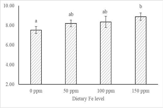
The results showed that the egg index, albumen and yolk indices, percentage of white and yolk weight, eggshell thickness, and Haugh Unit did not differ significantly (P>0.05) among treatments. However, the 150 ppm Fe group significantly increased (P<0.05) the yolk color score and percentage of eggshell weight compared to the control and 50 ppm Fe groups. Supplementation of 50 ppm had the lowest eggshell strength (P<0.05) (Table 3). Supplementation of 150 ppm Fe significantly increased egg Fe deposits compared to the control group (P<0.05) but was not different from the 50 ppm and 100 ppm groups (Figure 1).
Table 2: Productive performances of laying quail fed a diet supplemented with organic Fe.
Parameters
Fe level
P-Value
0 ppm
50 ppm
100 ppm
150 ppm
Feed intake (g/bird/day)
25.4±1.28
25.0±0.75
26.7±0.88
26.1±0.90
0.27
Egg weight (g/egg)
10.7± 0.09
10.7±0.11
10.7± 0.21
10.6±0.22
0.84
Quail day production (%)
64.3±5.04b
67.7±4.12b
69.7±5.75b
77.8±4.83a
0.01
Egg mass (g/bird/day)
6.49 ± 1.01b
7.23±0.37ab
6.96±1.32b
8.27±0.62a
0.04
Feed conversion ratio
3.58±0.26b
3.30±0.10ab
3.28±0.31ab
2.98±0.24a
0.01
Note: Lowercase superscripts on the same row represent significant differences among treatments.
Table 3: Egg quality of laying quail fed a diet supplemented with organic Fe.
Parameters
Fe level
P-Value
0 ppm
50 ppm
100 ppm
150 ppm
Egg index
81.2±2.86
83.4±5.54
83.3±8.00
81.4±4.05
0.96
Albumen index
0.12±0.01
0.13±0.03
0.13±0.02
0.13±0.02
0.67
Egg yolk index
0.44±0.03
0.44±0.03
0.45±0.02
0.44±0.02
0.59
Egg yolk weight (%)
36.6±5.25
33.9±4.69
33.9±2.93
33.4±6.63
0.76
Albumen weight (%)
49.9±4.85
52.5±4.23
52.0±2.83
51.9±6.39
0.65
Eggshell weight (%)
13.6±0.06b
13.6±0.79b
14.1±0.30ab
14.7±0.77a
< 0.01
Eggshell Thickness (mm)
0.22±0.01
0.21±0.00
0.21±0.02
0.22±0.01
0.34
Haugh unit
90.6±1.28
92.4±3.12
90.9±1.50
91.5±1.97
0.56
Yolk color score
6.50±0.31b
6.80±0.27b
7.20±0.48a
7.25±0.01a
<0.01
Eggshell strength (N)
10.8±1.27a
9.27±0.90b
10.8±1.23a
11.1±1.01a
<0.01
Note: Lowercase superscripts on the same row represent significant differences among treatments.
Table 4: Blood hematology of laying quail fed a diet supplemented with organic Fe
Parameters
Fe level
P-values
0 ppm
50 ppm
100 ppm
150 ppm
Erythrocytes (106/ mm3)
3.19±0.84
3.30±0.18
3.33±0.96
3.61±0.94
0.95
Leukocytes (103/ mm3)
22.0±3.97b
22.5±3.79b
16.6±2.19a
16.3±3.95a
0.03
Hemoglobin (g%)
12.2±0.91
13.1±0.93
11.7±2.23
12.4±1.19
0.40
Packed cell volume (%)
37.2±4.76
43.2±2.17
39.8±4.76
42.0±3.24
0.12
Leukocyte differentiation (%)
Lymphocytes
49.2±1.17
48.7±2.70
48.7±4.45
46.7±2.62
0.58
Heterophils
32.1±3.52
33.1±3.39
34.9±2.62
33.6±2.95
0.59
Eosinophils
9.62±1.85
9.23±1.24
9.56±2.18
10.5±1.83
0.73
Monocytes
8.27±1.92
8.17±2.40
6.04±1.59
8.37±1.89
0.17
Basophils
0.84±0.06
0.81±0.04
0.84±0.03
0.81±0.05
0.80
Note: Lowercase superscripts on the same row represent significant differences among treatments.
Table 5: Reproductive organs of laying quail fed a diet supplemented with organic Fe.
Parameters
Fe level
P-value
0 ppm
50 ppm
100 ppm
150 ppm
Infundibulum
Weight (g)
0.15±0.03
0.14±0.03
0.13±0.03
0.15±0.04
Length (cm/100g)
2.66±0.42
2.54±0.27
2.54±0.89
2.24±1.20
0.85
Magnum
Weight (g)
1.75±0.16
1.74±0.07
1.78±0.13
1.59±0.11
Length (cm/100g)
8.47±1.02
8.23±1.04
8.94±0.25
8.26±1.09
0.65
Isthmus
Weight (g)
0.46±0.09
0.41±0.03
0.38±0.09
0.42±0.07
Length (cm/100g)
3.54±0.59
3.15±0.43
3.22±0.36
3.66±0.66
0.41
Uterus
Weight (g)
1.02±0.17
0.97±0.08
0.94±0.07
0.87±0.17
Length (cm/100g)
2.45±0.28
2.44±0.65
2.33±0.30
2.52±0.40
0.92
Vagina
Weight (g)
0.33±0.11
0.33±0.13
0.35±0.16
0.29±0.10
Length (cm/100g)
1.89±0.33
2.13±0.36
1.68±1.01
1.68±0.24
0.63
Blood hematology
The erythrocytes, hemoglobin, PCV, and leukocyte differentiation (lymphocytes, heterophils, eosinophils, monocytes, basophils) did not show significant differences among treatments (P > 0.05), but the inclusion of 100 ppm and 150 ppm Fe in the feed reduced the number of leukocytes (P < 0.05) (Table 4).
Visceral and reproductive organs
The weight and length of the reproductive organ (infundibulum, magnum, isthmus, uterus, and vagina) were not significantly different among treatments (P>0.05) (Table 5). Similarly, the weight and relative percentage of the duodenum, jejunum, ileum, cecum, and colon did not differ significantly, except that the relative length of the duodenum increased significantly (P< 0.05) at a dose of 100 ppm Fe (Table 6).
Table 6: Visceral organs of laying quail fed a diet supplemented with organic Fe
Parameters
Fe level
P value
0 ppm
50 ppm
100 ppm
150 ppm
Liver
(g)
7.33±0.63
7.35±0.71
6.94±0.76
6.66±1.31
(%)
4.05±0.75
3.75±0.48
3.70±0.34
3.46±0.55
0.45
Heart
(g)
1.29±0.17
1.29±0.14
1.24±0.22
1.35±0.09
(%)
0.71±0.09
0.65±0.05
0.66±0.11
0.71±0.06
0.65
Kidney
(g)
0.73±0.26
0.87±0.12
0.59±0.28
0.62±0.26
(%)
0.40±0.16
0.45±0.04
0.32±0.16
0.33±0.16
0.38
Ventriculus
(g)
4.30±1.00
4.48±0.62
3.89±1.03
3.94±1.23
(%)
2.32±0.38
2.26±0.31
2.08±0.53
2.02±0.51
0.95
Proventriculus
(g)
0.76±0.17
0.86±0.10
0.87±0.10
0.80±0.11
(%)
0.41±0.07
0.47±0.06
0.46±0.05
0.42±0.03
0.18
Pancreas
(g)
0.44±0.20
0.32±0.15
0.36±0.05
0.42±0.11
(%)
0.23±0.09
0.16±0.02
0.18±0.03
0.22±0.05
0.32
Spleen
(g)
0.14±0.06
0.17±0.02
0.14±0.04
0.18±0.08
(%)
0.08±0.03
0.09±0.01
0.07±0.02
0.09±0.05
0.80
Thymus
(g)
0.15±0.09
0.17±0.07
0.10±0.03
0.09±0.06
(%)
0.08±0.05
0.08±0.03
0.05±0.02
0.04±0.02
0.11
Duodenum
(g)
1.86±0.39
2.08±0.67
1.98±0.33
1.56±0.13
(%)
1.00±0.15
1.04±0.30
1.04±0.18
0.82±0.09
0.21
(cm /100 g)
7.58±0.41ab
7.40±0.75ab
8.65±0.83b
7.02±0.31a
0.004
Jejunum
(g)
2.09±0.50
2.29±0.21
1.82±0.27
2.39±0.27
(%)
1.12±0.21
1.16±0.11
0.97±0.15
1.25±0.17
0.09
(cm /100 g)
15.2±1.89
15.9±2.70
14.7±2.03
17.7±1.20
0.14
Ileum
(g)
1.26±0.09
1.33±0.11
1.29±0.13
1.32±0.10
(%)
0.69±0.10
0.68±0.08
0.70±0.08
0.70±0.08
0.89
(cm /100g)
15.6±0.79
15.8±1.11
17.2±1.38
15.6±2.07
0.26
Cecum
(g)
0.88±0.08
0.88±0.05
0.90±0.09
0.90±0.11
(%)
0.48±0.04
0.45±0.04
0.48±0.04
0.47±0.05
0.54
(cm /100g)
6.48±0.83
6.06±0.70
6.16±0.58
6.08±0.94
0.81
Colon
(g)
0.33±0.04
0.36±0.06
0.38±0.05
0.35±0.03
(%)
0.18±0.03
0.18±0.02
0.20±0.02
0.18±0.02
0.15
(cm/100 g)
2.36±0.19
2.18±0.52
2.34±0.20
2.26±0.27
0.65
Note: Lowercase superscripts on the same row represent significant differences among treatments.
DISCUSSION
Fe acts as an essential cofactor for various enzymes involved in the transport and storage of oxygen in the body. In addition, this mineral functions in protein and energy metabolism, and contributes to increasing the capacity of the antioxidant and immune system ; . The present study revealed that Fe inclusion significantly increased egg production and egg mass and reduced FCR. Similarly, adding 120 ppm Fe-Glycine and 75 ppm Fe-Glycine improved egg production, egg weight, and feed efficiency. The beneficial effects of Fe are due to Fe as a hemoglobin component, which plays a role in oxygen availability, ATP production, and hormonal functions that regulate follicle development and ovulation ; . Moreover, Fe has a crucial function in the antioxidant defense system through antioxidant enzymes such as catalase and peroxidase , which help protect oocytes and oviduct tissue from oxidative damage . This protection may allow for greater energy allocation toward the egg formation process, which impacts the increased feed utilization efficiency in this study. In addition, the 150 ppm Fe-group differed significantly from the control group in egg iron deposits (Figure 1), which was in line with Xie et al. (2019). This may be due to chelation with amino acids, which increases the permeability of iron in the form of Fe-glycine into the membrane, thereby enhancing iron absorption and accumulation in the eggs. Indeed, organic minerals have high bioavailability as they are absorbed through the amino acid transport pathway, which reduces competition with other nutrients and enhances mineral retention in the body .The inclusion of up to 150 ppm organic Fe in the diet had no negative effect on reproductive organs. This was evident from the weight and length of the reproductive tract (infundibulum, magnum, isthmus, uterus, and vagina), which did not differ among treatments (Table 5). To our knowledge, no studies have discussed the effects of Fe on the reproductive organ parameters of laying birds; however, the great egg production in the 150 ppm group in our study suggested that the response of laying quails to Fe was more evident in physiological function than in changes in reproductive organ size. Furthermore, the reproductive organs play a role in the formation and transport of eggs, and their development depends on adequate nutrition, including minerals that play a crucial role in enzymatic activity, hormonal regulation, and the development of the reproductive tract. However, the development of the reproductive organs reaches its maximum at sexual maturity and tends to stabilize thereafter . Similarly, the absence of differences in reproductive organ size in this study indicated that Fe supplementation did not affect the development of reproductive organs in sexually mature quails. The benefits of Fe on physiological function may be a key mechanism explaining how Fe improves reproductive performance in quails, where Fe plays a prominent role in internal processes such as albumin synthesis, mineral deposition, oxygen transport, and metabolic efficiency .
In the present study, dietary 100 ppm and 150 ppm organic Fe increased eggshell weight, eggshell strength, and egg yolk color. This eggshell quality improvement was supported by the results of Tu et al. (2004), Xie et al. (2019), and Cao et al. (2023), which stated that organic Fe improved mineral deposition and strengthened the eggshell structure more efficiently than inorganic Fe. Indeed, Fe deficiency was known to reduce calcium deposition in eggshells, thereby decreasing their weight, thickness, and strength ; . Also, Collins et al. (2010) stated that Fe deficiency lowers Cu concentrations in tissues, while dietary Cu levels strongly influence eggshell strength . Conversely, sufficient organic trace minerals, including Fe, support the metabolic process of shell formation by increasing mineral density, strengthening the palisade layer, and stimulating the synthesis of organic matrix proteins such as osteopontin and ovocleidin-116, which play a role in mineral deposition . Meanwhile, egg yolk color also increased significantly at organic 100 to 150 ppm Fe supplementation, indicating increased pigment deposition . The positive effect of Fe on yolk color enhancement is due to organic Fe with high bioavailability, increasing lipid metabolism and xanthophyll absorption as the main pigment of yolk ; . Furthermore, Fe is an important component of the succinate dehydrogenase enzyme in the tricarboxylic acid cycle. An improvement in energy metabolism capacity supports the efficiency of physiological processes, including eggshell formation and lipid metabolism ; .
The treatment did not affect the weight of the liver, heart, kidneys, ventricles, proventriculus, pancreas, spleen, or thymus. Similarly, the weight and relative percentage of the duodenum, jejunum, ileum, cecum, and colon did not differ significantly, except that the relative length of the duodenum increased significantly at a dose of 100 ppm Fe. In line with Gou et al. (2020), Lin et al. (2020), and Akbari et al. (2025) stated that the weight of the thymus, spleen, and bursa of Fabricius was not affected by Fe supplementation up to a dose of 150 ppm. The thymus functions as the primary immune organ responsible for T lymphocyte formation and maturation, while the spleen acts as the primary peripheral immune organ in poultry, which is abundant in lymphocytes . Indeed, larger immune organs reflect stronger immune function, and Fe deficiency can interfere with their development and reduce disease resistance . Meanwhile, supplementation with 100 ppm Fe increased duodenum length in this study. Similar to previous studies, an increase in chicken duodenum length was accompanied by an increase in villi height and surface area with Fe sulfate supplementation in the diet . Also, Zhou et al. (2020) stated that Fe supplementation affected intestinal development by improving intestinal weight and length, supporting overall intestinal health, and stimulating intestinal epithelial maturation. The beneficial effects of Fe on duodenum morphology were due to the fact that the greatest Fe absorption occurred in the duodenum , which impacted mineral availability, metabolic activity, and promoted intestinal epithelial cell proliferation (Minor et al., 2010; .
The results indicated that the treatment did not affect erythrocytes, hemoglobin, PCV, and leukocyte differentiation, but the inclusion of 100 ppm and 150 ppm Fe in the diet reduced the number of leukocytes. This phenomenon is similar to the results of Olgun et al. (2022) that organic Fe supplementation can alter immune cells, specifically, number of leukocytes were lower at high dose (200 ppm) compared to the 150 ppm dose of Fe. In contrast, the administration of 100 ppm Fe in the laying hens’ diet did not affect the levels of white blood cells and serum monocytes . Organic minerals are superior due to their higher bioavailability compared to inorganic forms. Additionally, organic minerals chelated with amino acids form more stable complexes in the digestive tract and reduce antagonistic interactions with other nutrients . In this study, high-dose organic Fe supplementation may reduce leukocyte counts due to its very high bioavailability and may increase the risk of oxidative stress. High doses of chelated Fe have the potential to trigger pro-oxidative reactions and form hydroxyl radicals, which cause immunosuppression and leukocyte suppression . However, the decrease in leukocyte count did not affect the quail’s health status as evidenced by an increase in egg production.
CONCLUSIONS
Inclusion of up to 150 ppm organic Fe in feed increases egg production, feed efficiency, egg Fe content, eggshell weight, and yolk color without adversely affecting health status, visceral organs, and reproductive organs. These findings suggest the use of organic Fe as a nutritional strategy for improving the performance of laying quail.
ACKNOWLEDGEMENTS
We would like to thank all those who have contributed to our study, especially the Directorate General of Research and Development, Ministry of Higher Education, Science, and Technology, Indonesia, for providing funding under Research Contract No. 006/C3/DT.05.00/PL/2025.
NOVELTY STATEMENT
This study evaluates the effect of organic Fe supplementation on the diet on performance, egg quality, egg Fe content, blood hematology parameters, and visceral and reproductive organs in laying quails. Studies on the effects of organic Fe supplementation in laying quails, particularly on hematological parameters, reproductive organs, and egg iron deposition, are still limited and show inconsistent results. Therefore, this study provides new evidence on the potential of organic iron as a nutritional strategy to improve quail performance and egg quality.
AUTHOR’S CONTRIBUTION
AD, WH, RKR: Conceptualisation, methodology, data validation, draft writing, and review. FAR, MRR, NA, SNSM: Investigation, data collection, and draft writing. MUS, RN: Interpreting data, draft writing, and editing. All authors have read and approved the final version of the manuscript.
Ethical approval
All procedures in this study were approved by the animal ethics committee, IPB University, with the number 343-2025.
Generative AI and AI-assisted technology statement
The authors state that no generative AI and AI-assisted technology was used in writing the article.
Conflict of interest
The authors have declared no conflict of interest.
Figures
Figure 1:.
Egg Fe content (mg/100g, P=0.006).
References
Abou-Shehema BM, Rizk RE, El-Deken MR, Ahmed MR, Fares WA, Khalifah AM (2025). Assessing the role of organic iron supplementation in enhancing productive and reproductive performance in chickens at two different ages. J. Anim. Health Prod., 13: 1127–1139. https://doi.org/10.17582/journal.jahp/2025/13.4.1127.1139[Full text][CrossRef]
Akbari N, Shahir MH, Tzschentke B, Jafari Anarkooli I (2025). Effects of dietary iron concentrations on growth performance, ascites incidence, blood biochemical parameters, and antioxidant status of broiler chickens in ascites-inducing condition. Anim. Feed Sci. Technol., 319: 116185. https://doi.org/10.1016/j.anifeedsci.2024.116185[CrossRef]
AOAC (2005). International official methods of analysis. 18th ed. Association of Official Analytical Chemists, Maryland, USA.
Cao J, Zhu J, Zhou Q, Zhao L, Zou C, Guo Y, Curtin B, Ji F, Liu B, Yu D (2023). Efficacy evaluation of novel organic iron complexes in laying hens: effects on laying performance, egg quality, egg iron content, and blood biochemical parameters. Anim. Biosci., 36: 498–505. https://doi.org/10.5713/ab.22.0086[CrossRef]
Collins JF, Prohaska JR, Knutson MD (2010). Metabolic crossroads of iron and copper. Nutr. Rev., 68: 133–147. https://doi.org/10.1111/j.1753-4887.2010.00271.x[CrossRef]
Corrêa NG, de Vitt MG, Deolindo GL, da Silva GB, Manica D, Bagatini MD, Bajay MM, da Silva AS (2025). Organic iron supplementation in cows and its impacts on animal health and production. Animals, 15: 3373. https://doi.org/10.3390/ani15233373[CrossRef]
da Silva JVC, de Lima AV, de Brito ANEF, Ferreira TDS, Kaneko IN, Pascoal LAF, de Lima MR, Guerra RR, Ribeiro AG, Muniz JCL, Carvalho LRRA, Costa FGP (2025). Effect of supplementation with different sources and levels of zinc, copper, and manganese on the reproductive development of laying hens. Poult. Sci., 104(8): 105306. https://doi.org/10.1016/j.psj.2025.105306[CrossRef]
Fu Y, Lu M, Liu D, Wang J, Zhang H, Qi G, Wang J (2025). Redox status regulates eggshell color by modulating protoporphyrin IX biosynthesis via the SIRT1/PGC-1α/ALAS1 axis in brown-shelled hens. J. Anim. Sci. Biotechnol., 16: 157. https://doi.org/10.1186/s40104-025-01292-9[CrossRef]
Gou Z, Fan Q, Li L, Jiang Z, Lin X, Cui X, Wang Y, Zheng C, Jiang S (2020). Effects of dietary iron on reproductive performance of Chinese yellow broiler breeder hens during the egg-laying period. Poult. Sci., 99: 3921–3929. https://doi.org/10.3382/ps/pez006[CrossRef]
Hanif N, Rout P, Anwer F (2026). Iron deficiency and microcytic hypochromic anemia. StatPearls. Treasure Island (FL): StatPearls Publishing. https://www.ncbi.nlm.nih.gov/books/NBK560876/
Elnesr SS, Mahmoud BY, da Silva Pires PG, Moraes P, Elwan HAM, El-Shall NA, El-Kholy MS, Alagawany M (2024). Trace minerals in laying hen diets and their effects on egg quality. Biol. Trace Elem. Res., 202: 5664–5679. https://doi.org/10.1007/s12011-024-04121-8[CrossRef]
Esteban-Amo MJ, Jiménez-Cuadrado P, Serrano-Lorenzo P, de la Fuente MA, Simarro M (2024). Succinate dehydrogenase and human disease: Novel insights into a well-known enzyme. Biomedicines, 12: 2050. https://doi.org/10.3390/biomedicines12092050[CrossRef]
Hidayat C, Sadarman S, Adli DN, Rusli RK, Bakrie B, Ginting SP, Asmarasari SA, Brahmantiyo B, Darmawan A, Zainal H, Fanindi A, Rusdiana S, Herdiawan I, Sutedi E, Yanza YR, Jayanegara A (2024). Comparative effects of dietary zinc nanoparticle and conventional zinc supplementation on broiler chickens: A meta-analysis. Vet. World, 17: 1733–1747. https://doi.org/10.14202/vetworld.2024.1733-1747[CrossRef]
Jarosz Ł, Marek A, Grądzki Z, Kwiecień M (2020). Effects of dietary supplementation of iron as sulphates or glycine chelates on productive performance and concentrations of acute-phase proteins and iron in serum and liver tissues of broiler chickens. Ann. Anim. Sci., 21: 267–290. https://doi.org/10.2478/aoas-2020-0069[CrossRef]
Lin X, Gou Z, Wang Y, Li L, Fan Q, Ding F, Zheng C, Jiang S (2020). Effects of dietary iron level on growth performance, immune organ indices and meat quality in Chinese yellow broilers. Animals, 10: 670. https://doi.org/10.3390/ani10040670[CrossRef]
Lee Y, Lee R, Kim J, Han YH, Hunter C, Park J (2024). Comparative analysis of changes in immune cells in the chicken spleen across different ages using flow cytometry. BMC Vet. Res., 20: 429. https://doi.org/10.1186/s12917-024-04287-2[CrossRef]
Laledashti MA, Saki AA, Rafati AA, Abdolmaleki M (2020). Effect of in-ovo feeding of iron nanoparticles and methionine hydroxy analogue on broiler chickens small intestinal characteristics. Acta Sci. Anim. Sci., 42: e46903. https://doi.org/10.4025/actascianimsci.v42i1.46903[CrossRef]
Minor EA, Kupec JT, Nickerson AJ, Narayanan K, Rajendran VM (2020). Increased DMT1 and FPN1 expression with enhanced iron absorption in ulcerative colitis human colon. Am. J. Physiol. Cell Physiol., 318: C263–C271. https://doi.org/10.1152/ajpcell.00128.2019[CrossRef]
Mu Q, Chen L, Gao X, Shen S, Sheng W, Min J, Wang F (2021). The role of iron homeostasis in remodeling immune function and regulating inflammatory disease. Sci. Bull., 66: 1806–1816. https://doi.org/10.1016/j.scib.2021.02.010[CrossRef]
Noaman HA, Mohammed DJ (2025). Organic vs. inorganic mineral supplements in poultry: Availability and physiological effects. Am. J. Biol. Nat. Sci., 2(7): 165–175. https://doi.org/10.51699/ajbns.v2i7.1238[CrossRef]
Obeagu EI (2025). Iron homeostasis and health: understanding its role beyond blood health: A narrative review. Ann. Med. Surg., 87: 3362–3371. https://doi.org/10.1097/MS9.0000000000003100[CrossRef]
Olgun O, Gül E, Yildiz A (2022). Influence of organic iron enriched diets on performance, egg quality, and blood parameters in quails (Coturnix coturnix japonica). J. Hellenic Vet. Med. Soc., 70: 1629–1636.
Olgun O, Yıldız A, Şentürk ET (2020). Effect of organic copper supplementation in commercial quail diets on performance, egg quality and haematological parameters. Turk. J. Agric. Food Sci. Technol., 8: 1517–1521. https://doi.org/10.24925/turjaf.v8i7.1517-1521.3390[CrossRef]
Pollack AZ, Schisterman EF, Goldman LR, Mumford SL, Albert PS, Jones RL, Wactawski-Wende J (2011). Cadmium, lead, and mercury in relation to reproductive hormones and anovulation in premenopausal women. Environ. Health Perspect., 119: 1156–1161. https://doi.org/10.1289/ehp.1003284[CrossRef]
Öztürk E, Darmawan A, Özlü Ş, Abacı SH (2024). Effects of dietary local hemp seed meal as soybean meal alternative on productive performance, egg quality and yolk fatty acid composition of laying hens. Arch. Anim. Nutr., 78: 178–191. https://doi.org/10.1080/1745039X.2024.2373485.[CrossRef]
Sarlak S, Tabeidian SA, Toghyani M, Foroozandeh Shahraki AD, Goli M, Habibian M (2025). Organic iron and iodine in laying hen diet: Performance, egg quality, thyroid hormones, antioxidant status, serum and yolk lipids, and egg iron and iodine contents. J. Trace Elem. Miner., 11: 100211. https://doi.org/10.1016/j.jtemin.2025.100211[CrossRef]
Sastradipraja D, Sikar SHS, Wijayakusuma R, Ungerer T, Maad A, Nasution H, Suriawinata R, Hamzah R (1989). Penuntun praktikum fisiologi veteriner. Institut Pertanian Bogor, Bogor, Indonesia.
Seo YM, Shin KS, Rhee AR, Chi YS, Han J, Paik IK (2010). Effects of dietary Fe–soy proteinate and MgO on egg production and eggshell quality in laying hens. Asian-Aust. J. Anim. Sci., 23: 1043–1048. https://doi.org/10.5713/ajas.2010.10076[CrossRef]
Tako E, Rutzke MA, Glahn RP (2010). Using the domestic chicken (Gallus gallus) as an in vivo model for iron bioavailability. Poult. Sci., 89: 514–521. https://doi.org/10.3382/ps.2009-00326[CrossRef]
Tang W, Zhu X, Chen Y, Yang S, Wu C, Chen D, Xue L, Guo Y, Dai Y, Wei S, Wu M, Wang S (2024). Towards prolonging ovarian reproductive life: Insights into trace elements homeostasis. Ageing Res. Rev., 97: 102311. https://doi.org/10.1016/j.arr.2024.102311[CrossRef]
Tu YJ, Zou XT, Tang SQ (2004). Effect of different iron sources on laying performance and egg quality in Roman hens. Agric. Life Sci., 5: 18.
Utama DT, Lee SG, Baek KH, Kim HK, Cho CY, Lee CK, Lee SK (2016). Correlation between antioxidant enzyme activity, free iron content and lipid oxidation in four lines of Korean native chicken meat. Korean J. Food Sci. Anim. Resour., 36: 44–50. https://doi.org/10.5851/kosfa.2016.36.1.44[CrossRef]
Xie C, Elwan HAM, Elnesr SS, Dong XY, Zou XT (2019). Effect of iron glycine chelate supplementation on egg quality and egg iron enrichment in laying hens. Poult. Sci., 98: 7101–7109. https://doi.org/10.3382/ps/pez421[CrossRef]
Yang S, Deng H, Zhu J, Shi Y, Luo J, Chen T, Sun J, Zhang Y, Xi Q (2024). Organic trace elements improve eggshell quality via regulation of eggshell formation during the late laying cycle. Animals, 14: 1637. https://doi.org/10.3390/ani14111637[CrossRef]
Yusufa GM, Sumiati, Mutia R, Wardani WW, Akbar I, Putri NDS (2023). Performance, egg quality, bone health, and immunity assessments of Lohmann laying hens supplemented with vitamin D3 in the diet. Trop. Anim. Sci. J., 46: 461–470. https://doi.org/10.5398/tasj.2023.46.4.461[CrossRef]
Zarbakhsh S (2021). Effect of antioxidants on preimplantation embryo development in vitro: A review. Zygote, 29: 179–193. https://doi.org/10.1017/S0967199420000660[CrossRef]
Zhou J, Dong Z, Wan D, Wang Q, Huang J, Huang P, Li Y, Ding X, Li J, Yang H, Yin Y (2020). Effects of iron on intestinal development and epithelial maturation of suckling piglets. J. Anim. Sci., 98: skaa213. https://doi.org/10.1093/jas/skaa213[CrossRef]