
A 10-year-old female unsterilized Chinese rural dog was found to have a lump in the groin area over the past two months. The dog visited the doctor due to inguinal mass, loss of appetite, dyspnea, fever, and other symptoms. Blood routine examination displayed declined MCV and lymphocytes, while augmentation of neutrophils. Serum biochemistry, SNAP cPL and blood gas analysis suggested that the dog have pancreatitis and chronic compensatory alkalosis. The X-ray examination revealed pleural effusion of the dog; B-ultrasound showed nodular lesions in the spleen and enlarged lymph nodes in the abdominal cavity. Subsequent cytological examination of smear made from lymph node FNA and peritoneal fluid revealed a large number of lymphoblasts. The above symptoms could be combined to make a preliminary diagnosis of lymphoma in dogs. To further determine the type of lymphoma, the H and E, and immunohistochemistry were done. The immunohistochemical test exposed the immunophenotype positive for CD79a. Finally, B-cell (CD79a) polycentric lymphoma was confirmed. This case report introduces the diagnostic methods of IHC and immunohistochemistry, rarely used in the diagnosis of lymphoma in clinical practice in China, which can provide a reference for veterinarians in clinical diagnosis.
Short Communication · Volume 54, Issue 2, 27 January 2022 · Open Access
Diagnosis of Canine Multicentric Lymphoma in Dog
· · · · · · · · · ·
Affiliations & Notes ⌄
- 1College of Veterinary Medicine, South China Agricultural University, Guangzhou 510642, China 2Faculty of Veterinary and Animal Sciences, Islamia University of Bahawalpur, Bahawalpur, Pakistan * Corresponding author: lying@scau.edu.cn
- 2hz236@scau.edu.cn
Article Info ⌄
- Journal:
- Pakistan Journal of Zoology
- Published:
- 27 January 2022

From the article
- In this case, X-ray revealed mediastinal lymph node enlargement and pleural effusion.
- Most cases of multicentric lymphoma have a large lymphoma cell, so the diagnosis can be established by cytology.
- In this study, the results showed normal population of T-lymphocytes, presenting a conventional polyclonal response pattern, while the monoclonal proliferation of B-lymphocytes, which suggested B-cell lymphoma.
- In conclusion, this case incorporates the diagnostic methods of immunohistochemistry, rarely used in the diagnosis of lymphoma in the clinical practice in China, which can provide a reference for veterinarians in clinical diagnosis.
Selected from the article's own text.
Abstract
Materials and methods
Informed consent was obtained from client and an animal utilization protocol was approved by the Animal Care Committee in South China Agricultural University.
A 10-year-old female unsterilized Chinese rural dog was found to have a lump in the groin area over the past two months. This change had gradually occurred over approximately 1-2 months’ time with the loss of appetite, reduced output of urine and feces. Abnormalities were not detected on physical examination. The dog had moderate to intense spirit, weak heart sound, 40oC body temperature. The inguinal lymph nodes were palpated, and obvious swelling was found. The popliteal, anterior shoulder, and cervical lymph nodes were also found swollen and firm.
The complete blood count (CBC) was performed at veterinary teaching hospital of South China Agricultural University, Guangzhou, China, using the automated hematology analyzer according to our previous study (Zhang et al., ). The smear is obtained by rapidly moving towards the spreader slide. Once smeared, the slides should be immediately air-dried. Then, Diff-Quik or Reiss-Giemsa staining was carried out. After staining, the sample was observed under microscope. The blood gas analysis was done by the blood gas analyzer according to the manufacturer’s instructions. The concentrations of alanine aminotransferase (ALT), alkaline phosphatase (ALP), gamma-glutamyl transferase (GGT), total bilirubin (TBIL), cholesterol (CHO), amylase (AMY), and lipase (LIPA), were measured in a dog using the semi-automated biochemistry analyzer machine (Coulter® LH 750, Guangdong).
The blood sample was centrifuged at 1500×g for 10 min to separate the serum and the canine pancreatitis was screened using a SNAP® cPL (canine pancreas-specific lipase) test kit according to the manufacturer’s instructions. The diagnostic imaging was obtained using digital radiography (DR) imaging system and color Doppler ultrasound instrument (APK525F Plus VET, Mylab 2.0) available in the veterinary teaching hospital.
For cytological examination initially, appearance, characteristics, texture, dissociation, and hardness of the tumor were evaluated. Subsequently, cytological sampling was performed using fine needle aspiration (FNA), and multiple sites of tumor and peritoneal fluid were punctured and smeared. After rapid air drying, Diff-Quik staining was carried out and examined under the microscope.
For histopathological and immunohistochemical examinations the tumor sample was obtained surgically and stored in a 10% formalin solution for histopathological and immunohistochemical examinations according to previous studies . The sample was dehydrated in graded ethanol solutions, cleared in xylene, and embedded in paraffin wax. Sections were cut with 4-5 μm thickness and stained with hematoxylin and eosin to prepare slides. Meanwhile, the sections were incubated with special antibody for CD3 and CD79a overnight at 4oC and then incubated with a secondary antibody in the dark for 2 h at 25 oC. The slips were observed by microscope (Leica DM500-TR).
Table I. Hematological parameters and blood gas analysis of dog blood.
| Parameters | Results | Normal values |
| Hematological parameters | ||
| WBC (109/L) | 16.9 | 6-17 |
| RBC (×1012/L) | 6.87 | 5.5-8.5 |
| HGB (g/L) | 138 | 120-180 |
| HCT | 0.421 | 0.37-0.55 |
| MCV (fL) | 61.3 | 66-77 |
| MCH (Pg) | 20.1 | 19.9-24.5 |
| MCHC (g/L) | 328 | 320-360 |
| PLT (×109/L | 252 | 200-500 |
| LYM (%) | 0.056 | 0.13-0.30 |
| OTHR (%) | 0.852 | 0.60-0.77 |
| EO (%) | 0.092 | 0.02-0.10 |
| LYM# (×109/L) | 0.9 | 1-4.8 |
| OTHR# (×109/L) | 14.4 | 3-11.5 |
| EO# (×109/L) | 1.6 | 0.1-1.25 |
| Blood gas analysis | ||
| pH | 7.41 | 7.31-7.42 |
| HCO3 (mmol/L) | 12.2 | 20.0-29.0 |
| PCO2 (mmol/L) | 21 | 34.0-49.0 |
| AnGap (mmol/L) | 30 | / |
| TCO2 (mmol/L) | 12.8 | 27.0-31.0 |
| Na+ (mmol/L) | 163 | 150.0-165.0 |
| K+ (mmol/L) | 3.3 | 3.5-5.8 |
| Cl- (mmol/L) | 124 | 109.0-122.0 |
Results
As routine blood parameters, the red blood cell (RBC) and white blood cell (WBC) counts were within the normal range, while MCV and LYM were decreased, and neutrophils were augmented (Table I). The blood smear examination showed normal. The blood gas analysis revealed decline of K+ while increment of Cl-, and strong ion difference (SID>30), indicating the presence of alkalemia. Since the animal had shortness of breath and lasts longer than 1 day, there could be chronic compensatory alkalosis (Table I). Biochemical results presented augmentation of ALP, AMY, and LIPA, representing possible pancreatitis (Table II). The SNAP cPL was found positive confirming pancreatitis.
The X-ray results showed blurry heart contour, enhanced soft tissue opacity in the lung area, and separation of pulmonary lobes from each other, indicating a possible pleural effusion (Supplementary Fig. S1A). The examination of abdominal ultrasonography revealed two tuberous lesions in the spleen of sizes 0.31×0.46 cm and 1.07×0.81 cm, respectively (Supplementary Figs. S1B and S1C). In addition, multiple enlarged lymph nodes are seen in the abdominal cavity.
Table II. Biochemical criterion of blood samples analysis in dogs.
| Items | Results | Normal values |
| ALT (U/L) | 19 | 10-100 |
| ALKP (U/L) | 286 | 23-212 |
| GGT (U/L) | 0 | 0-7 |
| TBIL (mg/dL) | 0.3 | 0-0.9 |
| CHOL (mg/dL) | 216 | 110-320 |
| AMYL (U/L) | >2500 | 500-1500 |
| LIPA (U/L) | 5850 | 200-1800 |
The smear of lymph node FNA showed the abundant cells especially lymphocytoblast. The nucleoli in the nucleus are large, the sizes and numbers of nucleoli were different. The nuclei were located mostly in the eccentric position of the cell. Normal and abnormal mitotic phases were also seen (Figs. 1A and 1B). Cytological examination of pleural effusion showed a large number of lymphoblasts in the background of red blood cells, accounting for more than 70%. A few NEU and small LYM were also observed (Figs. 1C and 1D).
Histopathological examination showed diffuse and intensive proliferation of the tumor cells (Supplementary Fig. S2A), and the lymph nodes lost their intrinsic structure. The tumor cells were large and showed a pleomorphic appearance. The boundaries between the cells were obvious and showed a patchy pattern. Most proliferating cells had abundant cytoplasm and were slightly eosinophilic, nuclear roundness, quasi-roundness, or pleomorphism. At low power microscopes, the diffuse and Starry-Sky-like appearance with macrophage proliferation and partial lymphocytic necrosis was seen. Immunohistochemical examination exhibited most of the tumor cells, positive for CD79a (Supplementary Fig. S2B) while a few were positive for CD3 (Supplementary Fig. S2C), which confirmed as the B cell lymphoma.
Discussion
Canine lymphoma is the most common canine malignancy, accounting for up to 24% of all reported neoplasms . The clinical symptoms of lymphoma are mostly marked by superficial lymph node enlargement . In this case, the animal’s inguinal lymph nodes gradually enlarged over a short period of time, which is a more obvious manifestation. In addition, the breathing difficulty was caused by pleural effusion as a result of tumor invasion to mediastinal lymph nodes or pleura. Other possible symptoms include loss of appetite, depression, weight loss, vomiting, fever, and/or diarrhea, were also very common.
The physical examination can usually detect the abnormality of superficial lymph nodes, providing an idea for diagnosis; then CBC and biochemical analysis are recommended . The CBC can indicate the presence of infection in order to understand the basic body condition before chemotherapy, and eliminate other problems. Biochemical changes are usually consistent with invasive diseases, such as elevated ALP, ALT, bilirubin, hypercalcemia, and nitrogenemia . Thoracic and abdominal radiographs are widely employed for the purpose of differential diagnosis with multicentric lymphoma. Meanwhile, the ultrasonography of the abdomen and (peripheral) lymph nodes is helpful to assess accurately their size and architecture . The X-ray can help in the detection of mediastinal or tracheobronchial lymph node enlargement, pneumonia, etc. . In this case, X-ray revealed mediastinal lymph node enlargement and pleural effusion. The pleural effusion is generally caused by tumor invasion to the pleura, which increases the permeability of the capillary wall, and facilitates entry of more proteins in the chest, which in turns raise pleural osmotic pressure, leading to effusion. In addition, it is recommended to perform B-ultrasound examination that may help greatly to explore intra-abdominal tumors . B-ultrasound of this case revealed nodular lesions located in the spleen and abdominal cavity.
Cytological examination of the neoplastic lymph node is considered as a quick, sensitive, and minimally invasive technique for diagnosing high-grade cL. Most cases of multicentric lymphoma have a large lymphoma cell, so the diagnosis can be established by cytology. However, the cells of rare small- or medium-lymphomas require molecular diagnosis, which is a drawback of cytological diagnosis. In the present study, the smears were cell rich and well readable. A population lymphoblasts was seen (cross sections of the nuclei >2x size of an erythrocyte). The cells showed moderate anisocytosis and anisokaryosis. Some cells showed a hand mirrow like appearance. The nuclei displayed large nucleoli in varying size and number. The nucleoli were predominantly located in the periphery of the mitotic figures. Atypical mitoses and a few lymphoglandular bodies were found.
Molecular diagnosis is recommended and the immunophenotype of the tumor is needed to determine, when cytology and histopathology cannot establish a diagnosis . The abnormal lymphocytes are present in the tissue; they are more likely to be tumors if they have homogenous lymphocytes of the same immunophenotype. The immunophenotype of lymphocytes is determined by detecting specific molecules expressed by B-cells (e.g. CD79a, CD20) and T-cells (e.g. CD3) ; . Although tumor cells have typical morphological characteristics that allow them to correspond to specific immunophenotypes but still exceptions are there. Therefore, it is not possible to determine immunophenotypes by morphological manifestations alone, so accurate molecular diagnosis is imperative. To accurately determine an immunophenotype, immunohistochemistry and immunocytochemistry were generally used to detect the antibodies to lymphocyte markers. The population of T-lymphocytes was normal and showed a regular polyclonal reaction pattern. In this study, the results showed normal population of T-lymphocytes, presenting a conventional polyclonal response pattern, while the monoclonal proliferation of B-lymphocytes, which suggested B-cell lymphoma.
In conclusion, this case incorporates the diagnostic methods of immunohistochemistry, rarely used in the diagnosis of lymphoma in the clinical practice in China, which can provide a reference for veterinarians in clinical diagnosis.
Supplementary material
There is supplementary material associated with this article. Access the material online at: https://dx.doi.org/10.17582/journal.pjz/20200415090435
Statement of conflict of interest
Figures
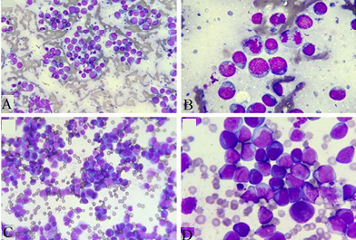
Microscopic pictures showing the typical cytological appearance of a high-grade lymphoma in lymph nodes and pleural effusion test (A and C, 40×; B and D, 100×). The smear of lymph node FNA showed the abundant cells especially lymphocytoblast.
References
- Allen, D.C., 2006. Springer London, London. pp. 367-387.
- Aptekmann, K.P., Okamoto, C.E., Campos, K.C.H. and Takahira, R.K., 2005. Arq. Bras. Med. Vet. Zoo., 57: 307-311. https://doi.org/10.1590/S0102-09352005000300005[CrossRef]
- Gavazza, A., Rossi, G., Lubas, G., Cerquetella, M., Minamoto, Y. and Suchodolski, J.S., 2018. Vet. Comp. Oncol., 16: E169-E175. https://doi.org/10.1111/vco.12367[CrossRef]
- Impellizeri, J.A., Gavazza, A., Greissworth, E., Crispo, A., Montella, M., Ciliberto, G., Lubas, G. and Aurisicchio, L., 2018. J. Transl. Med., 16: 349. https://doi.org/10.1186/s12967-018-1738-6[CrossRef]
- Jones, I.D., Daniels, A.D., Lara-Garcia, A., Peters, L.M. and Mantis, P., 2017. J. Small Anim. Pract., 58: 622-628. https://doi.org/10.1111/jsap.12714[CrossRef]
- Lindemann, D.M., Carpenter, J.W., Nietfeld, J.C., Gonzalez, E., Hallman, M. and Hause, B.M., 2015. J. Zoo. Wildl. Med., 46: 961-966. https://doi.org/10.1638/2015-0118.1[CrossRef]
- Matutes, E., Oscier, D., Montalban, C., Berger, F., Callet-Bauchu, E., Dogan, A., Felman, P., Franco, V., Iannitto, E., Mollejo, M., Papadaki, T., Remstein, E.D., Salar, A., Sole, F., Stamatopoulos, K., Thieblemont, C., Traverse-Glehen, A., Wotherspoon, A., Coiffier, B. and Piris, M.A., 2008. Leukemia, 22: 487-495. https://doi.org/10.1038/sj.leu.2405068[CrossRef]
- Nyman, H.T., Lee, M.H., McEvoy, F.J., Nielsen, O.L., Martinussen, T. and Kristensen, A.T., 2006. Am. J. vet. Res., 67: 978. https://doi.org/10.2460/ajvr.67.6.978[CrossRef]
- Oskouizadeh, K., Oskouizadeh, K., Esmaeilzadeh, S., Esmaeilzadeh, S., Ghadiri, A., Ghadiri, A., Naddaf, H. and Naddaf, H., 2011. Comp. clin. Pathol., 20: 273-275. https://doi.org/10.1007/s00580-010-1111-5[CrossRef]
- Ponce, F., Marchal, T., Magnol, J.P., Turinelli, V., Ledieu, D., Bonnefont, C., Pastor, M., Delignette, M.L. and Fournel-Fleury, C., 2010. Vet. Pathol., 47: 414-433. https://doi.org/10.1177/0300985810363902[CrossRef]
- Zandvliet, M. and Zandvliet, M., 2016. Vet. Quart., 36: 76-104. https://doi.org/10.1080/01652176.2016.1152633[CrossRef]
- Zhang, H., Wang, Y.J., Li, K., Rehman, M.U., Nabi, F., Gui, R., Lan, Y.F. and Luo, H.Q., 2017. Pakistan J. Zool., 49: 1033-1037. https://doi.org/10.17582/journal.pjz/2017.49.3.1033.1037[CrossRef]
- Zhang, H., Dong, H.L., Mehmood, K., Li, K., Nabi, F., Chang, Z.Y., Rehman, M.U., Ijaz, M., Wu, Q.X. and Li, J.K., 2018. Arch. Physiol. Biochem., 124: 458-460. https://doi.org/10.1080/13813455.2018.1423623[CrossRef]
Supplementary material
Cited by 1 article
In journals published by ResearchersLinks.
Services
Article usage statistics
Loading…