
This study was designed to undertake molecular characterization of an Infectious bronchitis virus (IBV) recovered from a suspected case of avian infectious bronchitis from commercial poultry. Initially the isolated IB-virus was characterized by RFLP using enzymes Alu I, Hae III, BstYI and XcmI. On the basis of its distinct RFLP pattern from other known IBV vaccine strains, the isolate was named as KU145467_NARC/786_Pakistan_2013 (also named as Pak-786) was subjected to Spike gene sequencing covering the amino-terminus region of 01 to 974 base pairs. The S protein sequence was submitted to the GenBank with accession number KU145467. Phylogenetic grouping and maximum nucleotide sequence identity values were used to identify the isolate that looked to be derived from recombination. It showed maximum nucleotide homology 99.5% with ck/CH/LHB/121010 (KP036503), India/IBV572 (KF809797) and Japan/JP/Wakayama-2/2004 (AB363951.2) and 99.3% with 4/91 vaccine (KF377577), Iran/491/08 (HQ842715) and 99.1% with India/NMK/72/VRI/10 (HM748585) and was least related to rest of IBV lineages compared in this study. It is concluded that Pak-786 isolate belongs to GI-13 lineage that include both the vaccine and virulent field strains, previously assigned to the 793B like. The study supports the concept that due to mass usage of live IBV vaccinal strains of diverse origin, variety of variants originate through random spontaneous mutation and genetic recombination which could lead to genetic drift. The emergent strain of IBV in this study points out the need to include such variants in killed-vaccine form in the vaccination program of the affected region.
Research Article · Volume 50, Issue 2, 21 March 2018 · Open Access
Determination of Genetic Variability in Avian Infectious Bronchitis Virus (AIBV) Isolated from Pakistan
· · · · · · ·
Affiliations & Notes ⌄
- 1National Reference Laboratory for Poultry Diseases, Animal Sciences Institute, National Agricultural Research Center, Park Road, Islamabad, Pakistan 2Quaid-i-Azam University, Islamabad, Pakistan , * Corresponding author: sabarafique@ymail.com
Article Info ⌄
- Journal:
- Pakistan Journal of Zoology
- Published:
- 21 March 2018

From the article
- Of the two IBV field isolate in this study, one isolate is selected on the basis of distinct RFLP pattern as compare to 793/B and some other reference strains.
- In conclusion the molecular characterization of new isolate of IBV leads us to believe that its glycoprotein have closest sequence identity with number of Indian, Chinese, Iranian, Japanese and 793/B or 4/91 vaccine strain as well.
- So we have to include these emerging strains of IBV in the locally manufactured killed vaccines.
- However more studies would be required for developing a homologous and potent vaccine against such new variants of IBV.
Selected from the article's own text.
Abstract
Introduction
Infectious bronchitis virus (IBV) mostly infects chicken (Gallus gallus) and is ubiquitous in Asia since 1967 , despite the use of live attenuated and killed vaccines. It is highly contagious disease (Colvero et al., 2017) and present everywhere especially where poultry are present and it can spread very quickly in non-vaccinated birds. Being a coronavirus, enveloped, pleomorphic, with the mean diameter of around 120nm and therefore a single- stranded RNA virus with heavily glycosylated spike (S) glycoprotein, normally creating novel antigenic types which leads to multiple serotypes of the virus that are not able to cross protect. The seroconversion against several American and European IBV serotypes have been confirmed in Pakistan, but further studies have still to be performed to isolate and characterize these viruses.
Being an RNA virus, IBV has a huge capacity to change both by mutation and by genetic recombination if they occur in the hypervariable region . More specifically, the spike protein gene mainly S1 subunit in this region is the most mutable component due to genetic drifts and recombination events happening in the environment . For the haemagglutination and infectivity the S1 part is responsible because it comprises of serotype specific virus neutralizing epitopes . This variability in nucleotide sequence, lower the cross protection among serotypes. It has been reported that sequence variations as little as 5% in the S1 region is responsible to modify the protection ability of the vaccine . Among the IBV serotypes S1 region has variability of 20 to 25% but in some serotypes, it is as low as 2% . As a result of this IBV may exist in the form of numerous different antigenic or genotypic types so the protection against such IBVs is made further tough to achieve, usually referred to as variants. Furthermore, as many new IBV variants survive on a long term basis, a few of them emerge which become to have worldwide distribution or only in the limited geographic areas.
Several IBV live and killed vaccines of classical Massachusetts strains especially M-41 and other European variant strains are used for vaccine manufacturing for poultry industry in Pakistan. In the past few years, multiple IBV vaccination failures have been recorded in Pakistan, indicative of different IBV variants circulating in the country. Though vaccination is required to increase the immunity of chickens against the circulating IBV strains, however, it has been made difficult to achieve this because of the lack of information regarding the type and number of existing IBV variants. This report reflects the evidence of the emergence of new IBV variant in commercial poultry, which requires further investigation to develop a new potent vaccine, accordingly.
The characteristic of continuous mutation among IB virus a number of distinct serotypes have evolved in nature. However, as very little field investigation and reporting is carried out regarding avian pathogen circulation in developing and under-developed countries, in most of the instances, so the information in literature regarding different strains of circulating IBV serotypes or their mutants is scanty. Therefore, most of the IBV vaccines available commercially are based on a few selected European or American strains. This has resulted in partial or no-protection against emerging IBVs under field conditions, despite having multiple shots of live and killed IBV vaccines of the available strains. Such reports of IBV vaccine failures in this country led us to undertake studies regarding the determination of the current status of prevailing strains of IBV and correlate their existence with the efficacy of the existing IB-vaccines being used here.
A flock of Broiler-breeder (BB) of 28 weeks of age, comprising of 50,000 birds, situated at district Attoct, was reported with signs of severe respiratory tract infection and decline in egg production. Initial mortality of 0.1% was reported in the flock along with high morbidity. The clinical signs included sneezing, coughing and rales. Other signs recorded in some birds included conjunctivitis and watery eyes. Within a period of one week post-infection (PI) the mortality rate increased to 0.3%. Post mortem examination of dead birds revealed congestion in the trachea and lungs. The kidneys were also inflamed.
The flock was earlier vaccinated with multiple shots of live IBV M-41 and 4/91 strains up to 22nd week of age and two shots of killed IB (IB M41 vaccine, Intervet) at 8-week and 12-week , along with one killed vaccine shot of IB (Nobilis 4/91 type, Intervet).
Materials and methods
Sample processing and virus isolation
The clinical specimens of trachea, lungs, spleen, cloacal and tracheal swabs were collected from the affected flock and brought to the National Reference Lab for Poultry Diseases (NRLPD) Islamabad. The samples were triturated @ 20% homogenate was prepared using phosphate buffered saline (PBS), and clarified at 2000xg for 15min and the supernatant was filtered via 0.45 um disposable cellulose nitrate filters, (Membrane Filter Products System, San Diego, CA 92121, USA) A part of the sample was further processed for the detection of Avian Influenza Virus (AIV) serotype H9, IBV and NDV by PCR using the procedure described elsewhere and inoculated in specific pathogen free (SPF) egg (Lohman, Germany) using the standard procedure (OIE, 2008). Briefly a total of 60, nine-day old embryonated SPF chicken eggs were inoculated with 200ul of the above filtered samples and eggs were incubated at 37oC. Eggs were candled daily for a week, considering the mortality within the first 24 h as nonspecific. Half of the eggs were left for incubation up to 7 days, whereas allantoic fluids of remaining half the eggs were harvested and pooled three days post inoculation; this pool was further passage into another set of SPF eggs for up to a total of four passages . The embryos were examined for any lesions and dwarfism.
RNA isolation and RT-PCR
For detection of IBV and confirmation of contaminants through RT-PCR, RNA extraction was carried out on swabs and organ samples using QIAamp Viral RNA Mini Kit (QIAGEN, Valencia, California, USA) according to the manufacturer’s instructions and stored at -20oC until use. RT PCR was performed on Genamp 9700 thermal cycler (ABI) using Superscript Tm One step RT-PCR with Platinum Taq kit (Invitrogen, USA) using RT-PCR protocols earlier described . In this study IBV 4/91 strain was used as positive control.
Restriction fragment length polymorphism (RFLP) of S1 region
Amplification of the S1 gene was carried out by RT-PCR using the forward primer: 5-TGAAACTGAACAAAAGACA-3 and the reverse primer 5-CATAACTAACATAAGGGCAA-3 for a final product of 1720 bp . PCR product was purified through QIAquick Spin Miniprep kit (QIAGEN, Valencia, California, USA). The restriction enzymes Alu I, Hae III, BstYI and XcmI (New England Biolabs) were used according to manufacturer’s specifications for digestion of the PCR products which were then separated by electrophoresis in 2% of agarose gel. The RFLP patterns of the field isolate was then compared with those of the digested vaccine reference strain.
Sequencing spike gene and data analysis
On the basis of RFLP pattern, the unique isolate was subjected to sequence analysis. For this purpose, RNA was extracted from egg fluids (Passage Level 1) with QIAamp Viral RNA Mini Kit (QIAGEN, Valencia, CA, USA) in accordance with the manufacturer’s instructions. Sequencing templates for partial spike gene were produced by amplifying through RT-PCR. Templates were then purified by agarose gel extraction with the QIA-quick gel extraction kit (QIAGEN, Valencia, CA, USA). The BigDye Terminator v3.1 Cycle Sequencing Kit (Applied Biosystems, Foster City, California, USA) was used for cycle sequencing and subsequently run on an ABI PRISM 3130 Genetic Analyzer (Applied Biosystems, Foster City, CA). The consensus sequence of the nucleotide sequences was generated using SeqScape software version 2.6 (ABI). GenBank accession number allotted is KU145467.
The S gene sequence of the new isolate of IBV was subjected to Blast by using NCBI website. S gene sequences of the IBV isolates were selected from the Genbank on the basis of sequence identity. Multiple nucleotide and amino acid sequence alignments for the spike gene were performed using Clustal W . Phylogenetic tree was made with the help of neighbor-joining analysis in the Mega program (version 4.0) and were evaluated by bootstrap of 1000 replicate .
Results
Avian Infectious Bronchitis Virus
The initial passaging of field isolate had very limited effect on the embryo, however, after 5th to 6th passages lesions, curling and dwarfism became more apparent (Fig. 1). As the virus became more egg adapted, the mortality of embryos at early stage of inoculation decreased.
Table I.- Comparison of restriction cleavage sites of KU145467_NARC/786_Pakistan_2013 with 4/91 vaccine strain.
| S No. | Restriction Enzymes | KU145467_NARC/ 786_Pakistan_2013 or (F1) (MW bp) | 4/91 vaccine strain (MW bp) |
| 1 | Alu I | 130, 200, 220, 400, 1000 | 120, 220, 280, 1000 |
| 2 | Hae III | 150, 180, 440, 482, 600, 1200 | 150, 180, 300, 500, 1200 |
| 3 | Bst Y | 155, 500, 1500 | 150, 550, 1200 |
| 4 | Xcm I | 120, 320, 380, 1200, 1500 | 120, 1200 |
MW, molecular weight in base pair.
The isolate KU145467_NARC/786_Pakistan_2013 or (F1) was detected by using RT-PCR and the results showed a product of 1720 bp. This product was subjected to further characterization through RFLP analysis using restriction enzymes Alu I, Hae III, BstYI and XcmI (Fig. 2). On the basis of RFLP characterization we have found a distinct pattern of F1 isolate from IBV 4/91. Here the isolate F1 had showed the distinct restriction sites with all four enzymes when compare to control 4/91 vaccine strain and reference IBV strains including 793/B (Intervet live vaccine), M41 (Avipro, IB M48) & IT-02 (GD Lab, Netherland). While the F2 isolate had the similar restriction sites as 4/91 vaccine strain has. Specifically the RFLP pattern of isolate F1 with control 4/91 vaccine strain presented in tabular form (Table I). The isolate was further processed for the sequencing of spike glycoprotein.
Table II.- Highest nucleotide homology and cleavage site motifs of Pakistani IBV compared to sequences available in GenBank.
| S No. | Virus Nomenclature | Nucleotide homology % | Cleavage site motifs | Accession number |
| 1 | KU145467_NARC/786_Pakistan_2013 | - | R-R-S-R-R | KU145467 |
| 2 | ck/CH/LHB/121010_china_2012 | 99 % | R-R-S-R-R | KP036503 |
| 3 | IBV572_India_2013 | 99.5% | R-R-S-R-R | KF809797 |
| 4 | JP/Wakayama-2/2004_Japan | 99.5% | H-R-R-R-R | AB363951.2 |
| 5 | 4/91 vaccine | 99% | R-R-S-R-R | KF377577 |
| 6 | ck/CH/LZJ/111113_china_2011 | 99% | R-R-S-R-R | JX195176 |
| 7 | IR/491/08_2008 | 99.3% | R-R-S-R-R | HQ842715 |
| 8 | India/NMK/72/VRI/10 | 99.1% | No data | HM748585 |
| 9 | M41 | 75% | R-R-F-R-R | DQ834384 |
| 10 | Arkansas | 74.9% | R-R-S-R-R | GQ504721.2 |
| 11 | Connecticut | 74.9% | R-R-S-R-R | KF696629 |
| 12 | JMK | 74.2% | R-R-S-R-R | GU393338 |
| 13 | Gray | 74.2% | R-R-F-R-R | GU393334 |
| 14 | Holte | 73% | R-R-F-R-R | GU393336 |
| 15 | Iowa 97 | 72.9% | R-R-S-R-R | GU393337 |
| 16 | Cal 1995 | 72.4% | R-R-F-R-R | FJ904714 |
Amino acids abbreviation: R, Arginine; S, Serine; F, Phenylalanine; L, Leucine; T, Threonine; H, Histidine.
Phylogenetic analysis
Phylogenetic study was carried out on the basis of spike gene of IBV coronaviruses available in the GenBank. This includes the BLAST sequences including IBV Chinese’s 4/91 like strains, 4/91 vaccine strain, Indian IBV strain, Ukraine IBV strain, Mass type of IBV, Connecticut strain of IBV and 32 distinct viral lineages . The S gene of Pakistani IBV was supposed to have point mutations in terms of substitution. It showed a maximum sequence homology of 99.1%-99.5% with the Chinese isolates ck/CH/LHB/121010 and ck/CH/LSD/110410, Indian isolates India/IBV572 and India/NMK/72/VRI/10, Japanese isolate Japan/JP/Wakayama-2/2004, Iran/491/08 and 4/91 vaccine strain, respectively. It also showed nucleotide homology with Russian and Moroccan isolates of 793B origin. Moreover the phylogenetic comparison with the other earlier reported IBV reference strains reflects their sequence identity at the level of 75% with M41, 74.9% with Arkansas, Connecticut and Peafowl/GD/KQ6/2003 and others listed below (Table II).
Sequence analysis
A comparative analysis of Spike Sequence Pak-786 was performed using the MEGA Sequence Alignment Program. The tree was constructed using the neighbor joining program (Fig. 3). From the GenBank database IBV Spike sequences representative to genotypes were used for the alignment. They include Chinese strains (KP036503.1 and KP118893.1), Indian strain (KF809797.1 and HM748585.1) and with reference strains 4/91 (JN192154.1), M41 (DQ834384.1) and so on with other reference strains. The cleavage recognition site detected here was Arg-Arg-Ser-Arg-Arg, and it’s basically representing a number of different serotypes. It holds mutations at nucleotide level that caused amino acid substitutions, and has some deletions. It possesses 24 N-glycosylation sites.
The data regarding molecular evaluation Pak-786 indicates that it possesses a few unique amino acid substitutions. Here these mutations reported only with reference to closely related isolates. Aspartic acid at position 03 was found instead of glycine, Asparagine at position 23 instead of lysine, proline at position 38 instead of Glutamine, arginine at position 51 instead of lysine, glutamic acid at position 55 instead of glycine except indian/IBV/572, glycine position 60 instead of valine in some close related isolate and valine at position 179 instead alanine is present in all isolates except in ck/CH/LHB/121010.
Discussion
For IBV isolation, SPF eggs were used and it is found that as the number of passages increased embryo mortality and dwarfing also increases. But in other report it is found that some field isolates haven’t any marked effect on embryo during propagation so number of other methods used for detection . Of the two IBV field isolate in this study, one isolate is selected on the basis of distinct RFLP pattern as compare to 793/B and some other reference strains.
The S gene of the sequenced isolate (Pak-786) isolated in 2013 from Pakistan shared 99.1%-99.5% with the Chinese, Indian, Japanese, Iranian and 793/B vaccine strain, respectively. It also showed nucleotide homology with Russian and Moroccan isolates that basically belongs to IBV lineage GI-13. This high nucleotide homology basically showed a close genetic relationship and common origin.
The cleavage recognition site detected here was Arg-Arg-Ser-Arg-Arg, and it’s basically representing at least 11 different serotypes. Serotype doesn’t look to associate with cleavage recognition site, as it is common S1-S2 connecting peptide of IBV . It is also observed that this order does not correlate with pathogenicity because attenuated and pathogenic isolates comprise identical cleavage recognition site sequences .
24 N-glycosylation sites are present and it is found that these sites vary with serotypes. The difference in N-glycosylation sites may disturb survival and spread of the virus, as the small changes can lead to disruption in folding and conformation of the molecule . Changes in N-glycosylation sites lower the interaction with the receptor, so the variants become more vulnerable to host immune system that will finally affect the virus replication and infectivity ability.
Conclusion
In conclusion the molecular characterization of new isolate of IBV leads us to believe that its glycoprotein have closest sequence identity with number of Indian, Chinese, Iranian, Japanese and 793/B or 4/91 vaccine strain as well. This isolate possess few unique mutation in the hyper-variable region so it is assumed that this mutant could comfortably cross the IBV specific immunity developed as a result of set of vaccines used in the affected flocks. So we have to include these emerging strains of IBV in the locally manufactured killed vaccines. However more studies would be required for developing a homologous and potent vaccine against such new variants of IBV.
Acknowledgement
This study was partly funded by the grant No. ASI/124/2012/PARC, received from the Agri-Linkage Program of Pakistan Agricultural Research Council (ALP-PARC).
Statement of conflict of interest
The author(s) declare(s) that there is no conflict of interests regarding the publication of this article.
Figures
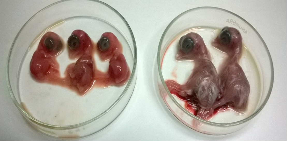
Significant dwarfism of the chicken embryo (L) at 7 dpi as compared to control group embryos (R). dpi, days post inoculation.
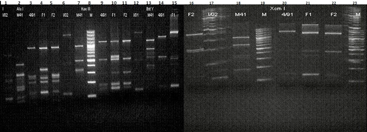
RFLP patterns of the PCR-amplified S1 glycoprotein genes from five IBV strains (3 Reference strains and 2 Field isolates) digested with Alu I, Hae III, BstY and Xcm I. Lane 1 to 5, Alu I; Lane 6 to 11, Hae III; Lane 12 to 16, BstY; Lane 17 to 22, Xcm I; Lanes 8, 19 and 23, molecular weight marker l kb DNA step ladder.
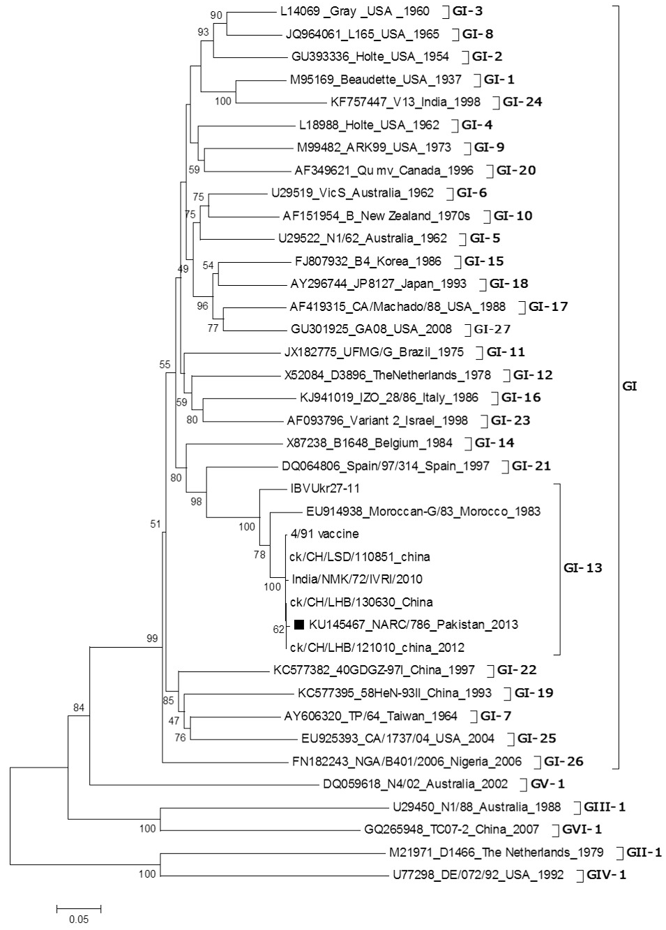
Phylogenetic tree based on S gene, showing inter-relationship among IBV isolate from Pakistan, commercial vaccines and selected reference strains. The Pakistani isolate is highlighted in Black Square.
References
- Abro, S.H., Ullman, K., Belak, S. and Baule, C., 2012. Bioinformatics and evolutionary insight on the spike glycoprotein gene of qx-like and massachusetts strains of infectious bronchitis virus. Virol. J., 9: 211. https://doi.org/10.1186/1743-422X-9-211[CrossRef]
- Ahmed, Z., Naeem, K. and Hameed, A., 2007. Detection and seroprevalence of infectious bronchitis virus strains in commercial poultry in pakistan. Poult. Sci., 86: 1329-1335. https://doi.org/10.1093/ps/86.7.1329[CrossRef]
- Casais, R., Dove, B., Cavanagh, D. and Britton, P., 2003. Recombinant avian infectious bronchitis virus expressing a heterologous spike gene demonstrates that the spike protein is a determinant of cell tropism. J. Virol., 77: 9084-9089. https://doi.org/10.1128/JVI.77.16.9084-9089.2003[CrossRef]
- Cavanagh, D., Davis, P.J., Cook, J.K., Li, D., Kant, A. and Koch, G., 1992. Location of the amino acid differences in the s1 spike glycoprotein subunit of closely related serotypes of infectious bronchitis virus. Avian Pathol., 21: 33-43. https://doi.org/10.1080/03079459208418816[CrossRef]
- Cavanagh, D., Davis, P.J., Pappin, D.J., Binns, M.M., Boursnell, M.E. and Brown, T.D., 1986. Coronavirus ibv: Partial amino terminal sequencing of spike polypeptide s2 identifies the sequence arg-arg-phe-arg-arg at the cleavage site of the spike precursor propolypeptide of ibv strains beaudette and m41. Virus Res., 4: 133-143. https://doi.org/10.1016/0168-1702(86)90037-7[CrossRef]
- Cavanagh, D. and Gelb, J.J., 2008. Diseases of poultry. Blackwell Publishing, Ames, Iowa, USA.
- Colvero, L.P., Brandão, P.E., Rosales, C.A.R., Swam, H. and Villarreal, R.W.B., 2017. Comparison of two inactivated vaccines against infectious bronchitis virus evidences the interference of vaccine formulations on vaccine performance. Hosts Viruses, 4: 92-95. http://dx.doi.org/10.17582/journal.hv/2017/4.6.92.95[CrossRef]
- De Wit, J.J., 2000. Detection of infectious bronchitis virus. Avian Pathol., 29: 71-93.
- Higgins, D.G., Thompson, J.D. and Gibson, T.J., 1996. Using clustal for multiple sequence alignments. Methods Enzymol., 266: 383-402. https://doi.org/10.1016/S0076-6879(96)66024-8[CrossRef]
- Jackwood, M.W., Hilt, D.A., Callison, S.A., Lee, C.W., Plaza, H. and Wade, E., 2001. Spike glycoprotein cleavage recognition site analysis of infectious bronchitis virus. Avian Dis., 45: 366-372. https://doi.org/10.2307/1592976[CrossRef]
- Kingham, B.F., Keele, C.L.JR., Nix, W.A., Ladman, B.S. and Gelb, Jr. J., 2000. Identification of avian infectious bronchitis virus by direct automated cycle sequencing of the s-1 gene. Avian Dis., 44: 325-335. https://doi.org/10.2307/1592547[CrossRef]
- Kwon, H.M., Jackwood, M.W. and Gelb, Jr. J., 1993. Differentiation of infectious bronchitis virus serotypes using polymerase chain reaction and restriction fragment length polymorphism analysis. Avian Dis., 37: 194-202. https://doi.org/10.2307/1591474[CrossRef]
- Lohr, J.E., 1981. Diagnosis of infectious bronchitis (ib) by examination of tracheal mucus for ib-precipitating antigens. Avian Dis., 25: 1058-1064. https://doi.org/10.2307/1590083[CrossRef]
- Muneer, M.A., Newman, J.A., Goyal, S.M. and Ajmal, M., 1987. Antibodies to avian infectious bronchitis virus in pakistani chickens. Poult Sci., 66: 765-767. https://doi.org/10.3382/ps.0660765[CrossRef]
- OIE, 2008. Manual of diagnostic tests and vaccines for terrestrial animals (mammals, birds and bees). OIE, Paris, France.
- Senne, D.A., 2008. A laboratory manual for the isolation, identification nd characterization of avian pathogens (eds. F.L. Jacksonville). American Association of Avian Pathologists, USA.
- Tamura, K., Dudley, J., Nei, M. and Kumar, S., 2007. Mega4: Molecular evolutionary genetics analysis (mega) software version 4.0. Mol. Biol. Evol., 24: 1596-1599. https://doi.org/10.1093/molbev/msm092[CrossRef]
- Valastro, V., Holmes, E.C., Britton, P., Fusaroa, A., Jackwood, M.W., Cattoli, G. and Monne, I., 2016. S1 gene-based phylogeny of infectious bronchitis virus: An attempt to harmonize virus classification. Infection Gene Evol., 39: 349-364. https://doi.org/10.1016/j.meegid.2016.02.015[CrossRef]
Cited by 2 articles
In journals published by ResearchersLinks.
Services
Article usage statistics
Loading…