Research Article · Volume 10, Issue 4, 17 March 2022 · Open Access
Molecular Identification of IBDV from Naturally Infected Chicken Flocks
· · · · · · ·
Affiliations & Notes ⌄
1Poultry Diseases Department, Veterinary Research Division, National Research Centre, P.O. 12622, Giza, Egypt
2Laboratory of Veterinary Vaccines Technology (VVT), Central Laboratories Network, National Research Centre, P.O. 12622, Giza, Egypt
3Department of Poultry Diseases, Faculty of Veterinary Medicine, Cairo University, P.O. 12211, Giza, Egypt. *Correspondence | Aly M. Ghetas, Poultry Diseases Department, Veterinary Research Division, National Research Centre, P.O. 12622, Giza, Egypt
In our study, we molecularly detected two vvIBDV strains from two vaccinated native broiler chicken flocks showing clinical signs of IBD.
Our IBDV isolated were closely related to vvIBDV detected in Egypt, France, Spain, South Africa, and Israel.
Therefore, continuous monitoring of genetic diversity of IBDV strains and the characteristics of the virulent IBDV as well as assessment of used vaccination programs are necessary to control IBDV infection more efficiently in Egyptian poultry farms.
Selected from the article's own text.
Abstract
Our study was performed to molecularly identify infectious bursal disease (IBD) virus (IBDV) from naturally infected native chickens. Thus, bursa specimens were collected from two IBD vaccinated native broiler chicken flocks showing clinical signs, mortality and lesions of IBD in this study. The collected bursae showed pathological changes related to those of IBD infection. Furthermore, the isolates were molecularly characterized using reverse transcription polymerase chain reaction (RT-PCR) technique and gene sequencing of partial portion of VP2 gene. The two isolates (Genbank accession numbers MW925051 and MW9250522) were characterized as very virulent IBDV (vvIBDV). The obtained nucleotide sequences of partial portion of VP2 gene of 2 isolates revealed 97.0 - 100% and 91.2-92.5% identity with the Egyptian strains and vaccine strains respectively. Finally, signiï¬cant genetic changes in our IBDV isolates were observed as comparing to vaccine strains. Thus, continuous monitoring of IBDV outbreaks in vaccinated chickens and evaluation of the used vaccines are required.
Infectious bursal disease (IBD) is an acute and highly contagious disease in young chickens. The disease is characterized by massive destruction of B lymphocytes accompanied by immunosuppression and high morbidity and mortality rates in susceptible chickens (Eterradossi and Saif, 2013). Infectious bursal disease virus (IBDV) is the causative agent IBD. This virus is a double stranded, bi-segmented (A and B) RNA virus belonging to family Birnaviridae ; . IBDV segment B (2.8 Kb) encodes a viral RNA polymerase (VP1), while segment A (3.2 Kb) encodes two structural proteins (VP2 and VP3), a serine viral protease (VP4), and the nonstructural protein (VP5). The VP2 subunit is comprised of 3 distinct domains; the base (B), the shell (S) and the projection (P). The P domain is the most exposed and is more variable which contains the hypervariable region (HVR) in VP2 subunit . VP2 is the major antigenic protein stimulating neutralizing antibodies and is contribute to the pathogenicity of IBDV . Furthermore, IBDV strains were classified to different genogroups based on a VP2 gene sequencing .
Two distinct serotypes of IBDV have been described in which only viruses belong to serotype 1 are pathogenic to chickens. Furthermore, classical, variant, and very virulent IBDV strains have been detected within serotype 1 based on antigenic variation and virulence (Van den Berg et al., 2004). In Egypt 1974, IBDV was first described , while the first report of vvIBDV was in 1989 . Additionally, variant IBD strains were also reported in Egypt ; ; .
Despite extensive vaccination program, IBDV strains continue to cause economic losses in Egyptian chicken farms vaccinated with IBDV vaccines ; ; ; . Thus, continuous identification of the circulating IBDV to detect mutations especially in VP2 gene and to monitoring the virus evolution is essential. In this study, we molecularly identified IBDV from outbreaks in two native chicken flocks.
Materials and Methods
Ethical approval
The work was approved by the Ethics Committee of the National Research Centre under number 1478092021, Egypt.
Chicken flocks
Two native broiler chicken flocks suffering from infectious bursal disease were sampled. The first flock was 35 days old (n=5000). The second flock was 28 day old chickens (n=1200). Both flocks were vaccinated against IBDV using intermediate strain vaccine at the 12th day of live via drinking water. The clinical signs in both flocks were whitish diarrhea, ruffled feathers, anorexia, depression, and up to 5% to 10% mortality rate.
Samples
Bursa specimens were collected aseptically in 10% neutral buffered formalin for histopathological examination and others were kept at -20 for further molecular identification. The number of collected bursae were 5 per chicken flock. Moreover, we did pooling of bursae of each flock for molecular identification of IBDV.
Molecular identification of IBDV
RNA extraction
The QIAamp viral Mini kit (Qiagen, Germany, GmbH) was used for RNA extraction according to the manufacture procedures.
Oligonucleotide primers
Primers used in this study (Table 1) were supplied from metabion, Germany.
Amplification of VP2 gene using RT-PCR
The 25 µl reaction mixture was used. This reaction included 12.5 µl of RT-PCR kit buffer (Qiagen, Germany, GmbH), 1 µl of each primer (20 pmol), 0.25 µl of RT-enzyme, 5.25 µl of RNase-free water, and 5 µl of RNA template. The RT-PCR conditions were mentioned in Table 2 . The products of PCR were separated by horizontal gel electrophoresis in 1.5% agarose. The fragment size was determined by comparison with a 100 bp DNA ladder (Fermentas, Germany). Reference IBDV strain was used as positive control .
Gene sequencing and phylogeny
Purification of PCR products were done using QIAquick PCR Product extraction kit. (Qiagen, Valencia). Applied Biosystems 3130 genetic analyzer (HITACHI, Japan) was used in DNA sequencing. Then, a BLAST® analysis was initially completed to establish sequence identity to Genbank accessions. Furthermore, the MegAlign module of Lasergene DNA Star version 12.1 was used to create the phylogenetic tree and phylogenetic analysis was done using maximum likelihood, neighbor joining and maximum parsimony in MEGA7 software .
Histopathological examination
Bursae were collected and were fixed in 10% neutral buffered formalin. Several steps (dehydration, clearing, wax
Table 1: Primers sequences, target gene, and amplification size.
Target gene
Primers sequences
Amplified segment (bp)
Reference
IBD VP2
TCACCGTCCTCAGCTTACCCACATC
620
Metwally et al., 2009
GGATTTGGGATCAGCTCGAAGTTGC
Table 2: RT-PCR reaction conditions.
Target
RT reaction
Primary denaturation
Amplification cycles
Final extension
Secondary denaturation
Annealing
Extension
IBD VP2
50 ˚C 20 min
94˚C 15 min.
94˚C 30 sec
59˚C 40 sec.
72˚C 1 min
72˚C 10 min
infiltration, and blocking out) were preformed to obtain paraffin tissue sections at 4-6 μm thickness. Then, these sections were stained with hematoxylin and eosin .
Results and Discussion
Gross lesions
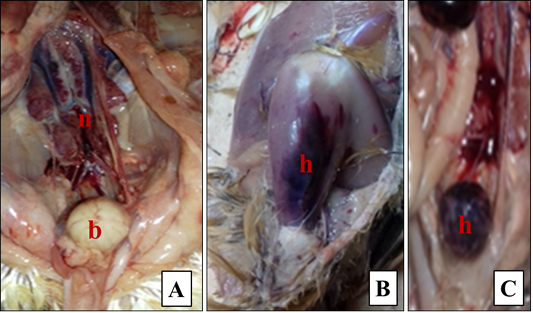
Enlarged bursa with gelatinous transudate covering its serosal surface, hemorrhagic bursae, hemorrhage on thigh muscle, and hemorrhage between gizzard and proventriculus were observed (Figure 1).
Histopathological findings
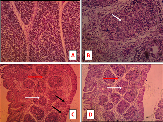
As shown in Figure 2, sever depletion of the lymphoid follicle germinal center (Figure 1B) deposition of interfollicular connective tissues (Figure 1B), depletion of the lymphoid follicle tissue (Figure 1C, D) accompanied with deposition of interfollicular connective tissues (Figure 1C, D), and hypertrophy of the epithelial lining making finger like projection (Figure 1C) were detected.
Molecular identification of IBDV
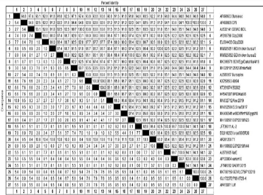
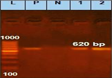
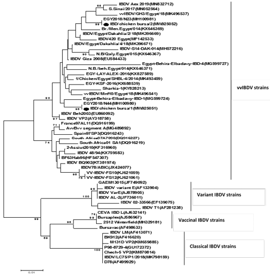
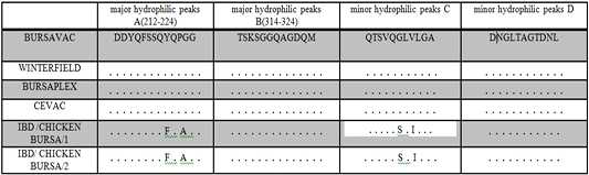
The partial portion of VP2 gene was amplified using RT-PCR method (Figure 3). The PCR products of IBDV VP2 gene were sequenced and submitted to Genbank accession numbers MW925051 and MW925052. Phylogenetic analysis revealed that our two isolates are related to vvIBDV isolates rather than classical or variant strains (Figure 4). As shown in Table 3, the IBDV isolate (MW925051) was closely related to Giza2008 (EU584433) with 99.0% nucleotide identity comparing to vaccine strains D78 (AF499929), BURSAVAC (AF498633), CEVAC IBDL (AJ632141) with nucleotide identity 91.2%, 91.8%, and 91.8% respectively. Furthermore, the IBDV isolate (MW925052) showed 100% nucleotide identity with EGY2018N23 (MH100981) while the lowest nucleotide identity (highest divergence) was detected with the three vaccine strains BURSAVAC (AF498633), D78 (AF499929), and CEVAC IBDL (AJ632141) with nucleotide identity 91.8%, 92.2% and 92.5%, respectively. Finally, alignment of deduced amino acid sequences located in the HVR of VP2 of the our IBDV isolates compared to amino acid sequence of other IBDV vaccine strains from amino acid (aa) position (212 to 224) was performed. The two isolates contained amino acid of vvIBD at position F220, A222 at major hydrophilic peak A (Table 4). In addition, our IBDV isolates were also different from vaccine strains at the minor hydrophilic peaks, C (S254G).
IBDV causes high economic losses in poultry industry due to high mortality rate and immunosuppression in Egypt ; ; ; and worldwide . This study was accomplished to assess pathological and molecular characterization of vvIBDV isolated from naturally infected native chickens as well as comparing with IBD vaccine strain used in Egyptian chicken farms. The native chicken flocks were vaccinated with intermediate vaccine at 12th day of age via drinking water. The signs and mortality started at 28 days of life and lasted for 7 days with mortality rate 10%.
In our study, IBDV was collected from suspected naturally infected 28 and 35 day old native chickens showed whitish diarrhea, ruffled feathers, anorexia, depression, similar observations were previously reported (Eterradossi and Saif, 2013). The low mortality rate (5% to 10%) in chicken flocks in this study comparing to mortality rate reviewed by others may be due partial protection in these flocks as a result of vaccination with intermediate vaccine. Different pathological lesions were reported varying from enlarged bursa to severe hemorrhagic bursa. Additionally, a hemorrhage on thigh muscle and sever nephrosis were detected. These pathological findings indicate the high pathogenicity of IBDV . Furthermore, results of histopathological examination of bursae showed severe depletion of the lymphoid follicle germinal center, deposition of interfollicular connective tissues, depletion of the lymphoid follicle tissue accompanied with deposition of interfollicular connective tissues, and hypertrophy of the epithelial lining making finger like projection in intestine, these results agree with findings previously reported .
IBDV VP2 is an essential determinant of antigenic variation, virulence, and cell tropism ; . Thus, our RT-PCR amplified partial portion of VP2 gene (620). As shown in Figure 4, our strains have close genetic relationship to vvIBDV which clustered with Egyptian strains and closely related with strain isolated from France, Spain, South Africa, and Israel. However, we found our strains segregated out vaccine strain in separate distinct genogroup. In addition, the nucleotide sequences of the VP2 of our isolate shared 97.0-100% homology with Egyptian strains, but the nucleotide identity with vaccine strains ranged from 91.2-92.5% Table 3.
Indeed, the HVR of VP2 between amino acid positions 206 and 350 is very important because it contains four hydrophilic regions determining virulence and antigenic variation ; ; . Two major hydrophilic peaks, A (212–224) and B (314–324) have been reported ; . Furthermore, two minor hydrophilic peaks C (249–254) and D (279–289) have also been identified . This region has been extensively used in studying the molecular epidemiology of IBD ; ; . In this study, the conserved virulence markers for vvIBDV (222A, 242I, 249Q, 253Q, 256I, 272I, 279D, 284A, 294I, 299S) were detected which agreed with others but different from vaccine strains. We found our strains different from vaccine strain in major hydrophilic peaks A, (F 220Y and A222P) and minor hydrophilic peaks, C (S254G) as shown in Table 4. Amino acid substitution at position 254 was reported in vvIBDV strains detected from chickens vaccinated with classical intermediate IBDV vaccines ; ; . Furthermore, amino acid substitution at this position contributing to antigenic drift which allowed IBDV to escape from immune response produced by IBDV vaccines ; . This suggested that very virulent IBDV has been mutated and still leads to several outbreaks in Egypt which recorded also previously by others ; . Effective vaccination of IBD remains a challenge in Egypt which does not offer total protection. Thus pathological and molecular characterization of IBDV field strains are essential for better prevention and control of the spread of vvIBDV.
Conclusions and Recommendations
In our study, we molecularly detected two vvIBDV strains from two vaccinated native broiler chicken flocks showing clinical signs of IBD. Our IBDV isolated were closely related to vvIBDV detected in Egypt, France, Spain, South Africa, and Israel. The vvIBDV was still circulated in chicken farms in Egypt. Therefore, continuous monitoring of genetic diversity of IBDV strains and the characteristics of the virulent IBDV as well as assessment of used vaccination programs are necessary to control IBDV infection more efficiently in Egyptian poultry farms.
Novelty Statement
In our study, two vvIBDV strains were molecularly identified from vaccinated native chickens. Our results can help to further understanding the IBDV genetic diversity for efficient control of this disease in Egypt.
Author’s Contribution
MMA, KMA and HMM designed and planned this study. AMG, DMS, and HSF collect samples, and performed all laboratory work. MAS carried out pathological examination. AMG and AM collected research data for writing. All authors shared samples collection, performing the tests, and manuscript writing. All authors also revised and approved the final manuscript.
Conflict of interest
The authors have declared no conflict of interest.
Figures
Figure 1:.
Pathological finding from chickens suffering from infectious bursal disease. A: chicken shows enlarged bursa covered by gelatinous transudate (b) and severe nephrosis (n). B: chicken shows hemorrhage on thigh muscle (h). C: chicken shows hemorrhagic bursa (h).
Figure 2:.
Bursa tissue sections of naturally IBDV infected chicken stained by H and E. Severe depletion of the lymphoid follicle germinal center (B) deposition of interfollicular connective tissues (Fig. 1B), depletion of the lymphoid follicle tissue (C and D) accompanied with deposition of interfollicular connective tissues (C and D), and hypertrophy of the epithelial lining making finger like projection (C).
Table 3:.
Percentage of nucleotide identities for the VP2 genes of 2 IBDV strains named MW925051 and MW925052 as compared with 25 sequences published in Genbank.
Figure 3:.
Amplified VP2 gene of IBDV. Lane L = 100 bp marker; Lane P = Positive control. Lanes 1 and 2 = Examined samples; Lane N = Negative control.
Figure 4:.
Phylogenetic tree based on partial VP2 gene sequencing of IBDV. The tree was constructed using the maximum-likelihood method with 1000 bootstrap replicates using MEGA 7 software. Black dots refer to our isolated (MW925051 and MW925052).
Table 4:.
Amino acid substitution in VP2 hyper variable region of the IBD strains MW925051 and MW925052 in comparison to vaccine strains.
Table 4:.
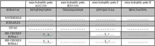
Amino acid substitution in VP2 hyper variable region of the IBD strains MW925051 and MW925052 in comparison to vaccine strains.
References
Brandt M, Yao K, Liu M, Heckert RA, Vakharia VN (2001). Molecular determinants of virulence, cell tropism, and pathogenic phenotype of infectious bursal disease virus. J. Virol., 75: 11974–11982. https://doi.org/10.1128/JVI.75.24.11974-11982.2001[CrossRef]
Camacho C, Coulouris G, Avagyan V, Ma N, Papadopoulos J, Bealer K, Madden TL (2009). BLAST+: Architecture and applications. BMC Bioinf., 10: 421. https://doi.org/10.1186/1471-2105-10-421[CrossRef]
Coulibaly F, Chevalier C, Gutsche I, Pous J, Navaza J, Bressanelli S, Delmas B, Rey FA (2005). The birnavirus crystal structure reveals structural relationships among icosahedral viruses. Cell 120, 761–772. doi: 10.1016/j.cell.2005.01.009[CrossRef]
Delmas B, Attoui H, Ghosh S, Malik YS, Mundt E, Vakharia VN, Ictv Report, C (2019). ICTV virus taxonomy profile: Birnaviridae. J. Gen. Virol. 100, 5–6. DOI: 10.1099/jgv.0.001185[CrossRef]
Dey S, Pathak DC, Ramamurthy N, Maity HK, Chellappa, MM (2019). Infectious bursal disease virus in chickens: prevalence, impact, and management strategies. Vet. Med. (Auckl.), 10: 85–97. https://doi.org/10.2147/VMRR.S185159[CrossRef]
El-Bagoury G, Elsamaloty M, El-Habbaa A, Haggag N (2018). Full VP2 sequence analysis of Infectious Bursal Disease Virus (IBDV) in broiler chicken in Egypt. Benha Vet. Med. J., 35(2): 559-567. https://doi.org/10.21608/bvmj.2018.111776[CrossRef]
El-Batrawi AM (1990). Studies on severe outbreaks of infectious bursal disease. The natural and experimental disease in Proceedings of the 2nd Science Conference of the Egyptian Veterinary Poultry Association, pp. 339–252.
El-Batrawi AM, El-Kady MF (1990). Studies on sever outbreaks of infectious bursal disease 3-deternination of the critical age of susceptibility in maternally immune chicks. Proc. Second Sci. Conf. Egypt. Vet. Poult., pp. 264–269.
Ellakany HF, Elbestawy AR, Sayed-Ahmed A, Elgamal SM, Gado AR, Abd El-Hamid HS (2019). Genetic point mutation inducing antigenic drift in hypervariable region of a very virulent IBDV isolate in chickens in Egypt during 2014-2016. Damanhour J. Vet. Sci., 2(1): 12-17. https://doi.org/10.5455/DJVS.57924[CrossRef]
El-Sergany HA, Moursi A, Saber MS, Mohamed MA (1974). A preliminary investigation on the occurrence of Gumboro disease in Egypt. Egypt. J. Vet. Sci., 11: 185-208.
Eteradossi N, Saif YM (2013). Infectious bursal disease. In: Swayne, D.E., Glisson, J.R., McDougald, L.R., Nolan, L.K., Suarez, D.L., Nair, V. (Eds.), Diseases of Poultry, XIIIth edition. John Wiley and Sons Inc, Ames, Iowa, USA, pp. 219–246. https://doi.org/10.1002/9781119421481.ch7[CrossRef]
Ezeibe MC, Okoye JO, Ogunniran TM, Animoke PC, Mbuko IJ, Nwankwo IA, Ngene AA (2013). Mortality rates from a Nigerian isolate of the Infectious Bursa Disease Virus and passive haemagglutination antibody titer that protects chicks against challenge with the virus isolate. Health, 5: 1355-1359. https://doi.org/10.4236/health.2013.59184[CrossRef]
Hassan MK, Afify M, Aly MM (2002). Susceptibility of vaccinated and unvaccinated Egyptian chickens to very virulent infectious bursal disease virus. Avian Pathol., 31: 149-156. https://doi.org/10.1080/03079450120118630[CrossRef]
Hussein AH, Aly AN, Sultan H, Al-Safty M (2003). Transmissible viral pronventriculitis and stunting syndrome in broiler chicken in Egypt. 1. Isolation and characterized of variant infectious bursal disease virus. Vet. Med. J. Giza, 51(3): 445–462.
Jackwood DJ, Sreedevi B, LeFever LJ, Sommer-Wagner SE (2008). Studies on naturally occurring infectious bursal disease viruses suggest that a single amino acid substitution at position 253 in VP2 increases pathogenicity. Virology, 377: 110–116. https://doi.org/10.1016/j.virol.2008.04.018[CrossRef]
Jackwood DJ, Sommer-Wagner SE (2011). Amino acids contributing to antigenic drift in the infectious bursal disease Birnavirus (IBDV). Virology, 409 (1):33–37. doi:10.1016/j.virol.2010.09.030[CrossRef]
Kasanga CJ, Yamaguchi T, Munang’andu, HM, Ohya K, Fukushi H (2013). Molecular epidemiology of infectious bursal disease virus in Zambia. J. South Afr. Vet. Assoc., 84: E1–E4. https://doi.org/10.4102/jsava.v84i1.908[CrossRef]
Kasanga CJ, Yamaguchi T, Wambura PN, Maeda Machang’u AD, Ohya K, Fukushi H (2007). Molecular characterization of infectious bursal disease virus (IBDV): Diversity of very virulent IBDV in Tanzania. Arch. Virol., 152: 783-790. https://doi.org/10.1007/s00705-006-0898-5[CrossRef]
Kumar S, Stecher G, Tamura K (2016). MEGA 7: Molecular evolutionary genetics analysis version 7.0 for bigger datasets. Mol. Biol., 33(7): 1870–1874. https://doi.org/10.1093/molbev/msw054[CrossRef]
Leong JC, Brown D, Dobos P, Kibenge F, Ludert JE, Müller H, Mundt E, Nicholson B (2000). Birnaviridae. In: Regenmortel MHV, Fauquet CM, Bishop DHL, Carstens EB, Estes MK, Lemon SM, Manilof J, Mayo MA, McGeoch DJ, Pringle C R, ickner RB (eds) Virus taxonomy classification and nomenclature of viruses. Academic Press, New York, pp. 481–490.
Letzel T, Coulibaly F, Rey FA, Delmas B, Jagt E, van Loon AA, Mundt E (2007). Molecular and structural bases for the antigenicity of VP2 of infectious bursal disease virus. J. Virol., 81: 12827–12835. https://doi.org/10.1128/JVI.01501-07[CrossRef]
Martin AM, Fallacara F, Barbieri I, Tosi G, Rivallan G, Eterradossi N, Ceruti R, Cordioli P (2007). Genetic and antigenic characterization of infectious bursal disease viruses isolated in Italy during the period 2002-2005. Avian Dis., 51: 863-872. https://doi.org/10.1637/7904-020107-REGR.1[CrossRef]
Metwally AM, Sabry MZ, Sami AM, Omer MN, Yousif AA, Reda M (2003). Direct detection of variant infectious bursal disease virus in vaccinated Egyptian broiler flocks using antigen capture Elisa. Vet. Med. J. Giza, 51(1): 105–119.
Metwally AM, Yousif AA, Shaheed IB, Mohammed WA, Samy AM, Reda IM (2009). Re-emergence of very virulent IBDV in Egypt. Int. J. Virol., 5: 1–17. https://doi.org/10.3923/ijv.2009.1.17[CrossRef]
Michel LO, Jackwood DJ (2017). Classification of infectious bursal disease virus into genogroups. Arch. Virol., 162: 3661-3670. https://doi.org/10.1007/s00705-017-3500-4[CrossRef]
Mosad SM, Eladl AH, El-Tholoth M, Ali HS, Hamed MF (2020). Molecular characterization and pathogenicity of very virulent infectious bursal disease virus isolated from naturally infected turkey poults in Egypt. Trop. Anim. Health Prod., 52(6): 3819-3831. https://doi.org/10.1007/s11250-020-02420-5[CrossRef]
Müller H, Islam MR, Raue R (2003). Research on infectious bursal disease the past, the present and the future. Vet. Microbiol., 97: 153–165. https://doi.org/10.1016/j.vetmic.2003.08.005[CrossRef]
Ndashe K, Simulundu E, Hang’ombe BM, Moonga L, Ogawa H, Takada A, Mweene AS (2016). Molecular characterization of infectious bursal disease viruses detected in vaccinated commercial broiler flocks in Lusaka, Zambia. Arch Virol., 161: 513–519. https://doi.org/10.1007/s00705-015-2690-x[CrossRef]
Negash T, Gelaye E, Petersen H, Grummer B, Rautenschlein S (2012). Molecular evidence of very virulent infectious bursal disease viruses in chickens in Ethiopia. Avian Dis., 56(3): 605–610. https://doi.org/10.1637/10086-022012-ResNote.1[CrossRef]
Nwagbo IO, Shittu I, Nwosuh CI, Ezeifeka GO, Odibo, FJC, Michel LO, Jackwood DJ (2016). Molecular characterization of field infectious bursal disease virus isolates from Nigeria. Vet. World, 9: 1420–1428. https://doi.org/10.14202/vetworld.2016.1420-1428[CrossRef]
Qi X, Zhang L, Chen Y, Gao L, Wu G, Qin L, Wang X (2013). Mutations of residues 249 and 256 in VP2 are involved in the replication and virulence of infectious bursal disease virus. PLoS One, 8: e70982. https://doi.org/10.1371/journal.pone.0070982[CrossRef]
Rey FA (2005). The birnavirus crystal structure reveals structural relationships among icosahedral viruses. Cell, 120: 761–772. https://doi.org/10.1016/j.cell.2005.01.009[CrossRef]
Samy A, Courtillon C, Briand F, Khalifa M, Selim A, Arafa A, Hegazy A, Eterradossi N, Soubies SM (2020). Continuous circulation of an antigenically modified very virulent infectious bursal disease virus for fifteen years in Egypt. Infect. Genet. Evol., 78: 104099. https://doi.org/10.1016/j.meegid.2019.104099[CrossRef]
Shehata AA, Sultan H, Halami MY, Talaat S, Vahlenkamp TW (2017). Molecular characterization of very virulent infectious bursal disease virus strains circulating in Egypt from 2003 to 2014. Arch. Virol., 162: 3803-3815. https://doi.org/10.1007/s00705-017-3554-3[CrossRef]
Singh J, Banga HS, Brar RS, Singh ND, Sodhi S, Leishangthem GD (2015). Histopathological and immunohistochemical diagnosis of infectious bursal disease in poultry birds. Vet. World, 8(11): 1331-1339. https://doi.org/10.14202/vetworld.2015.1331-1339[CrossRef]
Suvarna K, Layton C, Bancroft JD (2018). Bancroft’s theory and practice of histological techniques, 8th Edition, London, Elsevier, pp. 672.
Thompson JD, Higgins DG, Gibson TJ (1994). Clustal W: Improving the sensitivity of progressive multiple sequence alignment through sequence weighting position-specific gap penalties and weight matrix choice. Nucl. Acids Res., 22(22): 4673-4680. https://doi.org/10.1093/nar/22.22.4673[CrossRef]
Van den Berg TP, Morales D, Eterradossi N, Rivallan G, Toquin D, Raue R, Zierenberg K, Zhang MF, Zhu YP, Wang CQ, Zheng HJ, Wang X, Chen GC, Lim BL, Muller H (2004). Assessment of genetic, antigenic and pathotypic criteria for the characterization of IBDV strains. Avian Pathol., 33(5): 470-476. https://doi.org/10.1080/03079450400003650[CrossRef]
Wagari A (2021). A review on infectious bursal disease in poultry. Health Econ. Outcome Res. Open Access, 7(2): 167(018-023).
Yilmaz A, Turan N, Bayraktar E, Gurel A, Cizmecigil UY, Aydin O, Bamac OE, Cecchinato M, Franzo G, Tali HE, Cakan B, Savic V, Richt JA, Yilmaz H (2019). Phylogeny and evolution of infectious bursal disease virus circulating in Turkish broiler flocks. Poult. Sci., 98: 1976–1984. https://doi.org/10.3382/ps/pey551[CrossRef]
Zohair GAM, Amer MM, El-Shemy A, Bosila MA, Elbayoumi KM (2017). Diagnosis and molecular identification of virulent infectious bursal disease in naturally infected broiler chickens. E Int. J. Pharma. Phytopharmacol. Res., 7(5): 29-34.