
Crimean-Congo hemorrhagic fever (CCHF) is a severe tick-borne zoonotic disease caused by Crimean-Congo hemorrhagic fever virus (CCHFV) and remains a major public health concern across Africa, Asia, the Middle East, and parts of Europe. Its geographic distribution continues to expand because of climate change, ecological alterations, and increasing human–animal interactions. This review examines the current understanding of CCHFV virology, molecular biology, epidemiology, pathogenesis, clinical manifestations, laboratory diagnosis, treatment, prevention, and emerging research directions, with particular emphasis on translating virological and epidemiological knowledge into clinical diagnosis and patient management. CCHFV is maintained primarily through a Hyalomma tick–vertebrate transmission cycle and can cause illness ranging from a nonspecific febrile syndrome to severe hemorrhagic disease with high case fatality rates. Disease severity is closely associated with high viral load, dysregulated host immune responses, endothelial injury, and coagulation abnormalities. Real-time reverse transcription polymerase chain reaction (RT-PCR) remains the preferred diagnostic method during the acute phase of infection, whereas serological assays are most useful during later stages of disease. Clinical management is largely supportive, and the therapeutic benefit of ribavirin remains uncertain because high-quality randomized clinical trial evidence is limited. Although several vaccine platforms and antiviral candidates are under development, none has yet achieved universal clinical use. Continued investment in surveillance, rapid diagnostics, vaccine and therapeutic development, and integrated One Health strategies will be essential for reducing the global burden of CCHF and strengthening preparedness against future outbreaks.
Mini-Review · 15 September 2026 · Open Access
Crimean-Congo Hemorrhagic Fever (CCHF): A Comprehensive Review of Virology, Epidemiology, Pathogenesis, Clinical Features, Diagnosis, and Management
Affiliations & Notes ⌄
- 1Department of Veterinary & Animal Sciences, University of Rajshahi, Rajshahi-6205, Bangladesh
- 2Faculty of Veterinary & Animal Sciences, University of Rajshahi, Rajshahi-6205, Bangladesh. *Correspondence | Md. Rimon Bhuiyan, Department of Veterinary & Animal Sciences, University of Rajshahi, Rajshahi-6205, Bangladesh
- 3Email: mdrimonbhuiyan1999@gmail.com, s1911168127@ru.ac.bd
Article Info ⌄
- Journal:
- Hosts and Viruses
- Published:
- 15 September 2026

From the article
- Crimean-Congo hemorrhagic fever continues to represent a major global zoonotic threat owing to its extensive geographic distribution, high case fatality rate, complex tick–animal–human transmission cycle, and increasing potential for emergence in previously unaffected regions.
- Advances in molecular virology and immunopathogenesis have substantially improved our understanding of CCHFV biology; however, significant gaps remain in the development of effective antiviral therapies and internationally licensed vaccines.
- Early diagnosis using molecular assays, prompt supportive management, rigorous infection prevention and control measures, and strengthened surveillance remain the cornerstones of disease control.
- Future research should prioritize vaccine development, evaluation of novel antiviral agents, improved point-of-care diagnostics, and integrated One Health strategies that address human, animal, and environmental health simultaneously.
Selected from the article's own text.
Abstract
Introduction
Crimean-Congo hemorrhagic fever (CCHF) is one of the most geographically widespread tick-borne viral diseases affecting humans and carries one of the highest case-fatality rates among viral hemorrhagic fevers . First described in the Crimean Peninsula in 1944 during a military campaign and subsequently linked to a virus isolated in Congo in 1956, CCHF is now recognised as a significant global public health threat with an expanding geographic footprint ; . The causative agent, CCHF virus (CCHFV), is a tri-segmented, negative-sense RNA virus maintained in nature primarily through a tick-vertebrate-tick transmission cycle involving Hyalomma species ticks ; .
The disease is endemic in Africa, Asia, Eastern Europe, and the Middle East, and has recently emerged in Western Europe, underscoring the risk of geographic spread driven by climate change, globalisation, and changes in tick and host ecology ; ; . In humans, CCHFV causes a severe febrile illness that progresses to a haemorrhagic phase characterised by coagulopathy, multi-organ dysfunction, and high mortality ; . Healthcare workers are at particularly elevated risk owing to nosocomial transmission through exposure to infected body fluids .
Despite decades of research, no universally licensed vaccine or antiviral therapy exists for CCHF . Ribavirin, an antiviral nucleoside analogue, is widely employed but evidence from randomised controlled trials remains lacking . The purpose of this comprehensive review is to provide a consolidated and updated account of the virology, epidemiology, pathogenesis, clinical presentation, diagnosis, and management of CCHF, integrating findings from a broad body of published literature. The aim of this review is to provide a mechanistically integrated, rather than purely descriptive, synthesis of CCHFV virology, epidemiology, pathogenesis, clinical features, diagnosis, and management, with the explicit objective of showing how molecular and epidemiological knowledge can be translated into improved clinical recognition, diagnosis, and patient management of CCHF.
This review is deliberately organised around a single unifying narrative: that the molecular biology and pathogenesis of CCHFV directly determine, and should directly inform, how the disease is recognised, diagnosed, and managed. Accordingly, the specific objectives of this review are to (i) summarise current understanding of CCHFV virology and molecular pathogenesis, (ii) describe the epidemiological drivers that determine where and when human disease occurs, (iii) link these virological and epidemiological features mechanistically to clinical presentation and outcome, and (iv) critically evaluate the diagnostic and therapeutic tools available for translating this knowledge into patient care. Rather than treating virology, epidemiology, pathogenesis, clinical features, diagnosis, and management as independent topics, each subsequent section is written to build explicitly on the one preceding it, and areas of scientific controversy, methodological limitation, and unresolved research priority are highlighted throughout the review rather than confined to a single section.
Virology and molecular biology
Taxonomy and classification
Because the clinical and public health significance of CCHF ultimately stems from the molecular properties of its causative agent, this review begins with the virus itself. Crimean-Congo hemorrhagic fever virus (CCHFV) belongs to the species Orthonairovirus haemorrhagiae, genus Orthonairovirus, family Nairoviridae, and order Bunyavirales ; . The virus possesses a tri-segmented, negative-sense, single-stranded RNA genome consisting of three segments: small (S), medium (M), and large (L) ; . The S segment encodes the nucleocapsid (N) protein, the M segment encodes the surface glycoproteins (Gn and Gc), and the L segment encodes the RNA-dependent RNA polymerase .
Genomic organisation and glycoproteins
The S segment encodes the nucleocapsid (N) protein, which encapsidates the viral genomic RNA to form ribonucleoprotein (RNP) complexes essential for viral replication and transcription . Beyond its structural scaffold role, the N protein actively interacts with host factors and plays a key role in modulating cellular immune responses .
The M segment of CCHFV encodes a large polyprotein that is co- and post-translationally cleaved to generate the two envelope glycoproteins, Gn and Gc . These glycoproteins are critical for viral entry into host cells and are primary targets of the host humoral immune response . The unique mucin-like domain and GP38 fragment, derived from M-segment processing, distinguish CCHFV from other nairoviruses; while GP38 functions primarily as a molecular chaperone for PreGn maturation and trafficking, the mucin-like domain is implicated in immune evasion .
The L segment encodes a large RNA-dependent RNA polymerase (RdRp) responsible for viral genome replication and transcription. Within the L protein, the N-terminal ovarian tumour (OTU) domain functions as a deubiquitinase and deISGylase, enabling CCHFV to suppress host innate immune signalling and evade type I interferon responses- a critical factor contributing to viral pathogenesis and virulence ; (Figure 1).
Genetic diversity and phylogenetics
CCHFV exhibits remarkable genetic diversity and can be grouped into at least nine distinct lineages (clades I–IX), which largely correspond to geographic origin ; ; WOAH, 2024). Phylogenetic analyses demonstrate that reassortment events between different genome segments contribute significantly to CCHFV evolution and the emergence of genetically novel variants ; . The highest genetic diversity is observed in African isolates, consistent with the ancient evolutionary origin of the virus on the African continent .
Furthermore, the geographic clustering and long-distance dispersal of CCHFV lineages are strongly influenced by tick vector distribution and migratory bird flyways . Although most avian species are generally refractory to CCHFV infection and do not develop systemic viremia, migratory birds serve as long-distance transport hosts for CCHFV-infected ticks (e.g., Hyalomma spp.), facilitating the introduction and spread of novel viral genotypes into distant, non-endemic regions ; .
Epidemiology and global distribution
Global burden and geographic range
Building directly on the tick-vertebrate-tick maintenance cycle described above, this section examines the epidemiological factors that determine the geographic and temporal distribution of human CCHF cases. CCHF is the most geographically widespread tick-borne viral disease in the world . Messina et al. (2015) mapped the
global distribution of CCHF and estimated that approximately 13 million km² of land across more than 50 countries falls within the geographic risk area of the disease. The endemic range spans sub-Saharan Africa, Central Asia, the Middle East, the Balkans, the Indian subcontinent, and parts of China. Recent decades have witnessed an expansion into Western Europe, with autochthonous cases confirmed in Spain and seroevidence detected in tick populations in Portugal and France ; (Figure 2).
Epidemiology in europe
Turkey represents the largest single-country burden of CCHF in Europe, reporting thousands of confirmed cases since the disease re-emerged in 2002 ; . Vatansever et al. (2007) documented that the proliferation of Hyalomma marginatum ticks in Turkey, associated with changes in land use, livestock farming practices, and climate, has driven the explosive increase in cases. Leblebicioglu et al. (2016) identified significant seasonal clustering of cases between April and September, coinciding with peak tick activity.
In the Balkans, CCHF is endemic in Bulgaria, Kosovo, and Albania ; ; . The first autochthonous CCHF case in Western Europe was confirmed in Spain in 2016, representing a landmark in the westward expansion of the disease . CCHFV-infected ticks had been detected in southwestern Spain since at least 2010 , and a subsequent survey confirmed an established enzootic focus spanning multiple Spanish provinces . Portillo et al. (2021) discussed future scenarios for CCHF emergence in Western Europe, noting that changing climatic conditions are likely to expand the range of Hyalomma ticks northward .
Epidemiology in africa
Africa hosts the greatest genetic diversity of CCHFV and is considered the ancestral home of the virus . However, human cases in Africa have historically been underreported due to limited surveillance and diagnostic capacity. South Africa and Mauritania have the longest documented histories of CCHF in the continent ; Nabeth et al., 2004a). Dunster et al. (2002) described the first documented human case in Kenya, while Nabeth et al. (2004b) confirmed human CCHF in Senegal. Temur et al. (2021) demonstrated that CCHF in Africa has been significantly underestimated for decades, with modelling suggesting far greater transmission than official case counts indicate.
Epidemiology in the middle east and central asia
Iran is among the most affected countries in the Middle East, with multiple endemic foci identified across its provinces ; Keshtkar-Jahromi etal., 2013; .
Mardani and Keshtkar (2007) reviewed the Iranian CCHF experience and noted high case fatality rates in hospitalised patients. The disease is also endemic in Pakistan, Afghanistan, Iraq, the United Arab Emirates, and neighbouring states ; . Across Central Asian republics of the former Soviet Union, CCHFV is maintained in large livestock populations managed in close proximity to Hyalomma tick-infested pastures .
Transmission routes
The principal route of transmission to humans is through the bite of infected ticks, predominantly Hyalomma species, which serve as both the reservoir and the vector of CCHFV ; . Occupational exposure during slaughter and processing of viremic livestock- particularly cattle, sheep, and goats- represents an important secondary route, as slaughterhouse workers can acquire infection through contact with animal blood and tissues . Nosocomial transmission through contact with infected blood, secretions, and bodily fluids has been responsible for numerous healthcare-associated outbreaks worldwide . Spengler et al. (2018) further summarised additional emerging evidence of airborne droplet transmission in specific nosocomial settings .
Critical appraisal: Surveillance gaps and underestimation of burden
A critical limitation of the epidemiological literature is the substantial discrepancy between modelled estimates of geographic risk and the number of cases actually reported. Messina et al. (2015) estimated that approximately 13 million km² across more than 50 countries lies within the CCHF risk area, yet reported case numbers from large parts of this area, particularly sub-Saharan Africa, remain very low. Temur et al. (2021) and Ince et al. (2014) attribute this discrepancy chiefly to weak surveillance and diagnostic capacity rather than genuinely low transmission, implying that the true burden of CCHF in Africa and parts of Asia is substantially underestimated. This mismatch between modelled and reported burden is a recurring and unresolved problem in the CCHF literature: it limits the comparability of case-fatality and incidence figures across regions, and it represents a clear research gap, namely the need for standardised, prospective, population-based surveillance in under-resourced endemic settings rather than continued reliance on retrospective case series or passive reporting systems such as ProMED.
Pathogenesis
Viral entry and immune evasion
The geographic and occupational exposure patterns described above translate into disease only once CCHFV establishes infection in the human host; the mechanisms by which it does so, and the reasons some infections progress to severe disease, are considered next. Following entry into the host, CCHFV primarily infects monocytes, macrophages, dendritic cells, and hepatocytes . The virus replicates rapidly in these cells, generating a high viral load that correlates strongly with disease severity and outcome ; . CCHFV suppresses the type I interferon (IFN) response through multiple mechanisms, including the OTU deubiquitinase activity of the L protein, which disrupts ubiquitin-dependent innate immune signalling pathways ; .
Cytokine storm and endothelial dysfunction
A hallmark of severe CCHF is a dysregulated and exaggerated cytokine response- commonly referred to as a cytokine storm- which contributes significantly to vascular injury, coagulopathy, and multi-organ failure . Papa et al. (2006) demonstrated markedly elevated serum levels of TNF-α, IL-6, IL-10, and IFN-γ in fatal versus non-fatal CCHF cases, highlighting the immunopathological nature of severe disease . Connolly-Andersen et al. (2011) demonstrated in vitro that CCHFV directly activates endothelial cells, inducing expression of adhesion molecules and pro-inflammatory mediators, contributing to vascular permeability and the haemorrhagic diathesis characteristic of severe disease .
Coagulopathy and haemorrhagic diathesis
Disseminated intravascular coagulation (DIC) is a central feature of severe CCHF and results from the interplay of direct viral endothelial injury, hepatocyte destruction causing impaired synthesis of coagulation factors, and platelet consumption ; . Thrombocytopenia, prolonged prothrombin time, elevated D-dimer, and decreased fibrinogen are consistent laboratory hallmarks of this coagulopathic state . Karanam et al. (2025) provided an updated mechanistic account of CCHFV-mediated coagulopathy, emphasising the role of hepatic injury as an amplifier of haemorrhagic manifestations . Hawman and Feldmann (2018) further highlighted the role of platelet-activating factor and thrombin generation in sustaining the haemorrhagic cycle .
Controversy: Direct viral injury versus immune-mediated pathology
The relative contribution of direct viral cytopathic injury versus dysregulated host immune responses to severe CCHF outcome remains a genuine point of scientific disagreement rather than settled fact. Duh et al. (2007) and Çevik et al. (2007) found that viral load on admission was the strongest single predictor of mortality, favouring a model in which disease severity is driven primarily by the extent of viral replication. In contrast, Papa et al. (2006) and Connolly-Andersen et al. (2011) demonstrated that markedly elevated pro-inflammatory cytokines and direct endothelial activation, independent of measured viral load, correlate with fatal outcome, supporting an immune-mediated model of pathogenesis analogous to the cytokine storm described in other viral haemorrhagic fevers. These two explanatory frameworks are not mutually exclusive, but the literature has rarely attempted to reconcile them within a single cohort using both quantitative viral load and cytokine profiling, and this remains an important research gap that limits the design of targeted host-directed therapies.
Clinical features
Clinical phases
The pathogenic events described above, namely high viral replication, cytokine dysregulation, and coagulopathy, are directly reflected in the clinical phases and severity patterns observed in patients, described below. The clinical course of CCHF has been classically divided into four phases: incubation, pre-hemorrhagic, hemorrhagic, and convalescence ; . The incubation period ranges from 1 to 13 days following tick bite (typically 1–3 days) or 5–6 days following exposure to infected blood or tissues . The pre-hemorrhagic phase (days 1–3) is characterised by the abrupt onset of fever, chills, severe headache, myalgia, and malaise. Nausea, vomiting, abdominal pain, and diarrhoea are also common. Flushing of the face and neck, conjunctival injection, and petechiae on the palate are characteristic early findings .
The hemorrhagic phase (days 3–6) is the most severe stage and encompasses ecchymoses, petechiae, purpura, and frank bleeding from multiple sites including the gastrointestinal tract, nose, gingiva, and venepuncture sites ; . Hepatomegaly, splenomegaly, and signs of hepatitis are frequently observed. Death, when it occurs, typically results from multi-organ failure during or shortly after the hemorrhagic phase, usually between days 5–14 of illness . Survivors enter a convalescent phase characterised by gradual clinical and laboratory recovery, though debilitating fatigue and neurological sequelae may persist for weeks to months .
Severity predictors and case fatality
Case fatality rates (CFR) for CCHF vary widely, from approximately 5% in population-based surveillance studies to greater than 80% in small hospital-based series, with most large case series reporting CFRs between 15% and 40% ; . Çevik et al. (2008) identified elevated AST, thrombocytopenia (< 20×10⁹/L), prolonged activated partial thromboplastin time, and high viral load as independent predictors of fatal outcome. Duh et al. (2007) confirmed that viral load on admission is the strongest single predictor of mortality, with fatal cases demonstrating significantly higher viraemia than survivors. Children appear to manifest a distinct clinical spectrum, with Tezer et al. (2010) reporting that the clinical course in paediatric CCHF is generally milder with lower mortality than in adults. This wide range in reported CFR is itself a methodological limitation rather than a true reflection of variable virulence: hospital-based case series, which enrol only the most severely ill patients, systematically inflate fatality estimates relative to population-based surveillance that captures mild and subclinical infections. Comparisons of CFR between countries and studies should therefore be interpreted cautiously unless case ascertainment methods are explicitly comparable, a caveat that is frequently overlooked in the wider literature.
Laboratory diagnosis
Molecular detection
Because the clinical features described above are frequently nonspecific, particularly early in illness, laboratory confirmation is essential and, as discussed below, must be matched to the stage of disease and to the underlying virological and immunological events described earlier. Reverse transcription polymerase chain reaction (RT-PCR) targeting the S or L segment of CCHFV is the gold standard for diagnosis during the acute febrile and hemorrhagic phases, when viral titres are high ;. RT-PCR is highly sensitive and specific and can detect CCHFV RNA within 24–48 hours of specimen collection, making it invaluable for early clinical management and isolation decisions . Real-time RT-PCR platforms offer quantitative viral load estimation, which has prognostic as well as diagnostic utility ; .
Serological methods
Serological diagnosis employing enzyme-linked immunosorbent assay (ELISA) is the primary method for diagnosis in the convalescent phase, when RT-PCR sensitivity declines due to viral clearance ; . IgM antibodies typically become detectable 7–9 days after illness onset, whereas IgG antibodies appear somewhat later and may persist for years . Neutralisation assays, though labour-intensive and requiring biosafety level 4 (BSL-4) containment, offer high specificity for confirmatory testing and seroprevalence studies .
Laboratory findings
The haematological and biochemical profile of CCHF is characteristic, though not pathognomonic. Thrombocytopenia is an early and near-universal finding, typically falling below 150×10⁹/L by day 3–5 of illness . Leukopenia with lymphopenia is also common in the early febrile phase . Elevated aminotransferases (AST > ALT, which is characteristic), elevated creatine kinase, and prolonged coagulation parameters- including prothrombin time and activated partial thromboplastin time- become increasingly pronounced as the disease progresses ; .
Critical comparison of diagnostic approaches and research gaps
RT-PCR and serology are best understood as complementary rather than competing tools, but each carries limitations that are not always made explicit in the literature. RT-PCR offers high sensitivity only within a narrow early window of illness ; , so a negative result obtained later in the clinical course does not exclude infection, whereas ELISA-based serology is unreliable in the first week of symptoms and in immunosuppressed patients who may fail to mount a measurable antibody response. Neither method is well suited to point-of-care use in the resource-limited, rural settings where most CCHF transmission occurs, since RT-PCR requires laboratory infrastructure and cold-chain reagent transport, while confirmatory neutralisation assays require BSL-4 containment . The development and field validation of rapid, low-cost, point-of-care molecular or antigen-based assays therefore remains an important and largely unmet research priority for CCHF-endemic regions.
Treatment and clinical management
Supportive care
Diagnosis alone does not alter patient outcome; translating the virological and pathogenic understanding outlined above into clinical benefit ultimately depends on the therapeutic and preventive options discussed in this and the following section. The cornerstone of CCHF management remains intensive supportive care, including fluid replacement, correction of electrolyte imbalances, transfusion of platelets and fresh frozen plasma for bleeding management, and meticulous monitoring of multi-organ function ; . Strict isolation precautions with appropriate personal protective equipment are mandatory to prevent nosocomial transmission .
Ribavirin
Ribavirin, a broad-spectrum antiviral nucleoside analogue with inhibitory activity against CCHFV in vitro, is the only antiviral agent widely recommended for CCHF treatment ; . Intravenous ribavirin is generally preferred over the oral route in severe cases. Observational data from Turkey and other endemic regions suggest a survival benefit when ribavirin is administered early in the clinical course ; . However, no randomised controlled trial has been completed, and evidence remains observational. Spengler et al. (2018) reviewed emerging antiviral candidates, including favipiravir and monoclonal antibodies, which show promise in animal models but require clinical evaluation. This divergence between observational data suggesting benefit and the continued absence of confirmatory randomised evidence constitutes one of the longest-standing controversies in CCHF clinical management: observational studies are inherently vulnerable to confounding by indication, since patients treated earlier may simply have presented with milder disease, yet the ethical and logistical difficulty of conducting placebo-controlled trials during sporadic, geographically dispersed outbreaks has prevented this uncertainty from being resolved for over three decades. Until adequately powered randomised controlled trials are conducted, the routine use of ribavirin will continue to rest on clinical consensus rather than definitive evidence, a limitation that should be explicitly acknowledged in clinical guidelines.
Vaccines and future therapeutics
No universally licensed vaccine is available for CCHF, though several candidates are in various stages of development ; . An inactivated CCHFV vaccine developed in Bulgaria and Russia has been used on a limited basis in endemic regions but lacks international regulatory approval. DNA vaccines, virus-like particle (VLP)-based platforms, and recombinant subunit vaccines targeting the glycoprotein antigens have demonstrated protective efficacy in rodent and non-human primate models . The prioritisation of CCHF by the World Health Organisation (WHO) on its Blueprint list of priority pathogens has accelerated vaccine development efforts globally .
Public health and prevention
Prevention of CCHF rests on integrated strategies addressing the tick vector, animal reservoirs, and human exposure pathways . Personal protective measures- including wearing protective clothing, use of tick repellents, and prompt tick removal- are central to individual protection in endemic areas. Occupational risk reduction in abattoirs and agricultural settings through appropriate personal protective equipment and animal dipping programmes to reduce tick burdens is equally important ; .
Strengthening laboratory diagnostic capacity, training healthcare workers in infection prevention and control, and establishing rapid alert and response systems are critical public health interventions ; . Ince et al. (2014) analysed global CCHF outbreak reports submitted to ProMED and identified significant underreporting, particularly from Africa and parts of Asia, highlighting the need for enhanced surveillance infrastructure. Kuehnert et al. (2021) described CCHFV as a ‘silent but widespread threat’, emphasising that its cryptic spread in many regions poses a serious unrecognised public health burden.
Research gaps and future directions
Drawing together the critical discussion threaded through the preceding sections, four cross-cutting research gaps stand out. First, surveillance in Africa and parts of Asia is insufficient to distinguish genuinely low transmission from underdiagnosis, and standardised population-based studies are needed to resolve the discrepancy between modelled risk and reported case numbers ; . Second, the relative contribution of direct viral injury versus immune-mediated pathology to severe disease has not been resolved within single, adequately characterised patient cohorts, which constrains the rational design of host-directed adjunctive therapies ; . Third, no rapid, field-deployable point-of-care diagnostic exists for use in the rural, resource-limited settings where most transmission occurs, leaving both RT-PCR and serology dependent on laboratory infrastructure that is frequently unavailable where it is most needed ; . Fourth, and most importantly for clinical practice, the therapeutic benefit of ribavirin remains unconfirmed by randomised controlled trials despite more than three decades of use, and no licensed vaccine has yet reached broad clinical deployment, although candidate platforms are in earlier-phase development ; . Addressing these four gaps through internationally coordinated, adequately funded research, rather than continued reliance on retrospective and observational data, should be regarded as the principal priority for the CCHF research community over the coming decade.
Conclusions and Recommendations
Crimean-Congo hemorrhagic fever continues to represent a major global zoonotic threat owing to its extensive geographic distribution, high case fatality rate, complex tick–animal–human transmission cycle, and increasing potential for emergence in previously unaffected regions. Advances in molecular virology and immunopathogenesis have substantially improved our understanding of CCHFV biology; however, significant gaps remain in the development of effective antiviral therapies and internationally licensed vaccines. Early diagnosis using molecular assays, prompt supportive management, rigorous infection prevention and control measures, and strengthened surveillance remain the cornerstones of disease control. Climate change, ecological alterations, expanding Hyalomma tick populations, and globalization are expected to further influence the epidemiology of CCHF, highlighting the need for continuous monitoring and international collaboration. Future research should prioritize vaccine development, evaluation of novel antiviral agents, improved point-of-care diagnostics, and integrated One Health strategies that address human, animal, and environmental health simultaneously. Such multidisciplinary efforts will be critical for reducing the burden of CCHF and improving global preparedness against this high-priority emerging infectious disease.
Acknowledgements
The author would like to express his sincere gratitude to the Bhuiyan Center for Interdisciplinary Research & Innovation (BCIRI) for its support and encouragement during the preparation of this manuscript.
Novelty Statement
This review integrates current knowledge of CCHFV virology, epidemiology, and pathogenesis with their practical implications for clinical diagnosis and management, providing a unified perspective rather than a purely descriptive overview. It also critically identifies major evidence gaps and future research priorities in therapeutics, diagnostics, surveillance, and One Health approaches.
Author Contributions
Md. Rimon Bhuiyan: Conceptualisation, literature search, writing- original draft, Data curation, writing- review and editing, Supervision, critical revision. The author approved the final manuscript.
Generative AI and AI-assisted technology statement
The author declares that no generative AI and AI assisted technology was used in the creation of this manuscript.
Conflict of interest
The author declares no conflicts of interest.
Figures
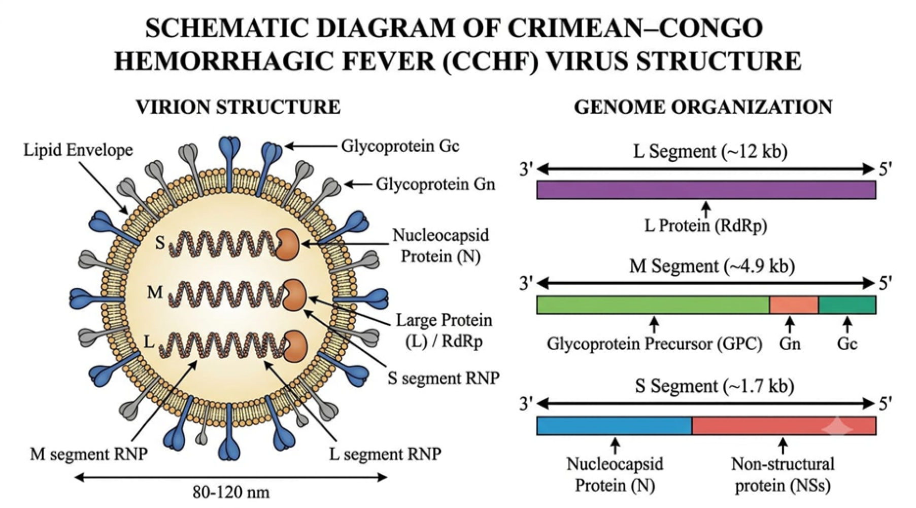
A schematic diagram of the CCHFV structure.
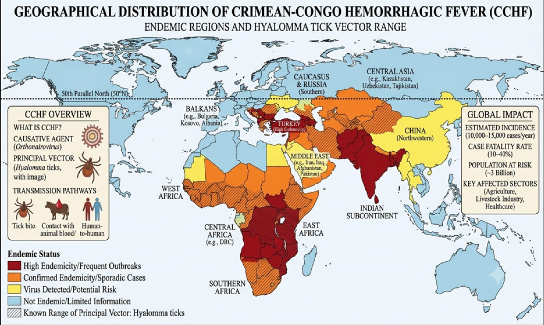
A world map showing the geographical distribution/endemic regions of CCHFV
References
- Akıncı, E., H. Bodur and H. Leblebicioglu. 2013. ‘Pathogenesis of Crimean-Congo hemorrhagic fever’, Vector-Borne Zoon. Diseas., 13(7): pp. 429–437. https://doi.org/10.1089/vbz.2012.1061[CrossRef]
- Anagnostou, V. and A. Papa. 2009. ‘Evolution of Crimean-Congo hemorrhagic fever virus’, Infection, Genet. Evolut., 9(5): pp. 948–954. https://doi.org/10.1016/j.meegid.2009.06.018[CrossRef]
- Appannanavar, S.B. and B. Mishra. 2011. ‘An update on Crimean Congo hemorrhagic fever’, J. Glob. Infect. Diseas., 3(3): pp. 285–292. https://doi.org/10.4103/0974-777X.83537[CrossRef]
- Carroll, S.A., B.H. Bird, P.E. Rollin, and S.T. Nichol. 2010. ‘Ancient common ancestry of Crimean-Congo hemorrhagic fever virus’, Molecul. Phylogenet. Evolut., 55(3): pp. 1103–1110. https://doi.org/10.1016/j.ympev.2010.01.006[CrossRef]
- Çevik, M.A., A. Erbay, H. Bodur, E. Gülderen, A. Baştuğ, A. Kubar, and E. Akıncı. 2008. ‘Clinical and laboratory features of Crimean-Congo hemorrhagic fever: predictors of fatality’, Int. J. Infect. Diseas., 12(4): pp. 374–379. https://doi.org/10.1016/j.ijid.2007.09.010[CrossRef]
- Çevik, M.A., A. Erbay, H. Bodur, S.S. Eren, E. Akinci, K. Şener, P. Öngürü, and A. Kubar. 2007. ‘Viral load as a predictor of outcome in Crimean-Congo hemorrhagic fever’, Clin. Infect. Diseas., 45(7): pp. e96–e100. https://doi.org/10.1086/521244[CrossRef]
- Chinikar, S., S.M. Ghiasi, R. Hewson, M. Moradi, and A. Haeri. 2010. ‘Crimean-Congo hemorrhagic fever in Iran and neighboring countries’, J. Clin. Virol., 47(2): pp. 110–114. https://doi.org/10.1016/j.jcv.2009.10.014[CrossRef]
- Chinikar, S., S.M. Ghiasi, A. Ghalyanchi Langeroudi, M.M. Gouya, M.R. Shirzadi, M. Zeynali, and A. Haeri. 2009. ‘An overview of Crimean-Congo hemorrhagic fever in Iran’, Arch. Iran. Med., 12(5): pp. 474–481.
- Connolly-Andersen, A.M., G. Moll, C. Andersson, S. Åkerström, H. Karlberg, I. Douagi, and A. Mirazimi. 2011. ‘Crimean-Congo hemorrhagic fever virus activates endothelial cells’, J. Virol., 85(15): pp. 7766–7774. https://doi.org/10.1128/JVI.02469-10[CrossRef]
- Deyde, V.M., M.L. Khristova, P.E. Rollin, T.G. Ksiazek, and S.T. Nichol. 2006. ‘Crimean-Congo hemorrhagic fever virus genomics and global diversity’, J. Virol., 80(17): pp. 8834–8842. https://doi.org/10.1128/JVI.00752-06[CrossRef]
- Drosten, C., D. Minnak, P. Emmerich, H. Schmitz, and T. Reinicke. 2002. ‘Crimean-Congo hemorrhagic fever in Kosovo’, J. Clin. Microbiol., 40(3): pp. 1122–1123. https://doi.org/10.1128/JCM.40.3.1122-1123.2002[CrossRef]
- Duh, D., A. Saksida, M. Petrovec, S. Ahmeti, I. Dedushaj, M. Panning, C. Drosten, and T. Avšič-Županc. 2007. ‘Viral load as predictor of Crimean-Congo hemorrhagic fever outcome’, Emerg. Infect. Diseas., 13(11): p. 1769. https://doi.org/10.3201/eid1311.070222[CrossRef]
- Dunster, L., M. Dunster, V. Ofula, D. Beti, F. Kazooba-Voskamp, F. Burt, R. Swanepoel, and K.M. DeCock. 2002. ‘First documentation of human Crimean-Congo hemorrhagic fever, Kenya’, Emerg. Infect. Diseas., 8(9): p. 1005. https://doi.org/10.3201/eid0809.010510[CrossRef]
- Elaldi, N. and S. Kaya. 2014. ‘Crimean-Congo hemorrhagic fever’, J. Microbiol. Infect. Diseas., 4(5): pp. 1–9. https://doi.org/10.5799/ahinjs.02.2014.S1.0135[CrossRef]
- Ergönül, Ö. 2006. ‘Crimean-Congo haemorrhagic fever’, The Lancet Infect. Diseas., 6(4): pp. 203–214. https://doi.org/10.1016/S1473-3099(06)70435-2[CrossRef]
- Ergonul, O. 2007. ‘Clinical and pathologic features of Crimean-Congo hemorrhagic fever’, in Ergonul, O. and Whitehouse, C.A. (eds.) Crimean-Congo Hemorrhagic Fever: A Glob. Perspect. Dordrecht: Springer Netherlands, pp. 207–220. https://doi.org/10.1007/978-1-4020-6106-6_16[CrossRef]
- Estrada-Peña, A., A.M. Palomar, P. Santibáñez, N. Sánchez, M.A. Habela, A. Portillo, L. Romero, and J.A. Oteo. 2012. ‘Crimean-Congo hemorrhagic fever virus in ticks, Southwestern Europe, 2010’, Emerg. Infect. Diseas., 18(1): p. 179. https://doi.org/10.3201/eid1801.111040[CrossRef]
- Flick, R. and C.A. Whitehouse. 2005. ‘Crimean-Congo hemorrhagic fever virus’, Curr. Molecul. Med., 5(8): pp. 753–760. https://doi.org/10.2174/156652405774962335[CrossRef]
- Frias-Staheli, N., Giannakopoulos, N.V., Kikkert, M., Taylor, S.L., Bridgen, A., Paragas, J., Richt, J.A., Rowland, R.R., Schmaljohn, C.S., Lenschow, D.J. and Snijder, E.J., 2007. Ovarian tumor domain-containing viral proteases evade ubiquitin-and ISG15-dependent innate immune responses. Cell host & microbe, 2(6), pp.404-416.
- Hawman, D.W. and H. Feldmann. 2018. ‘Recent advances in understanding Crimean–Congo hemorrhagic fever virus’, F1000 Res., 7: pp. F1000–Faculty. https://doi.org/10.12688/f1000research.16189.1[CrossRef]
- Hawman, D.W. and H. Feldmann. 2023. ‘Crimean–Congo haemorrhagic fever virus’, Nature Rev. Microbiol., 21(7): pp. 463–477. https://doi.org/10.1038/s41579-023-00871-9[CrossRef]
- Hewson, R., Gmyl, A., Gmyl, L., Smirnova, S.E., Karganova, G., Jamil, B., Hasan, R., Chamberlain, J. and Clegg, C., 2004. Evidence of segment reassortment in Crimean-Congo haemorrhagic fever virus. Journal of General Virology, 85(10), pp.3059-3070.
- Hoogstraal, H. 1979. ‘The epidemiology of tick-borne Crimean-Congo hemorrhagic fever in Asia, Europe, and Africa’, J. Med. Entomol., 15(4): pp. 307–417. https://doi.org/10.1093/jmedent/15.4.307[CrossRef]
- Ince, Y., C. Yasa, M. Metin, M. Sonmez, E. Meram, B. Benkli, and O. Ergonul. 2014. ‘Crimean-Congo hemorrhagic fever infections reported by ProMED’, Int. J. Infectious Diseas., 26: pp. 44–46. https://doi.org/10.1016/j.ijid.2014.04.005[CrossRef]
- Karanam, S.K., K. Nagvishnu, P.K. Uppala, S. Edhi, and S.R. Varri. 2025. ‘Crimean-Congo hemorrhagic fever: Pathogenesis, transmission and public health challenges’, World J. Virol., 14(1): p. 100003. https://doi.org/10.5501/wjv.v14.i1.100003[CrossRef]
- Karti, S.S., Z. Odabasi, V. Korten, M. Yilmaz, M. Sonmez, R. Caylan, E. Akdogan, N. Eren, I. Koksal, E. Ovali, and B.R. Erickson, 2004. ‘Crimean-Congo hemorrhagic fever in Turkey’, Emerg. Infect. Diseas., 10(8): p. 1379. https://doi.org/10.3201/eid1008.030928[CrossRef]
- Keshtkar-Jahromi, M., J.H. Kuhn, I. Christova, S.B. Bradfute, P.B. Jahrling, and S. Bavari. 2011. ‘Crimean-Congo hemorrhagic fever: current and future prospects of vaccines and therapies’, Antivir. Res., 90(2): pp. 85–92. https://doi.org/10.1016/j.antiviral.2011.02.010[CrossRef]
- Keshtkar-Jahromi, M., M.M. Sajadi, H. Ansari, M. Mardani, and K. Holakouie-Naieni. 2013. ‘Crimean–Congo hemorrhagic fever in Iran’, Antivir. Res., 100(1): pp. 20–28. https://doi.org/10.1016/j.antiviral.2013.07.007[CrossRef]
- Kuehnert, P.A., C.P. Stefan, C.V. Badger, and K.M. Ricks. 2021. ‘Crimean-Congo hemorrhagic fever virus (CCHFV): a silent but widespread threat’, Curr. Trop. Med. Repor., 8(2): pp. 141–147. https://doi.org/10.1007/s40475-021-00235-4[CrossRef]
- Kuhn, J.H., Alkhovsky, S.V., Avšič-Županc, T., Bergeron, É., Burt, F., Ergünay, K., Garrison, A.R., Marklewitz, M., Mirazimi, A., Papa, A. and Pawęska, J.T., 2024. ICTV virus taxonomy profile: Nairoviridae 2024. Journal of General Virology, 105(4), p.001974.
- Leblebicioglu, H., R. Ozaras, H. Irmak, and I. Sencan. 2016. ‘Crimean-Congo hemorrhagic fever in Turkey: Current status and future challenges’, Antiv. Res., 126: pp. 21–34. https://doi.org/10.1016/j.antiviral.2015.12.003[CrossRef]
- Leblebicioglu, H., Eroglu, C., Erciyas-Yavuz, K., Hokelek, M., Acici, M. and Yilmaz, H., 2014. Role of migratory birds in spreading Crimean-Congo hemorrhagic fever, Turkey. Emerging infectious diseases, 20(8), p.1331.
- Lindeborg, M., Barboutis, C., Ehrenborg, C., Fransson, T., Jaenson, T.G., Lindgren, P.E., Lundkvist, Å., Nyström, F., Salaneck, E., Waldenström, J. and Olsen, B., 2012. Migratory birds, ticks, and Crimean-Congo hemorrhagic fever virus. Emerging infectious diseases, 18(12), p.2095.
- Maltezou, H.C., L. Andonova, R. Andraghetti, M. Bouloy, O. Ergonul, F. Jongejan, N. Kalvatchev, S. Nichol, M. Niedrig, A. Platonov, and G. Thomson. 2010. ‘Crimean-Congo hemorrhagic fever in Europe: current situation calls for preparedness’, Euro Surveillance, 15(10): p. 19504. https://doi.org/10.2807/ese.15.10.19504-en[CrossRef]
- Maltezou, H.C. and A. Papa. 2010. ‘Crimean–Congo hemorrhagic fever: risk for emergence of new endemic foci in Europe?’, Trav. Med. Infect. Disease., 8(3): pp. 139–143. https://doi.org/10.1016/j.tmaid.2010.04.008[CrossRef]
- Mardani, M. and J.M. Keshtkar. 2007. ‘Crimean-Congo hemorrhagic fever’, Iran. J. Clin. Infect. Diseas., 2(1): pp. 43–49.
- Mertens, M., K. Schmidt, A. Ozkul, and M.H. Groschup. 2013. ‘The impact of Crimean-Congo hemorrhagic fever virus on public health’, Antiv. Res., 98(2): pp. 248–260. https://doi.org/10.1016/j.antiviral.2013.02.007[CrossRef]
- Messina, J.P., D.M. Pigott, N. Golding, K.A. Duda, J.S. Brownstein, D.J. Weiss, H. Gibson, T.P. Robinson, M. Gilbert, G.R. William Wint, and P.A. Nuttall. 2015. ‘The global distribution of Crimean-Congo hemorrhagic fever’, Transact. Roy. Soc. Trop. Med. Hyg., 109(8): pp. 503–513. https://doi.org/10.1093/trstmh/trv050[CrossRef]
- Mild, M., M. Simon, J. Albert, and A. Mirazimi. 2010. ‘Towards an understanding of the migration of Crimean–Congo hemorrhagic fever virus’, J. Gener. Virol., 91(1): pp. 199–207 https://doi.org/10.1099/vir.0.014878-0.[CrossRef]
- Morikawa, S., M. Saijo, and I. Kurane. 2007. ‘Recent progress in molecular biology of Crimean–Congo hemorrhagic fever’, Comparative Immunology, Microbiol. Infect. Diseas., 30(5–6): pp. 375–389. https://doi.org/10.1016/j.cimid.2007.07.001[CrossRef]
- Nabeth, P., D.O. Cheikh, B. Lo, O. Faye, I.O.M. Vall, M. Niang, B. Wague, D. Diop, M. Diallo, B. Diallo, and O.M. Diop. 2004a. ‘Crimean-Congo hemorrhagic fever, Mauritania’, Emerg. Infect. Diseas., 10(12): p. 2143. https://doi.org/10.3201/eid1012.040535[CrossRef]
- Nabeth, P., M. Thior, O. Faye, and F. Simon. 2004b. ‘Human Crimean-Congo hemorrhagic fever, Senegal’, Emerg. Infect. Diseas., 10(10): p. 1881. https://doi.org/10.3201/eid1010.040586[CrossRef]
- Negredo, A., F. de la Calle-Prieto, E. Palencia-Herrejón, M. Mora-Rillo, J. Astray-Mochales, M.P. Sánchez-Seco, E. Bermejo Lopez, J. Menárguez, A. Fernández-Cruz, B. Sánchez-Artola, and E. Keough-Delgado. 2017. ‘Autochthonous Crimean–Congo hemorrhagic fever in Spain’, New England J. Med., 377(2): pp. 154–161. https://doi.org/10.1056/NEJMoa1615162[CrossRef]
- Negredo, A., M.Á. Habela, E.R. de Arellano, F. Diez, F. Lasala, P. López, A. Sarriá, N. Labiod, R. Calero-Bernal, M. Arenas, and A. Tenorio. 2019. ‘Survey of Crimean-Congo hemorrhagic fever enzootic focus, Spain, 2011–2015’, Emerg. Infect. Diseas., 25(6): p. 1177. https://doi.org/10.3201/eid2506.180877[CrossRef]
- Öncü, S. 2013. ‘Crimean-Congo hemorrhagic fever: an overview’, Virologica Sinica., 28(4): pp. 193–201. https://doi.org/10.1007/s12250-013-3327-4[CrossRef]
- Papa, A., S. Bino, A. Llagami, B. Brahimaj, E. Papadimitriou, V. Pavlidou, E. Velo, G. Cahani, M. Hajdini, A. Pilaca, and A. Harxhi. 2002. ‘Crimean-Congo hemorrhagic fever in Albania, 2001’, European J. Clin. Microbiol. Infect. Diseas., 21(8): pp. 603–606. https://doi.org/10.1007/s10096-002-0770-9[CrossRef]
- Papa, A., S. Bino, E. Velo, A. Harxhi, M. Kota, and A. Antoniadis. 2006. ‘Cytokine levels in Crimean-Congo hemorrhagic fever’, J. Clin. Virol., 36(4): pp. 272–276. https://doi.org/10.1016/j.jcv.2006.04.007[CrossRef]
- Papa, A., I. Christova, E. Papadimitriou, and A. Antoniadis. 2004. ‘Crimean-Congo hemorrhagic fever in Bulgaria’, Emerg. Infect. Diseas., 10(8): p. 1465. https://doi.org/10.3201/eid1008.040162[CrossRef]
- Papa, A., A. Mirazimi, I. Köksal, A. Estrada-Pena, and H. Feldmann. 2015. ‘Recent advances in research on Crimean-Congo hemorrhagic fever’, J. Clin. Virol., 64: pp. 137–143. https://doi.org/10.1016/j.jcv.2014.08.029[CrossRef]
- Papa, A., K. Tsergouli, K. Tsioka, and A. Mirazimi. 2017. ‘Crimean-Congo hemorrhagic fever: tick-host-virus interactions’, Front. Cellul. Infect. Microbiol., 7: p. 213. https://doi.org/10.3389/fcimb.2017.00213[CrossRef]
- Portillo, A., A.M. Palomar, P. Santibáñez, and J.A. Oteo. 2021 ‘Epidemiological aspects of Crimean-Congo hemorrhagic fever in Western Europe: what about the future?’, Microorgan., 9(3): p. 649. https://doi.org/10.3390/microorganisms9030649[CrossRef]
- Raabe, V.N. 2020. ‘Diagnostic testing for Crimean-Congo hemorrhagic fever’, J. Clin. Microbiol., 58(4): pp. 10–1128. https://doi.org/10.1128/JCM.01580-19[CrossRef]
- Sanchez, A.J., M.J. Vincent, and S.T. Nichol. 2002. ‘Characterization of the glycoproteins of Crimean-Congo hemorrhagic fever virus’, J. Virol., 76(14): pp. 7263–7275. https://doi.org/10.1128/JVI.76.14.7263-7275.2002[CrossRef]
- Shahhosseini, N., G. Wong, G. Babuadze, J.V. Camp, O. Ergonul, G.P. Kobinger, S. Chinikar, and N. Nowotny, 2021. ‘Crimean-Congo hemorrhagic fever virus in Asia, Afric. Europe’, Microorgan., 9(9): p. 1907. https://doi.org/10.3390/microorganisms9091907[CrossRef]
- Shayan, S., M. Bokaean, M.R. Shahrivar, and S. Chinikar. 2015. ‘Crimean-Congo hemorrhagic fever’, Labor. Med., 46(3): pp. 180–189. https://doi.org/10.1309/LMN1P2FRZ7BKZSCO[CrossRef]
- Shepherd, A.J., R. Swanepoel, and P.A. Leman. 1989. ‘Antibody response in Crimean-Congo hemorrhagic fever’, Rev. Infect. Diseas., 11(Supplement 4): pp. S801–S806. https://doi.org/10.1093/clinids/11.Supplement_4.S801[CrossRef]
- Smego, R.A., A.R. Sarwari, and A.R. Siddiqui. 2004. ‘Crimean-Congo hemorrhagic fever: prevention and control limitations in a resource-poor country’, Clin. Infect. Diseas., 38(12): pp. 1731–1735. https://doi.org/10.1086/421093[CrossRef]
- Spengler, J.R., D.A. Bente, M. Bray, F. Burt, R. Hewson, G. Korukluoglu, A. Mirazimi, F. Weber, and A. Papa. 2018. ‘Second international conference on Crimean-Congo hemorrhagic fever’, Antivir. Res., 150: pp. 137–147. https://doi.org/10.1016/j.antiviral.2017.11.019[CrossRef]
- Spengler, J.R., É. Bergeron, and P.E. Rollin. 2016. ‘Seroepidemiological studies of Crimean-Congo hemorrhagic fever virus in domestic and wild animals’, PLoS Neglect. Trop. Diseas., 10(1): p. e0004210. https://doi.org/10.1371/journal.pntd.0004210[CrossRef]
- Spengler, J.R., É. Bergeron, and C.F. Spiropoulou. 2019. ‘Crimean-Congo hemorrhagic fever and expansion from endemic regions’, Current Opinion in Virology, 34, pp. 70–78. https://doi.org/10.1016/j.coviro.2018.12.002[CrossRef]
- Swanepoel, R. (1994) ‘Crimean-Congo hemorrhagic fever’, in Beran, G.W. (ed.) Handbook of Zoonoses, Section B: Viral Zoonoses, 2nd edn. Boca Raton: CRC Press, pp. 149–161.
- Swanepoel, R., D.E. Gill, A.J. Shepherd, P.A. Leman, J.H. Mynhardt, and S. Harvey. 1989. ‘The clinical pathology of Crimean-Congo hemorrhagic fever’, Rev. Infect. Diseas., 11(Supplement 4), pp. S794–S800. https://doi.org/10.1093/clinids/11.Supplement_4.S794[CrossRef]
- Swanepoel, R., A.J. Shepherd, P.A. Leman, S.P. Shepherd, G.M. McGillivray, M.J. Erasmus, L.A. Searle, and D.E. Gill. 1987. ‘Epidemiologic and clinical features of Crimean-Congo hemorrhagic fever in southern Africa’, Americ. J. Trop. Med. Hyg., 36(1): pp. 120–132. https://doi.org/10.4269/ajtmh.1987.36.120[CrossRef]
- Swanepoel, R., J.K. Struthers, A.J. Shepherd, G.M. McGillivray, M.J. Nel, and P.G. Jupp. 1983. ‘Crimean-Congo hemorrhagic fever in South Africa’, Americ. J. Trop. Med. Hygie., 32(6): pp. 1407–1415. https://doi.org/10.4269/ajtmh.1983.32.1407[CrossRef]
- Temur, A.I., J.H. Kuhn, D.B. Pecor, D.A. Apanaskevich, and M. Keshtkar-Jahromi. 2021. ‘Epidemiology of Crimean-Congo hemorrhagic fever (CCHF) in Africa—underestimated for decades’, Americ. J. Trop. Med. Hygie., 104(6): p. 1978. https://doi.org/10.4269/ajtmh.20-1413[CrossRef]
- Tezer, H., I.A. Sucaklı, T.R. Saylı, E. Celikel, I. Yakut, A. Kara, B. Tunc, and O. Ergonul. 2010. ‘Crimean-Congo hemorrhagic fever in children’, J. Clin. Virol., 48(3): pp. 184–186. https://doi.org/10.1016/j.jcv.2010.04.001[CrossRef]
- Vanhomwegen, J., M.J. Alves, T.A. Županc, S. Bino, S. Chinikar, H. Karlberg, G. Korukluoğlu, M. Korva, M. Mardani, A. Mirazimi, and M. Mousavi. 2012. ‘Diagnostic assays for Crimean-Congo hemorrhagic fever’, Emerg. Infect. Diseas., 18(12): p. 1958. https://doi.org/10.3201/eid1812.120710[CrossRef]
- Vatansever, Z., R. Uzun, A. Estrada-Pena, and O. Ergonul. 2007. ‘Crimean-Congo hemorrhagic fever in Turkey’, in Ergonul, O. and Whitehouse, C.A. (eds.) Crimean-Congo Hemorrhagic Fever: A Global Perspective. Dordrecht: Springer Netherlands, pp. 59–74. https://doi.org/10.1007/978-1-4020-6106-6_6[CrossRef]
- Vorou, R., I.N. Pierroutsakos, and H.C. Maltezou. 2007. ‘Crimean-Congo hemorrhagic fever’, Curr. Opin. Infect. Diseas., 20(5): pp. 495–500 https://doi.org/10.1097/QCO.0b013e3282a56a0a.[CrossRef]
- Watts, D.M., T.G. Ksiazek, K.J. Linthicum, and H. Hoogstraal. 2019. ‘Crimean-Congo hemorrhagic fever’, in Monath, T.P. (ed.) The Arboviruses: Epidemiol. Ecol. Boca Raton: CRC Press, pp. 177–222. https://doi.org/10.1201/9780429280245-9[CrossRef]
- Whitehouse, C.A. 2004. ‘Crimean–Congo hemorrhagic fever’, Antivir. Res., 64(3): pp. 145–160. https://doi.org/10.1016/S0166-3542(04)00163-9[CrossRef]
- World Organisation for Animal Health (WOAH). (2024). Crimean–Congo haemorrhagic fever. In Manual of diagnostic tests and vaccines for terrestrial animals (Chapter 3.1.5). https://www.woah.org/en/disease/crimean-congo-haemorrhagic-fever/
- Yilmaz, G.R., T. Buzgan, H. Irmak, A. Safran, R. Uzun, M.A. Cevik and M.A. Torunoglu. 2009. ‘The epidemiology of Crimean-Congo hemorrhagic fever in Turkey, 2002–2007’, Int. J. Infect. Diseas., 13(3): pp. 380–386. https://doi.org/10.1016/j.ijid.2008.07.021[CrossRef]
- Zivcec, M., F.E. Scholte, C.F. Spiropoulou, J.R. Spengler, and É. Bergeron. 2016. ‘Molecular insights into Crimean-Congo hemorrhagic fever virus’, Virus., 8(4): p. 106. https://doi.org/10.3390/v8040106[CrossRef]
Services
Article usage statistics
Loading…