
Convolvulus scammonia is a twining perennial plant with thick, fleshy roots, irregularly arrow-shaped leaves, and flowers resembling those of Convolvulus arvensis. The aim of this study was to determine the impact of C. scammonia on six genes expression (i.e., ACT1, BCR1, EFG1, AlS1, AlS3, and TEC1), known to be implicated in the development of Candida tropicalis biofilms, and define the minimum inhibitory concentration (MIC) of C. scammonia required for this purpose. The 2,3-bis(2-metoxy-4-nitro-5-sulphophenyl)-2H-tetrazolium-5-carboxanilide inner salt (XTT) assay was used to assess the anti-biofilm activity of C. scammonia in order to ascertain the formation of biofilms on C. tropicalis isolates obtained from thrush samples and evaluate the MIC of C. scammonia that inhibits the biofilms of Candida tropicalis during 24 and 48 h. Finally, the impact of C. scammonia on ACT1, AlS1, AlS3, BCR1, TEC1, and EFG1gene expression in C. tropicalis was examined using a real-time polymerase chain reaction (RT-PCR) and compared to the results obtained in the gene expression of the control C. tropicalis biofilms untreated with C. scammonia during 24 and 48 h. Through comparison, biofilms formation was found to decrease as C, scammonia concentration and time increased. Accordingly, the MIC of C. scammonia was 20 % w/v, and its minimum fungicidal - concentration (MFC) was 40 % (w/v) in biofilm-forming C. tropicalis. Additionally, gene expression level analysis revealed a decrease in ACT1, AlS1, AlS3, BCR1, EFG1, and TEC1 expression levels on treatment with C. scammonia during 24 and 48 h.
Research Article · Volume 9, Issue 3, 3 June 2025 · Open Access
Convolvulus scammonia Effect on Gene Expression of Candida tropicalis Biofilms
· ·
Affiliations & Notes ⌄
- 1Biology Department, College of Science, Al-Muthanna University, Iraq. *Correspondence | Mouna Akeel Hamed Al-Oebady, Biology Department, College of Science, Al-Muthanna University, Iraq
- 2Email: mouna@mu.edu.iq
Article Info ⌄
- Journal:
- Novel Research in Microbiology Journal
- Published:
- 3 June 2025
Abstract
Introduction
There are several species of Candida in nature, usually as part of commensal mammalian microbiota . Nevertheless, the commensal phase may give a way to the pathogenic phase due to modifications in the host environment, such as disruptions in the commensal microbiota . According to several studies, fungal infections in humans are neglected infectious diseases that haves emerged as a problem in the public health system in recent decades . Currently, over 200 species of Candida have been identified . Non-albicans Candida (NAC) species are becoming more prevalent in terms of mortality and morbidity, although C. albicans remains the most prevalent fungal pathogen . In addition to C. albicans, four other species of NAC have surfaced, mainly C. tropicalis, C. parapsilosis, C. glabrata, and C. krusei . Currently regarded as the most significant emerging fungal pathogen among these, C. tropicalis has been found to have multiple strains that are resistant to common empirical treatments, including fluconazole .
It is known that both C. albicans and C. tropicalis have a wide variety of commensal traits and virulence factors that enable them to colonize and infiltrate the host tissue . These include phenotypic switching and thigmotropism (contact sensing), production of hydrolytic enzymes, development of biofilms, expression of adhesins and invasins on the cell surface, and capacity to damage host cells . Although biofilms are the most prevalent way for microorganisms to grow and the formation of these biofilms among Candida species confers significant resistance to antifungal therapy, little is known about the life cycle of C. tropicalis biofilms . In 20٢٤, the efficacy of liposomal amphotericin B antifungal and the development of C. tropicalis biofilms were evaluated through time-lapse imaging, demonstrating that C. tropicalis is a fast-growing species capable of forming aggressive biofilms (Omelchuk et al., 20٢٤).
Wild plants have been used extensively by Aboriginal people to prevent nutritional and pathogen-related illnesses since the dawn of human history . Since the beginning of time, people have utilized plant-based products in a wide range of settings. Researchers have been examining the deliberate use of natural resources for more than a millennium . The medicinal plants have long been used to treat a variety of ailments, and they are still used as emerging new tools to combat these diseases .
Because of their bio-availability, biological properties, safety, and clinical efficacy, C. scammonia plant extracts and essential oils have drawn more attention . The abundance of the following compounds was observed in the genus Convolvulus: fatty acids, alkaloids, lipids, coumarins, lignans, alkaloids, terpenoids, steroids, flavonoids, amino acids, carbohydrates, anthraquinones, anthocyanidins, phenylpropanoids, and resins ; . Convolvulus species have been found to possess medicinal properties as the ability to lessen or completely eradicate the symptoms of several severe illnesses, including e fever, heart disease, sleeplessness, memory loss, and hair loss, in addition to urogenital disorders, severe purgatives, diarrhea, gastrointestinal irritation, animal stings; congestion, edema, gaseous distended intestine, and organ hemorrhages .
The objective of this study was to detect the effect of C. scammonia on the expression of several genes (ACT1, AlS1, AlS3, BCR1, EFG1, and TEC1) that are involved in C. tropicalis biofilm formation, in addition to the minimum inhibitory concentration of C. scammonia needed to accomplish this.
Materials and Methods
Candida tropicalis isolation
Candida tropicalis was isolated from thrush samples at the Samawah Teaching Hospital for pediatrics and Gynecology in AL-Muthanna governorates, Iraq, during the period from December 2021 to June 2023. Fifty oral swabs were obtained from children suffering from oral candidiasis and septicemia with oral thrush. Specimens were taken using sterile swabs, and then transported to the microbiology laboratory for diagnosis. C. tropicalis was isolated and identified using Sabouraud dextrose agar (SDA) and CHROM agar.
Analytical Profile Index (API) (HIMDIA/India) was employed as a biochemical assay for detection of C. tropicalis and to validate the morphological, cultural, and biochemical characteristics required to differentiate between Candida species, and demonstrate that Candida can utilize different types of sugars .
Convolvulus scammonia collection
The plnats samples (leaves, stems, and flowers) used in these experiments were gathered from general local gardens in Samawah city. Crude alkaloids extraction was conducted on the leafs of these plants as described by Hussein (2024).
Minimum inhibitory concentration (MIC) determination
The microdilution broth method was used to calculate the MICs of natural C. scammonia against the biofilms of C. tropicalis. C. scammonia was diluted in Yeast extract Peptone Dextrose (YPD) broth in steps of 5 % w/v, 10 % w/v, 20 % w/v, 40 % w/v, and 80 % w/v. Under vigorous shaking (120 rpm), the cultures were incubated at 35 °C for 36 h. After incubation, 1 ml of a broth (103 cfu/ ml) was aseptically injected into Petri plates of SDA. Following a 48-h of incubation, the resulting colonies’ growth was evaluated by ELISA, and the MIC of C. scammonia (w/v) was taken as the dilution that prevented C. tropicalis from making a significant growth .
Formation of biofilms
The biofilms were prepared in 96-well flat-bottomed polystyrene microtiter plates and their growth was evaluated using the XTT (2,3-bis(2-metoxy-4-nitro-5-sulphophenyl)-2H-tetrazolium-5-carboxanilide inner salt) reduction assay . The kinetics of biofilm inhibition were studied using various MIC dilutions of C. scammonia in YPD broth (5 % w/v, 10 % w/v, 20 % w/v, 40 % w/v, and 80 % w/v) in order to ascertain whether C. scammonia could prevent the formation of Candida biofilms and find the minimum concentration of C. scammonia that could do so. Every MIC dilution in a microtiter plate was analyzed in at least 7 wells. Each dilution (5 % w/v, 10 % w/v, 20 % w/v, 40 % w/v, and 80 % w/v) was added to aliquots that held 190 ml in the microtiter plate wells. A culture of 48 h of C. tropicalis was cultivated in 10 ml of YPD broth with 5 × 10⁸ cfu/ ml. On an orbital shaker running at 75 rpm, 10 µl of this 48-h culture were applied to each well and incubated for 1.5 h at 37 °C to guarantee even distribution and adherence to the well surfaces. In order to remove non-adherent cells, the adherent cells were carefully washed twice in sterile phosphate buffered saline (PBS) with a pH of 7.4, being cautious not to disturb the adherent cells. Following washing, 200 ml of a second aliquot of the same C. scammonia dilution in sterile YPD broth was added to each plate well. Afterward, the plates were kept in the same incubator for 48 h so that the biofilms could settle in and grow. As a control, 200 ml of an autoclaved YPD broth was added to each of the seven micro-titer plate wells. The broth could be positive (containing Candida) or negative (not containing Candida). The plate was incubated for 48 h at 37 °C .
Biofilm evaluation using the XTT reduction assay
The C. tropicalis cell concentrations in the biofilms were measured using the XTT assay following C. scammonia treatment. A filter of a pore size 0.22 mm was used to filter and sterilize the cells after making the XTT solution (1 mg/ml in PBS). Prior to the commencement of every assay, a menadione solution and XTT solution were mixed at a 5:1 (v/v). To stop the adherent biofilms from growing further, 2.5 % glutaraldehyde was applied for 5 min. to the microtiter plate wells after three PBS washings. Following the removal of the fixative, two PBS washes were applied to the wells. In all but the control well, which had no biofilm, 1 ml of PBS was added to the XTT-menadione solution. After that, the Metyrapones (MTPs) were maintained in the darkness for 2 h at 37 °C. After incubation, 75 ml of XTT-menadione solution were added to each well on a fresh microtiter plate, and spectrophotometry was used to measure the absorbance at 492 nm .
RNA isolation and RT-PCR
The PrestoTM Mini gDNA Yeast Kit was used to extract the total RNA from the C. tropicalis culture cells and co-culture them with C. scammonia in accordance with the manufacturer’s instructions. The concentration and degree of RNA presence were determined using a Nanodrop-1000 UV-VIS spectrophotometer. For the agarose gel electrophoresis, RNA templates were employed. The cDNA synthesis SuperMix Kit and EasyScript One-Step gDNA were used to perform the reverse transcription PCR cDNA synthesis. One cycle at 42 °C for 30 min. and 85 °C for 5 sec was the RT-PCR setup .
The mRNA transcripts were detected using real-time PCR with the Mx3000p (Agilent Technologies, USA). 10 mM primers were used to prepare the PCR components, which were then amplified by the genes presented in Table 1 . In this experiment, the housekeeping gene beta-actin was amplified in the forward direction 5’-CGTCGGTAGACCAAGACACC-3’ and the opposite direction 5’-CCCAGTTGGAGACAATACCGT-3’, A final volume of 20 μl was achieved by adding nuclease-free water, 200 ng of cDNA, and 10 μl of Luna universal qPCR master mix. The following parameters were used to run the PCR: 30 sec at 94 °C, 45 cycles of 94 °C for 5 sec, and 30 sec at 60 °C. Then, a dissociation curve was employed, with a single cycle lasting 1.0 min. at 95 °C, 30 sec at 55 °C, and 30 sec at 95 °C .
Table 1: The nucleotide sequence of the primers for real-time PCR amplification.
| Genes | Primer sequence (5`→3`) | GenBank accession no. | Product size (bp) |
| ACT1 | Forward TACGCTGGTTTCTCCTTGCC Reverse GCGGTGGTGGAGAAAGTGT | XM_002549283.1 | 116 |
| ALS1 | Forward TCTGTTGCCATTCCTGTCGAA Reverse AAACCAACCCAAGCAAGATCG | MK182724.1 | 70 |
| AlS3 | ForwardTGACTCCTAAGGAAGTATCGGGA Reverese GCCAAGCTGGATTAGCAGGA | MH753521.1 | 102 |
| BCR1 | Forward ATCCTTTACCTGCGTTGCGT Reverese GGACGAAGCTACTGACTCGG | XM_002545781.1 | 146 |
| EFG1 | Forward TCCTGCAGCTCCACCTATACC Reverese TTGGCTGGGTTTAACGTGTCT | KP314278.1 | 99 |
| TEC1 | Forward TTCAAGCAGTGCTCCAAGTTC Reverese AGGTGCAGAATGGAAACCAGT | XM_002547951.1 | 103 |
Statistical analysis
The analysis of variance (ANOVA) and t test were used to examine the various means of biofilm biomass (absorbance). GraphPad was used to analyze the data.
Results
Minimum inhibitory concentration determination
The growth of C. tropicalis was inhibited by C. scammonia in a concentration-dependent manner. The minimum fungicidal concentration (MFC) of C. scammonia on biofilm-forming C. tropicalis was 40 % (w/v), while its MIC was 20 % (w/v). Approximately, 99.9 % of the inoculum must be removed by the MFC. The value of MFC was typically higher than the MIC. Compared to cell growth without C. scammonia, the curves of growth for the C. tropicalis when exposed to 20 % (w/v) of the C. scammonia for 24 h showed a decrease in both the overall cells number and the growth rate (Figures 1 and 2). The treatment group and the control group differed significantly (p <0.05) according to the results of the one-way ANOVA test.
Effect of Convolvulus scammonia on biofilm formation
Convolvulus scammonia was added to YPD broth at several concentrations (5 % w/v, 10 % w/v, 20 % w/v, 40 % w/v, and 80 % w/v). It was shown that the amount of C. scammonia present affected the suppression of biofilm formation. Figure 2 shows that the formation of biofilm was neither disrupted nor stimulated by C. scammonia concentrations below 10 % w/v. However, biofilm development was greatly inhibited at concentrations greater than 10 % w/v. There was a significant difference between the control and treatment groups (p <0.05) according to the one-way ANOVA test analysis.
Gene expression analysis via RT-qPCR
The implicated genes (ACT1, AlS1, AlS3, BCR1, EFG1, and TEC1) were measured for relative expression in C. tropicalis over the course of the biofilm experiment for 24 and 48 h using RT-PCR.
In contrast to the genes encoding the biofilm treated with C. scammonia, the expression of the biofilm genes (ACT1, AlS1, AlS3, BCR1, EFG1, and TEC1) increased significantly over time in the absence of C. scammonia treatment. This is regarded as a measre of control (Figure 3). A p <0.05 for the expression of the biofilm genes was deemed statistically significant based on the t-test analysis.
Candida tropicalis cells exposed to C. scammonia had less biofilm formation during 24 h, according to results from a RT-PCR using the specific primers of ACT1, AlS1, AlS3, BCR1, EFG1, and TEC1 genes. Figure 4 shows the ACT1 (0.5), AIS1 (1), AIS3 (0.8), BCR1 (0.7), EFG1 (0.6), and TEC1 (0.8) genes that were treated with C. scammonia, respectively. This indicates the inhibitory role of C. scammonia on the C. tropicalis biofilm formation within 24 h compared to the control gene expression. According to the t-test analysis, the expression of the biofilm genes was p <0.05, which is considered statistically significant.
Results from RT-PCR using the particular primers ACT1, AlS1, AlS3, BCR1, EFG1, and TEC1 showed that C. tropicalis cells exposed to C. scammonia had less biofilm formation during 48 h. Figure 5 illustrates that C. scammonia was applied to the ACT1 (1), AlS1 (1.5), AlS3 (0.5), BCR1 (0.3), EFG1 (0.7), and TEC1 (0.9) genes, respectively. These results are conclusive evidence that C. scammonia is a highly efficient inhibitor of biofilm formation by C. tropicalis. It can be used as therapeutic alternatives for many diseases caused by C. tropicalis. These results display a positive impact of C. scammonia on the genes responsible for the formation of C. tropicalis biofilms (Figure 5). According to the t-test analysis, the expression of the biofilm genes was p < 0.05, which is considered statistically significant.
Discussion
Some intrinsic biofilm-associated factors, including immune system avoidance, antibiotic resistance, and mechanisms of horizontal gene transfer in multispecies biofilms, make biofilm formation a crucial component of pathogenicity in bacterial and fungal diseases ; ; . Candida biofilms have been found to form on a variety of surfaces, including blood, mucosal surfaces, and majority of medical devices, which are inanimate objects that come into contact with patients’ bodies ; ; . These indicate that C. tropicalis is the most common microbial species forming biofilms even more than C. albicans.
Thus, the purpose of this study was to determine C. scammonia effect on the expression of several genes (ACT1, AlS1, AlS3, BCR1, EFG1, and TEC1) involved in the formation of C. tropicalis biofilms. This experiment also aimed to determine the MIC of C. scammonia required to inhibit the growth of C. tropicalis forming biofilms. To the authors’ knowledge, this study is the first to employ these techniques to examine the biofilms of this Candida species throughout time, evaluate the precision of the biofilm formation evaluation, and determine the activity of C. scammonia affecting it.
These findings partially corroborate with previous studies showing that C. arvensis contains cadinene, carvacrol, and O-cymene . Additionally, the current results support the literature’s assertion that the Convolvulus contains oxygenated monoterpenes, oxygenated sesquiterpenes, and sesquiterpene hydrocarbons. It’s interesting to note that numerous authors have examined the phytochemical makeup of C. arvensis and found that the main constituents were lipids, sugar derivatives of quercetin and kaempferol, coumarins, saponins, flavonoids, phenolic acids, tannins, alkaloids, lactones, and steroids or terpenoids .
Importantly, C. arvensis organic extracts showed antifungal efficacy against A. fumigatus, C. albicans, C. tropicalis, Geotrichum candidum, Microsporum canis, and Trichophyton mentagrophytes, with MIC values ranging from 0.001 to 0.156 mg/ ml ; . Additionally, the antifungal results of this study are in line with those that reported that the extracts of C. althaeoides L. leaves with increasing polarity were effective against several dermatophytes (i.e., Microsporum canis, Trichophyton rubrum, and Trichophyton mentagrophytes), with 10 % inhibiting percentages at 50 mg/ml. Notably, C. althaeoides L. extracts significantly inhibited C. albicans , which is inconsistent with the current findings. Additionally, the antifungal findings in this study were consistent with earlier research on the antifungal effects of volatile fractions of C. althaeoides L. root . Furthermore, numerous studies have shown that essential oils and extracts from Convolvulus plants significantly suppress the growth of a variety of fungal pathogens such as Candida species .
The glycoproteins Als1 and Als3 of the cell wall of C. tropicalis are encoded by six biofilm-associated genes belonging to ALS gene family: ACT1, BCR1, ALS1, ALS3, TEC1, and EFG1. ALS1 expression can be found in both yeast and hyphae, whereas ALS3 transcription occurs only in germ tubes and hyphae . Sessile cells in C. tropicalis exhibited higher levels of expression for ALS1, ALS2, and ALS3 like genes, indicating their potential roles in biofilm formation . In C. tropicalis, the EFG1 ortholog showed a conserved effect on fungal filaments and biofilm formation . According to C. tropicalis deletion mutants of BRG1, TEC1, BCR1, EFG1, or NDT80 homologs have significantly reduced biofilm biomass, indicating that these genes have conserved functions in biofilm formation . C. albicans’s hyphae-specific gene expression is either fully or partially controlled by UME6, a common downstream target of the regulators Efg1, Chp1, and Ras1 .
The results of various analyses techniques of profile metabolites such as hydrogen peroxide activity, protease stability, thermal stability, and using liquid chromatography-mass spectrometry to might assist on identifying the active components and elucidate their mechanisms of action. An additional constraint pertains to the fact that we chose only a small number of target genes for the gene expression analysis out of the hundreds of genes and numerous signaling pathways that comprise the Candida biofilm regulatory network . Other well-known genes linked to biofilms were investigated by Nobile et al., (2006), including HWP1, which encodes adhesin in fungal cell wall necessary for the formation of biofilm and the negative regulator of filamentation NRG1.
Genomic-wide transcriptional profiling employing some technologies such as RNAsequence is a more efficient way to identify the elusive processes of C. scammonia’s activity .
Conclusions and Recommendations
The current results showed that C. scammonia could inhibit the expression of several genes in biofilm-forming C. tropicalis, including ACT1, BCR1, AlS1, AlS3, EFG1, and TEC1. Its MIC was 20 % (w/v) and its MFC was 40 % (w/v). Strong antifungal efficacies have been demonstrated for C. scammonia, suggesting that it contains compounds that could be useful in treating Candida infections. If these findings may be used to the treatment of biofilm-associated candidiasis; however, more in vivo studies are highly recommended.
Novelty Statement
This study enhances our understanding to determine the impact of C. scammonia on six gene expressions (i.e., ACT1, BCR1, EFG1, AlS1, AlS3, and TEC1); known to be implicated in the development of C. tropicalis biofilms, through the analysis of fifty oral swabs obtained from children suffering from oral candidiasis and septicemia with oral thrush. These findings offer valuable insights into the genetic dynamics of C. tropicalis biofilms, underscoring the necessity for continuous monitoring to address the C. scammonia impact.
Author’s Contribution
Mouna Akeel Hamed Al-Oebady: Investigation, validation of results, writing original draft, writing review & editing
Wafaa Ayad Al-Nuaimy: Investigation, writing original draft, writing review & editing
Noor Sami Al-Lebawy: Validation of results, writing original draft
Ethical approval
This study was approved by the ethical committee of Al-Muthanna University, Iraq, and the ethical approval code is 3102R5A5A2A11021350. Written consents of the participants were provided.
Funding source
None to declare.
Conflict of interests
The author have declared no conflicts of interest.
Figures
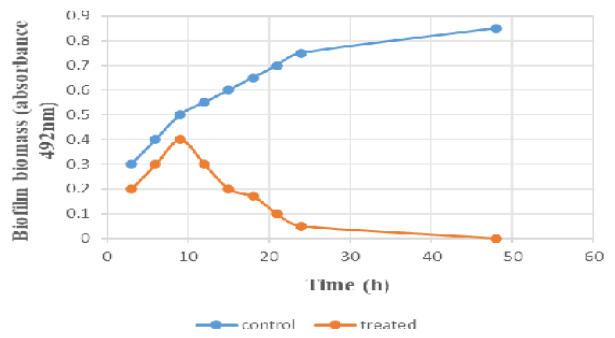
Growth evaluations of Convolvulus scammonia-treatment on established Candida tropicalis biofilms during 24 to 48 h (F=30.65941, p < 0.05).
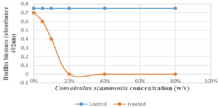
Convolvulus scammonia’s impact on the development of Candida tropicalis biofilms. Convolvulus scammonia’s MIC was 20 % (w/v), and its MFC was 40 % (w/v) in biofilm-forming Candida tropicalis during 24 to 48 h (F=12.36593, p < 0.05).
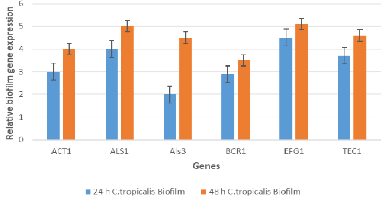
Relative expression of the implicated biofilm forming genes (ACT1, ALS1, AlS3, BCR1, EFG1, and TEC1) in Candida tropicalis during the biofilm experiment without treated of Convolvulus scammonia as a control for 24 and 48 h using reverse transcriptase real-time PCR. (t= 2.4868, df =10, standard error of difference = 0.442). p < 0.05 was considered statistically significant.
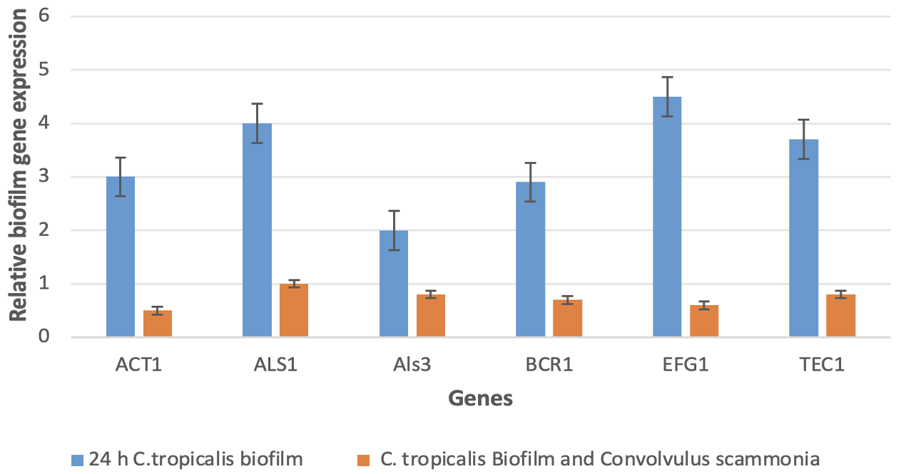
Relative expression of the implicated biofilm forming genes (ACT1, AlS1, AlS3, BCR1, EFG1, and TEC1) in Candida tropicalis during the biofilm experiment for 24 h and Candida tropicalis biofilm with Convolvulus scammonia using reverse transcriptase real-time PCR. (t =7.0198, df =10, standard error of difference= 0.373). p <0.05 was considered statistically significant.
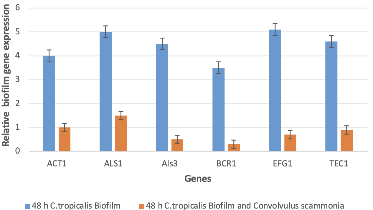
Relative expression of the implicated biofilm forming genes (ACT1, AlS1, AlS3, BCR1, EFG1, and TEC1) in Candida tropicalis during the biofilm experiment for 48 h and Candida tropicalis biofilm with Convolvulus scammonia using reverse transcription real-time PCR. (t= 12.0151, df = 10, standard error of difference= 0.302), p < 0.05 was considered statistically significant.
References
- Agarwal, V., Lal, P. and Pruthi, V., 2010. Effect of plant oils on Candida albicans. J. Microbiol. Immunol. Infect., 43: 447-451. https://doi.org/10.1016/S1684-1182(10)60069-2[CrossRef]
- Al-Rifai, A., Aqel, A., Al-Warhi, T., Wabaidur, S.M., Al-Othman, Z.A. and Badjah-Hadj-Ahmed, A.Y., 2017. Antibacterial, antioxidant activity of ethanolic plant extracts of some convolvulus species and their DART-ToF-MS profiling. Evid. Based Complement. Altern. Med., 1: 5694305. https://doi.org/10.1155/2017/5694305[CrossRef]
- Alves, R., Barata-Antunes, C., Casal, M., Brown, A.J.P., van Dijck, P. and Paiva, S., 2020. Adapting to survive: How candida overcomes host-imposed constraints during human colonization. PLoS Pathog., 16: e1008478. https://doi.org/10.1371/journal.ppat.1008478[CrossRef]
- Atiencia-Carrera, M.B., Cabezas-Mera, F.S., Tejera, E. and Machado, A., 2022. Prevalence of biofilms in Candida spp. bloodstream infections: A meta-analysis. PLoS One, 17: e0263522. https://doi.org/10.1371/journal.pone.0263522[CrossRef]
- Atriwal, T., Azeem, K., Husain, F.M., Hussain, A., Khan, M.N., Alajmi, M.F., and Abid, M., 2021. Mechanistic understanding of candida albicans biofilm formation and approaches for its inhibition. Front Microbiol., 12. https://doi.org/10.3389/fmicb.2021.638609[CrossRef]
- Bourhia, M., Laasri, F.E., Aourik, H., Boukhris, A., Ullah, R., Bari, A., Ali, S.S., El Mzibri, M., Benbacer, L. and Gmouh, S., 2019. Antioxidant and antiproliferative activities of bioactive compounds contained in rosmarinus officinalis used in the Mediterranean diet. Evid. Based Complement. Altern. Med., pp. 1–7. https://doi.org/10.1155/2019/7623830[CrossRef]
- Bourhia, M., Lahmadi, A., Achtak, H., Touis, A., Elbrahmi, J., Ullah, R., Shahat, A.A., Mahmood, H.M., Aboudkhil, S. and Benbacer, L., 2019. Phytochemical analysis and toxicity study of aristolochia paucinervis rhizomes decoction used in moroccan alternative medicine: Histopathological and biochemical profiles. Evid. Based Complement. Altern. Med., pp. 1–11. https://doi.org/10.1155/2019/1398404[CrossRef]
- Braun, B.R., Kadosh, D. and Johnson, A.D., 2001. NRG1, a repressor of filamentous growth in C. albicans, is down-regulated during filament induction. EMBO J., 20: 4753–4761. https://doi.org/10.1093/emboj/20.17.4753[CrossRef]
- Calderón-Hernández, A., Castro-Bonilla, N., and Cob-Delgado, M., 2024. Chromogenic, biochemical and proteomic identification of yeast and yeast-like microorganisms isolated from clinical samples from animals of Costa Rica. J. Fungi, 10: 218. https://doi.org/10.3390/jof10030218[CrossRef]
- Chen, G.T., Lu, Y., Yang, M., Li, J.L. and Fan, B.Y., 2018. Medicinal uses, pharmacology, and phytochemistry of convolvulaceae plants with central nervous system efficacies: A systematic review. Phytother. Res., 32: 823–864. https://doi.org/10.1002/ptr.6031[CrossRef]
- Chong, P.P., Chin, V.K., Wong, W.F., Madhavan, P., Yong, V.C. and Looi, C.Y., 2018. Transcriptomic and genomic approaches for unravelling Candida albicans biofilm formation and drug resistance. An update. Genes, 9: 540. https://doi.org/10.3390/genes9110540[CrossRef]
- de Barros, P.P., Rossoni, R.D., de Souza, C.M., Scorzoni, L., Fenley, J.D.C. and Junqueira, J.C., 2020. Candida biofilms: An update on developmental mechanisms and therapeutic challenges. Mycopathologia, 185: 415–424. https://doi.org/10.1007/s11046-020-00445-w[CrossRef]
- Eix, E.F. and Nett, J.E., 2020. How biofilm growth affects candida-host interactions. Front. Microbiol., 11. https://doi.org/10.3389/fmicb.2020.01437[CrossRef]
- Finkel, J.S. and Mitchell, A.P., 2011. Genetic control of Candida albicans biofilm development. Nat. Rev. Microbiol., 9: 109–118. https://doi.org/10.1038/nrmicro2475[CrossRef]
- Flores-Vargas, G., Bergsveinson, J., Lawrence, J.R. and Korber, D.R., 2021. Environmental biofilms as reservoirs for antimicrobial resistance. Front. Microbiol., 12. https://doi.org/10.3389/fmicb.2021.766242[CrossRef]
- Galán-Ladero, M.Á., Blanco-Blanco, M.T., Fernández-Calderón, M.C., Lucio, L., Gutiérrez-Martín, Y., Blanco, M.T. and Pérez‐Giraldo, C., 2019. Candida tropicalis biofilm formation and expression levels of the CTRG ALS-like genes in sessile cells. Yeast, 36: 107–115. https://doi.org/10.1002/yea.3370[CrossRef]
- Graf, K., Last, A., Gratz, R., Allert, S., Linde, S., Westermann, M., Gröger, M., Mosig, A.S., Gresnigt, M.S. and Hube, B., 2019. Keeping candida commensal: How lactobacilli antagonize pathogenicity of candida albicans in an in vitro gut model. Dis. Model. Mech. 12: 1–16. https://doi.org/10.1242/dmm.039719[CrossRef]
- Gupta, G.L. and Fernandes, J., 2019. Protective effect of Convolvulus pluricaulis against neuroinflammation associated depressive behavior induced by chronic unpredictable mild stress in rat. Biomed. Pharmacother., 109: 1698–1708. https://doi.org/10.1016/j.biopha.2018.11.046[CrossRef]
- Hrichi, S., Chaâbane-Banaoues, R., Alibrando, F., Altemimi, A.B., Babba, O., Majdoub, Y.O.E., Nasri, H., Mondello, L., Babba, H., Mighri, Z. and Cacciola, F., 2022. Chemical composition, antifungal and anti-biofilm activities of volatile fractions of Convolvulus althaeoides L. roots from Tunisia. Molecules, 27: 6834. https://doi.org/10.3390/molecules27206834[CrossRef]
- Hussein, A.A., 2024. The effects of crude alkaloid extracts of convolutes arvensis on tumour cell lines and their potential for toxicity. Academia Open, 9(2): 10.21070/acopen.9.2024.10314. https://doi.org/10.21070/acopen.9.2024.10314[CrossRef]
- Klotoe, J.R., Fanou, B.A., Agbodjento, E., Houehou, A., Fah, L., Dougnon, V. and Loko, F., 2021. Antifungal activity of Ocimum gratissimum L., Lantana camara L. and Pteleopsis suberosa Engl. and dies used in the treatment of vulvovaginal candidiasis in Benin. Future J. Pharma. Sci., 7: 1-11. https://doi.org/10.1186/s43094-021-00383-4[CrossRef]
- Kobayashi, T., Marra, A.R., Schweizer, M.L., Eyck, P.T., Wu, C., Alzunitan, M., Salinas, J.L., Siegel, M.O., Farmakiotis, D., Auwaerter, P. and Auwaerter, P., 2020. Impact of infectious disease consultation in patients with candidemia: A retrospective study, systematic literature review, and meta-analysis. Open Forum Infect. Dis., 7: 1–11. https://doi.org/10.1093/ofid/ofaa439.939[CrossRef]
- Lal, P., Sharma, D., Pruthi, P. and Pruthi, V., 2010. Exopolysaccharide analysis of biofilm-forming Candida albicans. J. Appl. Microbiol., 109: 128-136. https://doi.org/10.1111/j.1365-2672.2009.04634.x[CrossRef]
- Mancera, E., Porman, A.M., Cuomo, C.A., Bennett, R.J. and Johnson, A.D., 2015. Finding a missing gene: EFG1 regulates morphogenesis in Candida tropicalis. G3, 5: 849–856. https://doi.org/10.1534/g3.115.017566[CrossRef]
- Martins, M.D.R., Arantes, S., Candeias, F., Tinoco, M.T. and Cruz-Morais, J., 2014. Antioxidant, antimicrobial and toxicological properties of Schinus molle L. essential oils. J. Ethnopharmacol., 151: 485–492. https://doi.org/10.1016/j.jep.2013.10.063[CrossRef]
- Mastelic, J., Jerkovic, I., Blaževi´c, I., Poljak-Blaži, M., Borovi´c, S., Ivanˇci´c-Ba´ce, I., Smreˇcki, V., Žarkovi´c, N., Brˇci´c-Kostic, K. and Viki´ c-Topi´ c, D., 2008. Comparative study on the antioxidant and biological activities of carvacrol, thymol, and eugenol derivatives. J. Agric. Food Chem., 56: 3989–3996. https://doi.org/10.1021/jf073272v[CrossRef]
- Mishra, S.H. and Sethiya, N.K., 2010. Review on ethnomedicinal uses and phytopharmacology of memory boosting Herb’Convolvulus Pluricaulis’ Choisy. Aust. J. Med. Herbal., 22:19–25.
- Nobile, C.J., Nett, J.E., Andes, D.R. and Mitchell, A.P., 2006. Function of Candida albicans adhesin Hwp1 in biofilm formation. Eukaryot. Cell, 5: 1604–1610. https://doi.org/10.1128/EC.00194-06[CrossRef]
- Omelchuk, O., Bychkova, E., Efimova, S., Grammatikova, N., Zatonsky, G., Dezhenkova, L., Solovieva, S., Ostroumova, O., Tevyashova, A. and Shchekotikhin, A., 2024. Mono-N-alkylation of amphotericin B and nystatin A1 and its amides: Effect on the in vitro activity, cytotoxicity and permeabilization of model membranes. Antibiotics, 13(12): 1177. https://doi.org/10.3390/antibiotics13121177[CrossRef]
- Pinto, H., Simões, M. and Borges, A., 2021. Prevalence and impact of biofilms on bloodstream and urinary tract infections: A systematic review and metaanalysis. Antibiotics, 10: 1–24. https://doi.org/10.3390/antibiotics10070825[CrossRef]
- Pohl, C.H., 2022. Recent advances and opportunities in the study of candida albicans polymicrobial biofilms. Front. Cell. Infect. Microbiol., 12. https://doi.org/10.3389/fcimb.2022.836379[CrossRef]
- Ponde, N.O., Lortal, L., Ramage, G., Naglik, J.R. and Richardson, J.P., 2021. Candida albicans biofilms and polymicrobial interactions. Crit. Rev. Microbiol., 47: 91–111. https://doi.org/10.1080/1040841X.2020.1843400[CrossRef]
- Poon, Y., and Hui, M., 2023. Inhibitory effect of lactobacilli supernatants on biofilm and filamentation of Candida albicans, Candida tropicalis, and Candida parapsilosis. Front. Microbiol., 14: 1105949. https://doi.org/10.3389/fmicb.2023.1105949[CrossRef]
- Salamatullah, A.M., 2022. Convolvulus arvensis: Antioxidant, antibacterial, and antifungal properties of chemically profiled essential oils: An approach against nosocomial infections. Life, 12(12): 2138. https://doi.org/10.3390/life12122138[CrossRef]
- Salehi, B., Krochmal-Marczak, B., Skiba, D., Patra, J.K., Das, S.K., Das, G., Popovi´ c-Djordjevi´ c, J.B., Kosti´ c, A.Ž., Anil Kumar, N.V., Tripathi, A., 2020. Convolvulus plant. A Comprehensive review from phytochemical composition to pharmacy. Phytother. Res., 34: 315–328. https://doi.org/10.1002/ptr.6540[CrossRef]
- Sharma, S.B. and Gupta, R., 2015. Drug development from natural resource: A systematic approach. Mini Rev. Med. Chem. 15: 52–57. https://doi.org/10.2174/138955751501150224160518[CrossRef]
- Tsay, S.V., Mu, Y., Williams, S., Epson, E., Nadle, J., Bamberg, W.M., Barter, D.M., Johnston, H.L., Farley, M.M., Harb, S. and Thomas, S., 2020. Burden of candidemia in the united states 2017. Clin. Infect. Dis., 71: e449– e453. https://doi.org/10.1093/cid/ciaa193[CrossRef]
- Tseng, Y.K., Chen, Y.C., Hou, C.J., Deng, F.S., Liang, S.H., Hoo, S.Y., Hsu, C.C., Ke, C.L. and Lin, C.H., 2020. Evaluation of biofilm formation in Candida tropicalis using a silicone-based platform with synthetic urine medium. Microorganisms, 8: 660. https://doi.org/10.3390/microorganisms8050660[CrossRef]
- Vitális, E., Nagy, F., Tóth, Z., Forgács, L., Bozó, A., Kardos, G., Majoros, L. and Kovács, R., 2020. Candida biofilm production is associated with higher mortality in patients with candidaemia. Mycoses, 63: 352–360. https://doi.org/10.1111/myc.13049[CrossRef]
- Zeidler, U., Lettner, T., Lassnig, C., Müller, M., Lajko, R., Hintner, H., Breitenbach, M. and Bito, A., 2009. UME6 is a crucial downstream target of other transcriptional regulators of true hyphal development in Candida albicans. FEMS Yeast Res., 9: 126–142. https://doi.org/10.1111/j.1567-1364.2008.00459.x[CrossRef]
- Zhang, W., Song, X., Wu, H. and Zheng, R., 2020. Epidemiology, species distribution, and predictive factors for mortality of candidemia in adult surgical patients. BMC Infect. Dis., 20: 506. https://doi.org/10.1186/s12879-020-05238-6[CrossRef]
Services
Article usage statistics
Loading…