
Caffeic acid phenethyl ester (CAPE) is a key anticancer component of honeybees propolis (bee glue), however, its anticancer effect is limited due to its rapid degradation into caffeic acid. To get rid of this disadvantage and increase the anticancer effect of CAPE, CAPE-loaded chitosan nanoparticles (CNPs) were used. The anti-tumor effects of CAPE and chitosan CNPs on cancer cells have been separately studied but the precise epigenetic molecular mechanisms for the combined therapy are still unclear. This study aimed to investigate the epigenetic mechanism of CAPE and/or CNPs on human HepG2 cells. The results revealed a significantly higher cytotoxic effect for CAPE on HepG2 cells than CNPs. The combined therapy with CAPE and CNPs exhibited significantly higher expression of the apoptotic Bax gene and lower expression of the antiapoptotic Bcl2 gene than treatment with each alone. CAPE and CNPs co-treatment also inhibited global DNA methylation levels and downregulated the expression of DNA methylation-related genes (DNMT1 and Ube2e2) in HepG2 compared to cells treated with CAPE and CNPs each alone. These findings conclude that the cytotoxic impact of CAPE and CNPs combined therapy on HepG2 cells involved an epigenetic effect.
Research Article · Volume 56, Issue 2, 14 January 2024 · Open Access
Co-Treatment of Caffeic Acid Phenethyl Ester with Chitosan Nanoparticles Inhibits DNA Methylation in HepG2 Cells
·
Affiliations & Notes ⌄
- 1Department of Biochemistry, Faculty of Science, Embryonic Stem Cells Unit, King Fahd Medical Research Center, King Abdulaziz University, Jeddah, 21589, Saudi Arabia 2Department of Anatomy, Faculty of Veterinary Medicine, Kafrelsheikh University, Egypt. * Corresponding author: faahalzahani@kau.edu.sa, mohamed.abouelmagd@vet.kfs.edu.eg
Article Info ⌄
- Journal:
- Pakistan Journal of Zoology
- Published:
- 14 January 2024

From the article
- This study was conducted to check whether CAPE and CNPs apoptotic effects on HepG2 cells involved epigenetic changes.
- In the present study, we found that the cytotoxic effect of CAPE and/or CNPs was mediated though the induction of apoptosis as revealed by upregulation of the Bax gene and downregulation of the Bcl2 gene with best apoptotic effects for the cells co-treated with both CAPE and CNPs.
- Our results showed that treatment with CAPE and/or CNPs reduced the percentage of global DNA methylation and the mRNA levels of DNMT1 and Ube2e2) in HepG2 cells.
- Combined therapy with CAPE and CNPs had potent apoptotic effects accompanied by inhibitory effects on DNA methylation of HepG2 cells compared to individual therapy with either CAPE or CNPs alone.
Selected from the article's own text.
Abstract
Introduction
Hepatocellular carcinoma (HCC), a destructive liver cancer disease, leads to high morbidity and mortality all over the world. Infection with hepatitis C and B viruses is the main predisposing factor for HCC . Although liver transplantation can increase the survival time beyond 5 years, the prognosis is still poor due to the high rate of HCC recurrence . The application of natural products as alternative therapies for cancer remedies is quickly growing all over the world ; ; ; .
Among these natural products chitosan, which is a chitin polymer derivative, has anti-cancer potential on a large variety of cancer cells ; ; . However, due to poor bioaviliabity, the use of chitosan as an adjuvant to chemotherapeutics was limited . To conquer their limited uses, chitosan nanoparticles (CNPs) were commonly used as a carrier to deliver anti-cancer drugs to tumors . CNPs can also inhibit the HCC progression both in vitro ; and in vivo ; ; . The anti-cancer effects of CNPs are mediated by the induction of free radical scavenging activities ; ; , necrosis ; ; , apoptosis , and anti-angiogenesis effect .
Caffeic acid phenethyl ester (CAPE) is one of the main components which is derived from caffeic acid extracted from honeybee propolis . CAPE can also be prepared in the lab by mixing caffeic acid with phenethyl alcohols . CAPE exerts potent free radical scavenging, antimicrobial, and anti-inflammatory properties ; . Moreover, CAPE can ameliorate abamectin-induced hepatotoxicity . CAPE has an anti-cancer effect against a large variety of cell lines but with no cytotoxic effect on normal cells ; ; ; . This anti-cancer effect is mediated though induction of DNA damage and cell cycle arrest with a notable decline in expression of the oncosuppressor p53 gene ; ; ; . The chemopreventive potential of CAPE has also been attributed to its antioxidant activities that scavenge free radicals and reduce oxidative stress . It was also reported that CAPE plays a crucial role in the inhibition of angiogenesis, invasion, and metastasis of CT26 colon adenocarcinoma cells . Additionally, CAPE enhanced the efficacy of the radiation therapy of tumors though the modulation of the NF-kB pathway ; . However, the anticancer effect of CAPE is limited due to its rapid degradation into caffeic acid by secreted esterase enzymes ; . To overcome this disadvantage, CAPE was given in combination with other more stable molecules. Co-treatment with CAPE and γ-cyclodextrin (γCD) induced higher cytotoxicity in cancer cells ; .
Epigenetic changes, like histone modification, modulate gene expression by altering the accessibility of transcription factors to chomatin . HDAC inhibitors possess anti-cancer potential . CAPE, which is structurally related to the hydroxamic acid HDAC inhibitor, induces breast cancer apoptosis, and this effect is accompanied by epigenetic changes including aggregation of acetylated histone proteins that regulate the expression of oncogenes . However, it is still unclear whether CNPs could induce epigenetic changes in HCC. Also, the precise epigenetic molecular mechanisms for the combined therapy of CAPE and CNPs on HCC are still unclear. Therefore, this study was conducted to investigate the epigenetic mechanism of CAPE and/or CNPs on HepG2 cells.
Materials and Methods
Preparation and characterization of CNPs and CAPE
CNPs were prepared by dissolving chitosan powder (MW:340 KDa, 89% purity, Marine Hydrocolloids Company, Meron, India) in sodium tripolyphosphate (STPP) as previously detailed . Transmission electron microscope (TEM, JEM-2100, JEOL) was used to determine the size of the prepared CNPs as previously described but without using negative staining . Dynamic light scattering (DLS) was used to measure CNPs size distribution utilizing a Nano ZS zeta sizer system (Malvern Instruments). CAPE was purchased from Sigma-Aldrich (white powder, ≥97% purity as detected by HPLC, Saint Louis, MO, USA, Cas. No. 104594-70-9).
Detection of cell cytotoxicity by MTT assay
The human HCC cell line HepG2 was purchased from VACSERA (Egypt). MTT assay was performed to detect the cytotoxic potential of CAPE and CNPs on HepG2 cells. Approximately 10,000 cells per well of the 96-well plate were grown in a complete medium (DMEM, 10% fetal bovine serum, GIBCO, USA) at 37 °C, 5% CO2 for 24 h before the addition of variable concentrations of CAPE or CNPs (3.125–100 μg/ml). After incubation for 2 days, MTT (5 mg/ml) was added, and the cells were re-incubated for 4 h before the addition of 100 μl dimethyl sulfoxide (DMSO). The optical density (570 nm) was plotted against the concentrations to calculate the inhibition concentration of 50% by GraphPad Prism software.
Global DNA methylation assay
The genomic DNA was extracted from HepG2 treated with CAPE and/or CNPs at concentrations equal to their IC50 values using QIAamp DNA extraction kits (Qiagen, GmbH, Germany) following the manufacturer’s instructions and as previously detailed . Methylamp™ Global DNA Methylation Quantification Colorimetric Kit was used to detect the concentrations of global DNA methylation though the detection of a 5-mC antibody, rather than particular gene DNA methylation, following the manufacturer’s guidelines. The 5-Aza-dc is a powerful DNA demethylation compound and is utilized as a positive control. The levels of methylated DNA, which are proportionate to the optical density intensity, are calculated using an Elisa reader.
Real-time PCR
Real-time PCR (qPCR) was used to relatively quantify the expression of apoptosis-related genes (Bax and Bcl2) and DNA methylation-related genes (DNMT1 and Ube2e2) in HepG2 after treatment with CAPE and/or CNPs at concentrations equal to their IC50 values with incubation for 24 h at 37 °C and 5% CO2. Total RNA was extracted (Gene JET RNA Purification Kit, # K0731, Thermo Scientific, USA) and cDNA was obtained (Thermo Scientific, #EP0451). RNA integrity was determined by electrophoresis on 1.5 % agarose gels, and concentration and purity were evaluated by Quawell nanodrop Q5000 (USA). The qPCR mixture contained cDNA, 2XMaster Mix (QuantiTect SYBR Green, Germany), and the following primers:
Bax (sense 5’ CCTGTGCACCAAGGTGCCGGAACT 3’ and antisense 5’ CCACCCTGGTCTTGGATCCAGCCC3’); Bcl2 (sense 5’AGGAAGTGAACATTTCGGTGAC3’ and antisense 5’ GCTCAGTTCCAGGACCAGGC3’); DNMT1 (sense 5’ AGGTGGAGAGTTATGACGAGGC 3’ and antisense 5’ GGTAGAATGCCTGATGGTCTGC3’); Ube2e2 (sense 5’ CGTGAAAGTGTTCAGCAAGAACC3’ and antisense 5’ GGAGGGTCCAATGTGATTTCTGC 3’), and the housekeeping β actin gene as an internal control (sense 5’ CACCAACTGGGACGACAT 3’ and antisense 5’ ACAGCCTGGATAGCAACG 3’). The thermal conditions of 40 cycles included: denaturation at 94 °C for 40 s, annealing at 60 °C for 30 s, and extension at 72 °C for 30 s. These cycles were preceded by an initial denaturation cycle of 94 °C for 4 min. The melting curve condition and fold change calculation based on cycle theshold (Ct) of target genes and the housekeeping (β actin) gene using the Livak method (2−ΔΔCt) were performed as previously detailed ; ; .
Statistical analysis
Statistical analysis was performed using one-way ANOVA followed by the Duncan test as a post hoc test (GraphPad Prism software) to determine the difference between groups. Data were expressed as mean ± standard error of mean (SEM) and the significant values were detected at p≤ 0.05.
Results
Identification of CNPs
The shape and diameters of the prepared CNPs were determined by TEM and the results were shown in Figure 1. CNPs appeared spherical with variable diameters (150 to 300 nm). This size range was further confirmed using DLS.
Cytotoxic effect of CAPE and/or CNPs on HepG2 cells
The cytotoxic effect of CAPE and CNPs on HepG2 cells was determined using the MTT assay and the obtained results were presented in Figure 2. The results showed a significant inhibitory effect for CAPE and CNPs on HepG2 cells with IC50 values 14.26 ± 1.23 and 25.98 ± 1.62 µg/ml compared to the control cells (Fig. 2). These findings imply that both CAPE and CNPs had potent dose-dependent cytotoxic effects against HepG2 cells with better effect for CAPE.
Effect of CAPE and/or CNPs on apoptosis-related genes
Effects of CAPE and/or CNPs on the expression of apoptosis-related genes (Bax and Bcl2) in HepG2 cells were determined by real-time PCR (qPCR). Treatment with CAPE or CNPs significantly (P<0.05) upregulated the expression of Bax and significantly (P<0.05) downregulated the expression of Bcl2, with a better effect for CAPE, compared to the control group (Fig. 3). Co-treatment with CAPE and CNPs showed higher mRNA levels of Bax and lower mRNA levels of Bcl2 than individual treatment with either CAPE or CNPs. However, the treated groups (CAPE and/or CNPs) exhibited significantly higher Bax and significantly lower Bcl2 expression than the control group. These results inferred that the combined treatment with CAPE and CNPs caused a cytotoxic effect against HepG2 cells though induction of apoptosis.
Effect of CAPE and/or CNPs on global DNA methylation
To evaluate the influence of CAPE and/or CNPs on global DNA methylation in HepG2, the cells were treated with CAPE and/or CNPs at doses equal to their IC50 for 72 h and the obtained results were presented in Figure 4. The thee treated groups exhibited significantly lower DNA methylation levels than the control (untreated) cells. HepG2 co-treated with CAPE and CNPs showed a significant reduction in DNA methylation levels compared to cells individually treated with either CAPE or CNPs. However, the thee treated groups showed significantly higher DNA methylation levels than cells treated with the positive control 5-Aza-dc.
Effect of CAPE and/or CNPs on the expression of DNA methylation genes
To further confirm the effect of CAPE and/or CNPs on DNA methylation, the expression of DNA methylation-related genes (DNMT1 and Ube2e2) in HepG2 was detected using qPCR. Cells treated with CAPE and/or CNPs showed significantly (P<0.05) downregulated expression of DNMT1 and Ube2e2, with lowest expression in cells co-treated with CAPE and CNPs, compared to the control (untreated) group (Fig. 5). CAPE-treated cells exhibited lower expression than CNPs-treated cells. These results along with those of global DNA methylation implied that the combined treatment with CAPE and CNPs reduced DNA methylation in HepG2 cells.
Discussion
This study was conducted to check whether CAPE and CNPs apoptotic effects on HepG2 cells involved epigenetic changes. To the best of our knowledge, this is the first study to report that the co-treatment with CAPE and CNPs induced notable apoptosis accompanied by a reduction in global DNA methylation and the expression of DNA methylation-related genes (DNMT1 and Ube2e2) in Hepg2. CNPs prepared in the present study had a similar average size as those used in many studies ; ; but with a smaller size than CNPs prepared by Badawy et al. (2020). The cytotoxic effect of CNPs against HepG2 cells was consistent with that of Elkeiy et al. (2018) with a similar IC50 of 25 μg/ml. Similarly, the obtained IC50 of CAPE on Hepg2 cells was close to that reported by other studies on a large variety of cancer cells ; ; ; .
In the present study, we found that the cytotoxic effect of CAPE and/or CNPs was mediated though the induction of apoptosis as revealed by upregulation of the Bax gene and downregulation of the Bcl2 gene with best apoptotic effects for the cells co-treated with both CAPE and CNPs. In agreement with our results, other studies reported the similar apoptotic potential for CNPs on HepG2 and DENA-induced HCC in rats ; , and for CAPE on a large variety of cell lines ; ; ; . Apoptotic pathway involves many genes which divided into two main categories. The apoptotic genes comprise Bax, cytochome c, p53, caspase 3, 7, 8 and 9, while the anti-apoptotic genes consist of Bcl2 and survivin. There are two main types of apoptosis; extrinsic and intrinsic which both end with activation of caspase 3 (the end product of apoptosis). The intrinsic apoptotic pathway is subdivided into mitochondrial-dependent and mitochondrial independent subtype ; ; ; .
Epigenetic gene regulations have been known to play an important role in carcinogenesis. DNA methylation is one of the main epigenetic modifications that modulate gene expression though altering the accessibility of transcription factors to chomatin which could participate in cancer formation . DNA methylation occurs primarily in the promoter CpG islands of the genome though several DNA methyltransferases (DNMTs), such as DNMT1 . The expression and activities of DNMTs are increased in HepG2 cells . Our results showed that treatment with CAPE and/or CNPs reduced the percentage of global DNA methylation and the mRNA levels of DNMT1 and Ube2e2) in HepG2 cells. Consistent with our findings, CAPE induced breast cancer apoptosis, and this effect is accompanied by epigenetic changes including aggregation of acetylated histone proteins that regulate the expression of oncogenes . Additionally, CNPs have also been recognized as potent inhibitors for DNMT1 in HepG2 cells . As a methylase, DNMT1 is one of the main enzymes that plays a crucial role in DNA methylation . It also involves in regulation of the cell cycle and induction of apoptosis in many cancer cell lines ; . Other studies also reported a potent inhibitory effect for CAPE on HDAC enzymes that are involved in epigenetic modifications associated with apoptosis of breast cancer cells . Again, we found superior inhibitory effects on DNA methylation for CAPE and CNPs when given together compared to the individual therapy by each alone. As limitations, this study focused only on in vitro experiments. However, it is crucial to confirm these results on animal model before the clinical trials on human.
Conclusions
Combined therapy with CAPE and CNPs had potent apoptotic effects accompanied by inhibitory effects on DNA methylation of HepG2 cells compared to individual therapy with either CAPE or CNPs alone. Therefore, this combined therapy could be used as adjuvant therapy and/or chemoprevention. However, further investigations are required, especially clinical trials, to verify the clinical efficacy of this combination on liver cancer treatment and prevention.
Acknowledgments
This study was funded by the deanship of scientific research (DSR) at King Abdulaziz University, Jeddah, Saudi Arabia has funded this project, under grant number (FP-85-42).
Statement of conflict of interest
The authors have declared no conflict of interest.
Figures
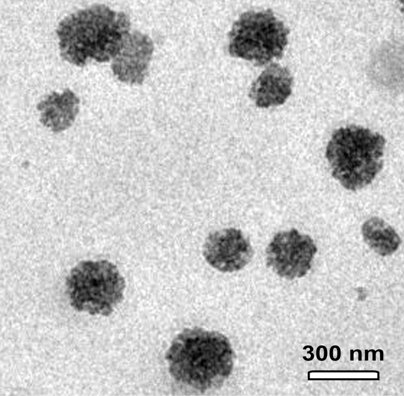
Transmission electron microscope showed the presence of CNPs with various sizes ranging from 150 to 300 nm. Scale bar = 300 nm.
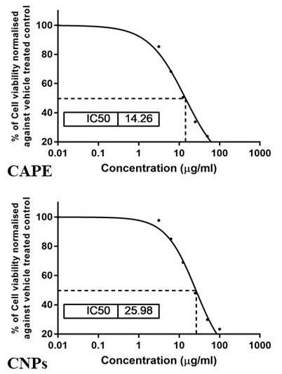
The cytotoxic potential of CAPE and CNPs on HepG2 cells. A representative graph displaying the IC50 value as revealed by the MTT assay. Cells were treated with CAPE or CNPs at serial concentrations from 3.125 to 100 μg/ml and were incubated for 24 h.
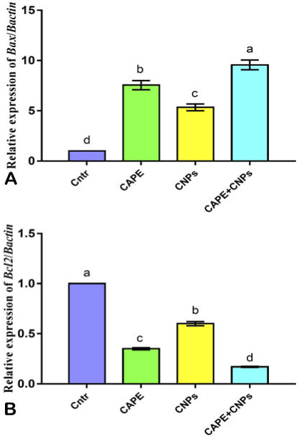
Effect of CAPE and/or CNPs on the expression of Bax (A) and Bcl2 (B) genes in HepG2 cells as detected by qPCR. Cells were treated with CAPE and CNPs alone or in combination (CAPE+CNPs) at doses of their IC50 and incubated for 24 h. Data were presented in the form of fold change mean ± SEM, n = 5/group. Different letters above means (as presented by columns plus error bars) refer to significant differences at P<0.05. All groups compared to each other.
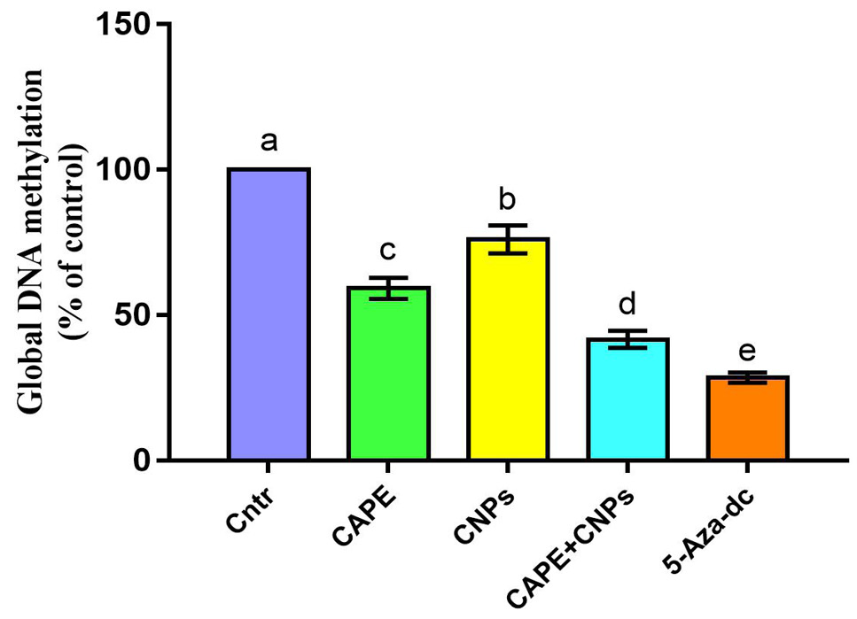
Effect of CAPE and/or CNPs on the Global DNA methylation quantities in HepG2 cells. Cells were treated with CAPE and CNPs alone or in combination (CAPE+CNPs) at doses of their IC50 and incubated for 72 h. Data were presented in the form of % mean ± SEM, n = 5/group. Different letters above means (as presented by columns plus error bars) refer to significant differences at P<0.05. All groups compared to each other. The control group was assigned a value of 100%.
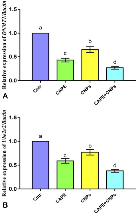
Effect of CAPE and/or CNPs on the expression of DNA methylation-related genes DNMT1 (A) and Ube2e2 (B) in HepG2 cells as detected by qPCR. Cells were treated with CAPE and CNPs alone or in combination (CAPE+CNPs) at doses of their IC50 and incubated for 24 h. Data were presented in the form of fold change mean ± SEM, n = 5/group. Different letters above means (as presented by columns plus error bars) refer to significant differences at P<0.05. All groups were compared to each other.
References
- Abbaszadeh, S., Rashidipour, M., Khosravi, P., Shahyarhesami, S., Ashafi, B., Kaviani, M., and Moradi S.M., 2020. Biocompatibility, cytotoxicity, antimicrobial and epigenetic effects of novel chitosan-based quercetin nanohydrogel in human cancer cells. Int. J. Nanomed., 15: 5963-5975. https://doi.org/10.2147/IJN.S263013[CrossRef]
- Abd-Allah, S.H., Shalaby, S.M., Abd-Elbary, E., Saleh, A.A., and El-Magd, M.A., 2015. Human peripheral blood CD34+ cells attenuate oleic acid-induced acute lung injury in rats. Cytotherapy, 17: 443-453. https://doi.org/10.1016/j.jcyt.2014.11.002[CrossRef]
- Abdel-Daim, M.M., and Abdellatief, S.A., 2018. Attenuating effects of caffeic acid phenethyl ester and betaine on abamectin-induced hepatotoxicity and nephotoxicity. Environ. Sci. Pollut. Res., 25: 15909-15917. https://doi.org/10.1007/s11356-018-1786-8[CrossRef]
- Abu Gazia, M., and El-Magd, M.A., 2018. Effect of pristine and functionalized multiwalled carbon nanotubes on rat renal cortex. Acta Histochem., 121: 207-217. https://doi.org/10.1016/j.acthis.2018.12.005[CrossRef]
- Ajun, W., Yan, S., Li, G., and Huili, L., 2009. Preparation of aspirin and probucol in combination loaded chitosan nanoparticles and in vitro release study. Carbohyd. Polym., 75: 566-574. https://doi.org/10.1016/j.carbpol.2008.08.019[CrossRef]
- Attia, A.A., Salama, A.F., Eldiasty, J.G., Mosallam, S.A.E.-R., El-Naggar, S.A., El-Magd, M.A., Nasser, H.M., and Elmetwalli, A., 2022. Amygdalin potentiates the anti-cancer effect of Sorafenib on Ehrlich ascites carcinoma and ameliorates the associated liver damage. Sci. Rep., 12: 6494. https://doi.org/10.1038/s41598-022-10517-0[CrossRef]
- Badawy, A., Hassanean, H., Ibrahim, A.K., Habib, E.S., El- Magd, M.A., and Ahmed, S.A., 2019. isolates from thymelaea hirsuta inhibit progression of hepatocellular carcinoma in vitro and in vivo. Natl. Prod. Res., 35: 1799-1807. https://doi.org/10.1080/14786419.2019.1643859[CrossRef]
- Badawy, M.E.I., Lotfy, T.M.R., and Shawir, S.M.S., 2020. Facile synthesis and characterizations of antibacterial and antioxidant of chitosan monoterpene nanoparticles and their applications in preserving minced meat. Int. J. Biol. Macromol., 156: 127-136. https://doi.org/10.1016/j.ijbiomac.2020.04.044[CrossRef]
- Chen, M.F., Wu, C.T., Chen, Y.J., Keng, P.C., and Chen, W.C., 2004. Cell killing and radiosensitization by caffeic acid phenethyl ester (CAPE) in lung cancer cells. J. Radiat. Res., 45: 253-260. https://doi.org/10.1269/jrr.45.253[CrossRef]
- Chen, Y.J., Shiao, M.S., and Wang, S.Y., 2001. The antioxidant caffeic acid phenethyl ester induces apoptosis associated with selective scavenging of hydrogen peroxide in human leukemic HL-60 cells. Anticancer Drugs, 12: 143-149. https://doi.org/10.1097/00001813-200102000-00008[CrossRef]
- Dan, J., and Chen, T., 2016. Genetic Studies on Mammalian DNA Methyltransferases. In: DNA methyltransferases-role and function (eds. A. Jeltsch and R. Jurkowska). pp. 123-150. https://doi.org/10.1007/978-3-319-43624-1_6[CrossRef]
- Dan, J., Chen, T. (٢٠١٦). In: DNA Methyltransferases - Role and Function. Advances in Experimental Medicine and Biology, vol ٩٤٥. Springer, Cham. https://doi.org/١٠.١٠٠٧/٩٧٨-٣-٣١٩-٤٣٦٢٤-١_٦
- Du, W.L., Niu, S.S., Xu, Y.L., Xu, Z.R., and Fan, C.L., 2009. Antibacterial activity of chitosan tripolyphosphate nanoparticles loaded with various metal ions. Carbohydr. Polym., 75: 385-389. https://doi.org/10.1016/j.carbpol.2008.07.039[CrossRef]
- El-Demerdash, F.M., El-Magd, M.A., and El-Sayed, R.A., 2021. Panax ginseng modulates oxidative stress, DNA damage, apoptosis, and inflammations induced by silicon dioxide nanoparticles in rats. Environ. Toxicol., 36: 362-1374. https://doi.org/10.1002/tox.23132[CrossRef]
- El-Denshary, E., Aljawish, A., El-Nekeety, A., Hassan, N., Saleh, R., Rihn, B., and Abdel-Wahhab, M., 2015. Possible synergistic effect and antioxidant properties of chitosan nanoparticles and quercetin against carbon tetrachloride-induce hepatotoxicity in rats. Soft Nanosci. Lett., 5: 36-51. https://doi.org/10.4236/snl.2015.52005[CrossRef]
- Elgazar, A.A., Selim, N.M., Abdel-Hamid, N.M., El-Magd, M.A., and El-Hefnawy, H.M., 2018. Isolates from Alpinia officinarum hance attenuate LPS induced inflammation in HepG2: Evidence from in silico and in vitro studies. Phytother. Res., 32: 1273-1288. https://doi.org/10.1002/ptr.6056[CrossRef]
- Elkeiy, M., Khamis, A., El-Gamal, M., Abo Gazia, M., Zalat, Z., and El-Magd, M., 2018. Chitosan nanoparticles from Artemia salina inhibit progression of hepatocellular carcinoma in vitro and in vivo. Environ. Sci. Pollut. Res. Int., 27: 19016-19028. https://doi.org/10.1007/s11356-018-3339-6[CrossRef]
- Erdemli, H.K., Akyol, S., Armutcu, F., and Akyol, O., 2015. Antiviral properties of caffeic acid phenethyl ester and its potential application. J. Int. Ethnopharmacol., 4: 344. https://doi.org/10.5455/jice.20151012013034[CrossRef]
- Feyzioglu, G.C., and Tornuk, F., 2016. Development of chitosan nanoparticles loaded with summer savory (Satureja hortensis L.) essential oil for antimicrobial and antioxidant delivery applications. LWT, 70: 104-110. https://doi.org/10.1016/j.lwt.2016.02.037[CrossRef]
- Forner, A., Llovet, J.M., and Bruix, J., 2012. Hepatocellular carcinoma. Lancet, 379: 1245-1255. https://doi.org/10.1016/S0140-6736(11)61347-0[CrossRef]
- Gailhouste, L., Liew, L.C., Yasukawa, K., Hatada, I., Tanaka, Y., Nakagama, H., and Ochiya, T., 2018. Differentiation therapy by epigenetic reconditioning exerts antitumor effects on liver cancer cells. Mol. Ther., 26: 1840-1854. https://doi.org/10.1016/j.ymthe.2018.04.018[CrossRef]
- Grunberger, D., Banerjee, R., Eisinger, K., Oltz, E., Efros, L., Caldwell, M., Estevez, V., and Nakanishi, K., 1988. Preferential cytotoxicity on tumor cells by caffeic acid phenethyl ester isolated from propolis. Experientia, 44: 230-232. https://doi.org/10.1007/BF01941717[CrossRef]
- Hsu, T.H., Chu, C.C., Hung, M.W., Lee, H.J., Hsu, H.J., and Chang, T.C., 2013. Caffeic acid phenethyl ester induces E2F-1-mediated growth inhibition and cell-cycle arrest in human cervical cancer cells. Fed. Eur. Biochem. Soc., 280: 2581-2593. https://doi.org/10.1111/febs.12242[CrossRef]
- Ishida, Y., Gao, R., Shah, N., Bhargava, P., Furune, T., Kaul, S.C., Terao, K., and Wadhwa, R., 2018. Anticancer activity in honeybee propolis: Functional insights to the role of caffeic acid phenethyl ester and its complex with γ-cyclodextrin. Integr. Cancer Ther., 17: 867-873. https://doi.org/10.1177/1534735417753545[CrossRef]
- Kabała-Dzik, A., Rzepecka-Stojko, A., Kubina, R., Jastrzębska-Stojko, Ż., Stojko, R., Wojtyczka, R.D., and Stojko, J., 2017. Comparison of two components of propolis: Caffeic acid (CA) and caffeic acid phenethyl ester (CAPE) induce apoptosis and cell cycle arrest of breast cancer cells MDA-MB-231. Molecules, 22: 1554. https://doi.org/10.3390/molecules22091554[CrossRef]
- Khoram, N.M., Bigdeli, B., Nikoofar, A., and Goliaei, B., 2016. Caffeic acid phenethyl ester increases radiosensitivity of estrogen receptor-positive and-negative breast cancer cells by prolonging radiation-induced DNA damage. J. Breast Cancer, 19: 18-25. https://doi.org/10.4048/jbc.2016.19.1.18[CrossRef]
- Kurata, A., Kitamura, Y., Irie, S., Takemoto, S., Akai, Y., Hirota, Y., Fujita, T., Iwai, K., Furusawa, M., and Kishimoto, N., 2010. Enzymatic synthesis of caffeic acid phenethyl ester analogues in ionic liquid. J. Biotechnol., 148: 133-138. https://doi.org/10.1016/j.jbiotec.2010.05.007[CrossRef]
- Liao, H.F., Chen, Y.Y., Liu, J.J., Hsu, M.L., Shieh, H.J., Liao, H.J., Shieh, C.J., Shiao, M.S., and Chen, Y.-J., 2003. Inhibitory effect of caffeic acid phenethyl ester on angiogenesis, tumor invasion, and metastasis. J. Agric. Fd. Chem., 51: 7907-7912. https://doi.org/10.1021/jf034729d[CrossRef]
- Loh, J.W., Yeoh, G., Saunders, M., and Lim, L.-Y., 2010. Uptake and cytotoxicity of chitosan nanoparticles in human liver cells. Toxicol. appl. Pharmacol., 249: 148-157. https://doi.org/10.1016/j.taap.2010.08.029[CrossRef]
- Loo, S.K., Hamid, S.S.A., Musa, M., and Wong, K.K., 2018. DNMT1 is associated with cell cycle and DNA replication gene sets in diffuse large B-cell lymphoma. Pathol. Res. Pract., 214: 134-143. https://doi.org/10.1016/j.prp.2017.10.005[CrossRef]
- Loutfy, S., Alam El-Din, H., Elberry, M., Allam, N., Hasanin, M., and Abdellah, A., 2016. Synthesis, characterization and cytotoxic evaluation of chitosan nanoparticles: in vitro liver cancer model. Adv. Natl. Sci. Nanosci. Nanotechnol., 7: 035008. https://doi.org/10.1088/2043-6262/7/3/035008[CrossRef]
- Mahfouz, D.H., EL-Magd, M.A., Mansour, G.H., Abdel Wahab, A.H., Abdelhamid, I.A., and Elzayat, E., 2021. Therapeutic potential of snake venom, l-amino oxidase and sorafenib in hepatocellular carcinoma. Mol. Cell. Toxicol., https://doi.org/10.1007/s13273-021-00151-8[CrossRef]
- Mansour, G.H., El-Magd, M.A., Mahfouz, D.H., Abdelhamid, I.A., Mohamed, M.F., Ibrahim, N.S., Hady A. Abdel Wahab, A., and Elzayat, E.M., 2021. Bee venom and its active component melittin synergistically potentiate the anticancer effect of Sorafenib against HepG2 cells. Bioorg. Chem., https://doi.org/10.1016/j.bioorg.2021.105329[CrossRef]
- Morin, P., St-Coeur, P.-D., Doiron, J.A., Cormier, M., Poitras, J.J., Surette, M.E., and Touaibia, M., 2017. Substituted caffeic and ferulic acid phenethyl esters: synthesis, leukotrienes biosynthesis inhibition, and cytotoxic activity. Molecules, 22: 1124. https://doi.org/10.3390/molecules22071124[CrossRef]
- Murtaza, G., Karim, S., Akram, M.R., Khan, S.A., Azhar, S., Mumtaz, A., and Bin Asad, M.H.H., 2014. Caffeic acid phenethyl ester and therapeutic potentials. BioMed. Res. Int., 2014. https://doi.org/10.1155/2014/145342[CrossRef]
- Omene, C., Kalac, M., Wu, J., Marchi, E., Frenkel, K., and O’Connor, O.A., 2013. Propolis and its active component, caffeic acid phenethyl ester (CAPE), modulate breast cancer therapeutic targets via an epigenetically mediated mechanism of action. J. Cancer Sci. Ther., 5: 334-342.
- Othman, R., Badawy, A., Alruwaili, M., and El-Magd, M., 2021. Camel milk exosomes potentiate the anticancer effect of doxorubicin on multidrug-resistant human leukemia Hl60 cells in vitro and in vivo. Pak. J. med. Hlth. Sci., 15: 3313-3320. https://doi.org/10.53350/pjmhs2115113313[CrossRef]
- Ozturk, G., Ginis, Z., Akyol, S., Erden, G., Gurel, A., and Akyol, O., 2012. The anticancer mechanism of caffeic acid phenethyl ester (CAPE): review of melanomas, lung and prostate cancers. Eur. Rev. med. pharmacol. Sci., 16: 2064-2068.
- Qi, L., Xu, Z., and Chen, M., 2007. In vitro and in vivo suppression of hepatocellular carcinoma growth by chitosan nanoparticles. Eur. J. Cancer, 43: 184-193. https://doi.org/10.1016/j.ejca.2006.08.029[CrossRef]
- Rzepecka-Stojko, A., Kabała-Dzik, A., Moździerz, A., Kubina, R., Wojtyczka, R.D., Stojko, R., Dziedzic, A., Jastrzębska-Stojko, Ż., Jurzak, M., and Buszman, E., 2015. Caffeic acid phenethyl ester and ethanol extract of propolis induce the complementary cytotoxic effect on triple-negative breast cancer cell lines. Molecules, 20: 9242-9262. https://doi.org/10.3390/molecules20059242[CrossRef]
- Saleh, A.A., Amber, K., El-Magd, M.A., Atta, M.S., Mohammed, A.A., Ragab, M.M., and Abd El-Kader, H., 2014. Integrative effects of feeding Aspergillus awamori and fructooligosaccharide on growth performance and digestibility in broilers: Promotion muscle protein metabolism. Biomed. Res. Int., 2014: 946859. https://doi.org/10.1155/2014/946859[CrossRef]
- Sarabi, M.M., and Naghibalhossaini, F., 2015. Association of DNA methyltransferases expression with global and gene-specific DNA methylation in colorectal cancer cells. Cell Biochem. Funct., 33: 427-433. https://doi.org/10.1002/cbf.3126[CrossRef]
- Selim, N.M., Elgazar, A.A., Abdel-Hamid, N.M., El-Magd, M.R.A., Yasri, A., Hefnawy, H.M.E., and Sobeh, M., 2019. Chysophanol, physcion, hesperidin and curcumin modulate the gene expression of pro-inflammatory mediators induced by LPS in HepG2: In silico and molecular studies. Antioxidants, 8: 371. https://doi.org/10.3390/antiox8090371[CrossRef]
- Song, S.H., Han, S.W., and Bang, Y.J., 2011. Epigenetic-based therapies in cancer. Drugs, 71: 2391-2403. https://doi.org/10.2165/11596690-000000000-00000[CrossRef]
- Subhapradha, N., and Shanmugam, A., 2017. Fabrication of β-chitosan nanoparticles and its anticancer potential against human hepatoma cells. Int. J. Biol. Macromol., 94: 194-201. https://doi.org/10.1016/j.ijbiomac.2016.10.016[CrossRef]
- Subhapradha, N., Shanmugam, V., and Shanmugam, A., 2017. Chitosan nanoparticles from marine squid protect liver cells against N-diethylnitrosoamine-induced hepatocellular carcinoma. Carbohydr. Polym., 171: 18-26.
- Torchilin, V.P., 2006. Micellar https://doi.org/10.1016/j.carbpol.2017.04.097 nanocarriers: Pharmaceutical perspectives. Pharm. Res., 24: 1. https://doi.org/10.1007/s11095-006-9132-0[CrossRef]
- Tseng, T.H., Shen, C.H., Huang, W.S., Chen, C.N., Liang, W.-H., Lin, T.H., and Kuo, H.C., 2014. Activation of neutral-sphingomyelinase, MAPKs, and p75 NTR-mediating caffeic acid phenethyl ester–induced apoptosis in C6 glioma cells. J. biomed. Sci., 21: 1-11. https://doi.org/10.1186/1423-0127-21-61[CrossRef]
- Wadhwa, R., Nigam, N., Bhargava, P., Dhanjal, J.K., Goyal, S., Grover, A., Sundar, D., Ishida, Y., Terao, K., and Kaul, S.C., 2016. Molecular characterization and enhancement of anticancer activity of caffeic acid phenethyl ester by γ cyclodextrin. J. Cancer, 7: 1755. https://doi.org/10.7150/jca.15170[CrossRef]
- Wagner, J.M., Hackanson, B., Lübbert, M., and Jung, M., 2010. Histone deacetylase (HDAC) inhibitors in recent clinical trials for cancer therapy. Clin. Epigenet., 1: 117-136. https://doi.org/10.1007/s13148-010-0012-4[CrossRef]
- Wang, W., Shi, J., and Xie, W.F., 2010. Transarterial chemoembolization in combination with percutaneous ablation therapy in unresectable hepatocellular carcinoma: A meta-analysis. Liver Int., 30: 741-749. https://doi.org/10.1111/j.1478-3231.2010.02221.x[CrossRef]
- Xu, Y., Su, D., Zhu, L., Zhang, S., Ma, S., Wu, K., Yuan, Q., and Lin, N., 2018. S-allylcysteine suppresses ovarian cancer cell proliferation by DNA methylation though DNMT1. J. Ovarian Res., 11: 39. https://doi.org/10.1186/s13048-018-0412-1[CrossRef]
- Xu, Y., Wen, Z., and Xu, Z., 2009. Chitosan nanoparticles inhibit the growth of human hepatocellular carcinoma xenografts though an antiangiogenic mechanism. Anticancer Res., 29: 5103-5109.
- Zedan, A.M.G., Sakran, M.I., Bahattab, O., Hawsawi, Y.M., Al-Amer, O., Oyouni, A.A.A., Nasr Eldeen, S.K., and El-Magd, M.A., 2021. Oriental hornet (Vespa orientalis) larval extracts induce antiproliferative, antioxidant, anti-inflammatory, and anti-migratory effects on MCF7 cells. Molecules, 26: 3303. https://doi.org/10.3390/molecules26113303[CrossRef]
Cited by 1 article
In journals published by ResearchersLinks.
Services
Article usage statistics
Loading…