
Cancer is considered as the second leading cause of death in the world. The decreased platelet count is one of the major problems that patients face through the treatment, which makes them prone to bleeding. PRP injection therapy is a therapeutic method in use to relieve this condition. It may lead to infections due to the injection, as well as platelet resistance. Therefore, an attempt was made to find an alternative treatment, and the recombinant Interleukin 11 (IL-11). In this study, IL-11 gene owning two restriction enzyme sites of BamHI and XbaI with a final size of 609 bp was synthesized by GenRay (China) and delivered to pGS vector. The vector was transferred to DH5α, then the gene was subcloned onto pHT43 shuttle vector and transformed into the Bacillus subtilis WB600. The vector containing the cloned gene was induced within the WB600 by IPTG. The SDS-PAGE also indicated the successful expression of IL-11 in B. subtilis. A band of about 22 kD corresponding to the recombinant protein was observed in positive sample. This research demonstrated the efficacy of using B. subtilis as an expression system for the production of IL-11 and it can be used as a candidate for the proteins without post-translational modification.
Short Communication · Volume 55, Issue 3, 20 April 2023 · Open Access
Cloning and Expression of Interleukin 11 in Bacillus subtilis
· · · ·
Affiliations & Notes ⌄
- 1Department of Microbiology, Islamic Azad University, Jahrom Branch, Jahrom, Iran 2Department of Biology, Islamic Azad University, Damghan Branch, Damghan, Iran * Corresponding author: dr.majidmoghbeli@yahoo.com
Article Info ⌄
- Journal:
- Pakistan Journal of Zoology
- Published:
- 20 April 2023

From the article
- As shown in Table I, the expression level in MFA medium was higher than LB medium (61.4 µg/ml in MFA and 21.8 µg/ml in LB).
- The recombinant protein expression after IPTG induction led to expression of a 22 KD protein like other researches.
- The recombinant IL-11 expression was extracellular, about 61.4 µg/ml and more than the expression yield in E. coli, so this host is suggested for the recombinant IL-11 production.
- This study demonstrated the efficacy of using B. subtilis as an expression system for production of IL-11 and it can be used as a candidate for the production of every recombinant protein which lacks post-translational modifications.
Selected from the article's own text.
Abstract
Materials and methods
The sequence of IL-11was obtained from NCBI database, the peptide signal sequence was removed, and also the BamHI site was placed at the beginning, and the XbaI site at the end of the gene. The sequence was then optimized for codon usage based on B. subtilis using J-CAT software to increase expression. The gene with a final size of 609 bp was synthesized (GenRay Co, China) and delivered on the pGS vector.
The IL-11 gene was cut from the pGS vector through digestion by BamHI and XbaI enzymes and purified from the agarose gel by DNA purification kit (Pouya Gene Azma Co. Iran). Then it was subcloned on the BamHI and XbaI sites of pHT43 B. subtilis shuttle vector (MoBiTec, Germany). The ligation reaction transferred to the E. coli (DH5α) using heat shock method. The presence of the gene on the plasmid was evaluated by enzymatic digestion and Polymerase Chain Reaction(PCR).
The recombinant plasmid carrying the gene was named pHT-MA52. The recombinant plasmid pHT-MA52 was isolated using plasmid purification kit (Pouya Gene Azma Co. Iran) from E. coli (DH5α) and transferred to B. subtilis (WB600) using the Optimized Transformation Buffer (OTB) containing: 0.75 g/l yeast extract; 8 g/l glucose; 1.0 mM CaCl2, and 10 mM MgCl2; pH 7.0. The recombinant bacteria were inoculated in 5 ml of MFA medium (1% glucose, 0.5% peptone, 0.5% yeast extract, 0.01%, MgSO4, 0.014% CaCl2, 0.001% FeSO4, 5 μg/ml chloramphenicol) and Luria-Bertani (LB) broth and incubated for 2.5-3 h at 37°C by shaking until the turbidity reached about 0.6-0.8 at wavelength of 600 nm. Recombinant protein expression was induced by adding Isopropyl β-D-1-thiogalactopyranoside (IPTG) to the final concentration of 1 mM. Prior to the induction and with an hour intervals after induction, 300 μl of the medium was withdrew for 6 h. The bacterial cells were separated centrifuging at 11400 g for 3 min and the supernatant was stored at 4°C until the next step. The protein concentration of each sample was evaluated using the Bradford protein assay . As the negative control (blank), the supernatant of B. subtilis carrying pHT43 plasmid without the gene was used. The recombinant protein band was examined by 12% SDS-PAGE.
Results
Figure 1 shows the sequence of the designed and synthesized interleukin 11 gene based on the used codons in B. subtilis.
Figure 2 shows the following enzymatic digestion of the gene from the pGS vector. The gene was cloned in B. subtilis using pHT43 vector. The presence of the gene in the vector was confirmed by double digestion using BamHI and XbaI as well as PCR.
The expression yield was measured both in LB and MFA media. As shown in Table I, the expression level in MFA medium was higher than LB medium (61.4 µg/ml in MFA and 21.8 µg/ml in LB). In addition, highest expression level was obtained in both media 4 h after IPTG induction. The MFA medium sample 4h after induction, and the medium sample of the pHT-43 without IL-11 gene were evaluated on SDS-PAGE. A band of about 22 kDa corresponding to the recombinant protein was observed in the test sample, whereas no bands were observed in the negative control (Fig. 3).
Table I. Interleukin 11 recombinant protein expressed in LB and MFA media 6h after IPTG
induction. The expression level is expressed as µg/ml of the culture medium.
| Hour | LB | MFA |
| First | 3.6 | 18.8 |
| Second | 16.3 | 35.6 |
| Third | 21.8 | 49.1 |
| Fourth | 24.1 | 61.4 |
| Fifth | 22.9 | 58.1 |
| Sixth | 22 | 54.7 |
Discussion
Chemotherapy-induced thrombocytopenia limits the therapeutic utilization of chemotherapeutic agents, and may adversely affect the outcome of potentially treatable malignancies. IL-11 is the first thrombocytopoietic cytokine made commercially available for the treatment of Chemotherapy-induced thrombocytopenia . While IL-11 appears to contribute to a host of regulatory pathways including the stimulation of myeloid and erythroid precursors as well as modulating macrophage and T cell inflammatory responses, stimulation of thrombopoiesis is regarded as the primary function of IL-11 . The thrombocytopoietic effect of IL-11 is exerted in all stages of megakaryocyte maturation . Previously presumed thrombopoietin (TPO) independent effect of IL-11 has been shown to fail in TPO- or TPO receptor-deficient subjects, implicating potential role of TPO signaling in IL-11 pathways ; . IL-11 induces the recovery of progenitor cells belonging to multiple hematopoietic lineages in myelosuppressed subjects .
The aim of this study is cloning and expression of Interleukin 11 in Bacillus subtilis in order to produce this valuable cytokine in lab scale. Till now, the gene of IL- 11 has been cloned and expressed in different pro- and eukaryotic hosts such as human endometrium, tobacco and E. coli ; ; . Here, for the first time the gene of IL-11 is cloned and expressed in B. subtilis. Every expression system has its own pros and cons and the suitable system has to be selected based on the target protein. High capacity of secretion, direct secretion of the recombinant protein in medium, as well as safe and easy culture condition of B. subtilis, made it an appropriate host for gene cloning and expression . In different studies, gene of IL-11 has been obtained using mRNA extraction, cDNA syntheses and PCR amplification. In this paper, IL-11 was designed and optimized for codon usage of B. subtilis by J-CAT software. The designed gene was then synthesized and cloned In pHT43. The recombinant protein expression after IPTG induction led to expression of a 22 KD protein like other researches. The recombinant IL-11 expression was extracellular, about 61.4 µg/ml and more than the expression yield in E. coli, so this host is suggested for the recombinant IL-11 production.
Conclusion
This study demonstrated the efficacy of using B. subtilis as an expression system for production of IL-11 and it can be used as a candidate for the production of every recombinant protein which lacks post-translational modifications.
Acknowledgements
Our sincere thanks to Pouya Gene Azma Co, Tehran, Iran, for providing facilities to carry out the experiments and Azad University of Jahrom branch for their help during this research.
Statement of conflict of interest
The authors have declared no conflict of interest.
Figures
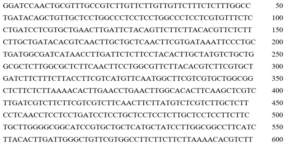
The sequence of the designed and synthesized interleukin 11gene based on the codon usage in B. subtilis: The underlined sequences are restriction sites: 5′ end and 3′ end respectively belongs to BamHI and XbaI.
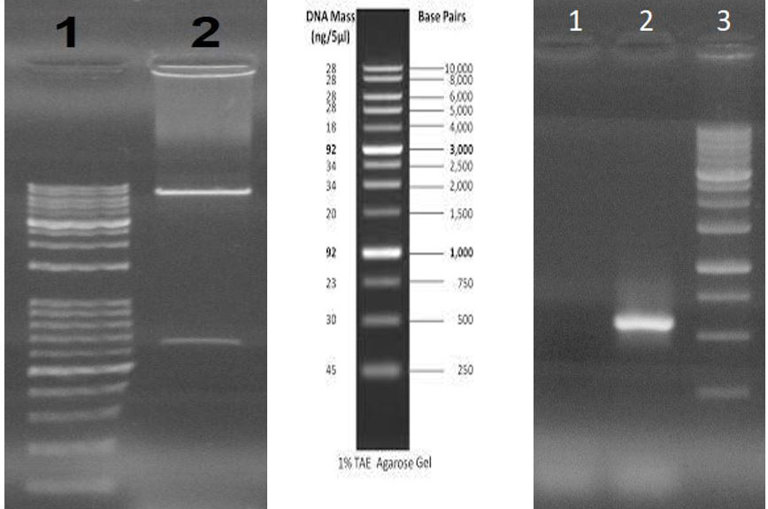
A, Double digestion of the plasmid pHT43 containing IL-11 gene: lane 1: 1 kb plus DNA ladder; lane 2: double digested recombinant plasmid. B, PCR of the recombinant plasmid pHT-MA52: lane 1: negative control. Lane 2: PCR product. Lane 3: 1 kb plus DNA ladder.
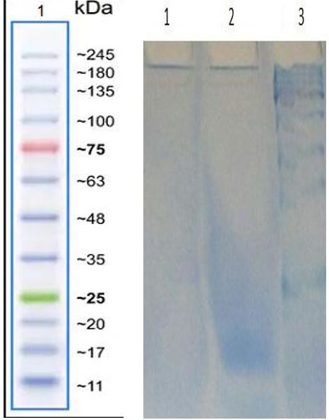
Evaluation of the recombinant protein band on SDS-PAGE: lane 1: negative control, lane 2: recombinant protein, lane 3: protein ladder.
References
- Avecilla, S.T., Hattori, K., Heissig, B., Tejada, R., Liao, F., Shido, K., Jin, D.K., Dias, S., Zhang, F. and Hartman, T.E.J.N., 2004. Nat. Med., 10: 64-71. https://doi.org/10.1038/nm973[CrossRef]
- Brocker, C., Thompson, D., Matsumoto, A., Nebert, D.W. and Vasiliou, V.J.H.G., 2010. BMC, 5: 1-26. https://doi.org/10.1186/1479-7364-5-1-30[CrossRef]
- Ciurea, S.O. and Hoffman, R., 2007. Cytokines for the treatment of thrombocytopenia, Cience direct. Elsevier, pp. 166-182. https://doi.org/10.1053/j.seminhematol.2007.04.005[CrossRef]
- Cremer, M., Sallmon, H., Kling, P.J., Bührer, C. and Dame, C., 2016. Thrombocytopenia and platelet transfusion in the neonate, seminars in fetal and neonatal medicine. Elsevier, pp. 10-18. https://doi.org/10.1016/j.siny.2015.11.001[CrossRef]
- Feinglass, S., Deodhar, A.J.A. and Rheumatology, R.O.J., 2001. Off. J. Am. Coll. Rheumatol., 44: 170-175. https://doi.org/10.1002/1529-0131(200101)44:1<170::AID-ANR22>3.0.CO;2-T[CrossRef]
- Ferreira, L., Ferreira, R.C. and Schumann, W., 2005. Annls Braz. Acad. Sci., 77: 113-124. https://doi.org/10.1590/S0001-37652005000100009[CrossRef]
- Fung, K.Y., Louis, C., Metcalfe, R.D., Kosasih, C.C., Wicks, I.P., Griffin, M.D. and Putoczki, T.L.J.C., 2022. Sci. Direct, pp. 155750. https://doi.org/10.1016/j.cyto.2021.155750[CrossRef]
- Guan, C., Cui, W., Cheng, J., Liu, R., Liu, Z., Zhou, L. and Zhou, Z.J.N.b., 2016. Sci. Direct, 33: 372-379. https://doi.org/10.1016/j.nbt.2016.01.005[CrossRef]
- He, F.J.B., 2011. Bradford protein assay. bio_protocol, pp. e45-e45. https://doi.org/10.21769/BioProtoc.45[CrossRef]
- Hill, G.R., Cooke, K.R., Teshima, T., Crawford, J.M., Keith, J.C., Brinson, Y.S., Bungard, D. and Ferrara, J.J.T.J., 1998. J. clin. Invest., 102: 115-123. https://doi.org/10.1172/JCI3132[CrossRef]
- Karpovich, N., Chobotova, K., Carver, J., Heath, J.K., Barlow, D.H. and Mardon, H., 2003. Basic Sci. Reprod. Med., 9: 75-80. https://doi.org/10.1093/molehr/gag012[CrossRef]
- Liu, R., Yang, Y., Yan, X. and Zhang, K.J.D., 2013. Eur. J. Dermatol., 23: 600-607. https://doi.org/10.1684/ejd.2013.2149[CrossRef]
- Lokau, J., Agthe, M., Flynn, C.M. and Garbers, M.C.R., 2017. Sci. Direct, 1864: 2105-2117. https://doi.org/10.1016/j.bbamcr.2017.06.008[CrossRef]
- Luan, C., Zhang, H.W., Song, D.G., Xie, Y.G., Feng, J. and Wang, Y.Z.J.A.M., 2014. Springer, 98: 3651-3658. https://doi.org/10.1007/s00253-013-5246-6[CrossRef]
- Murakami, M., Kamimura, D. and Hirano, T.J.I., 2019. Sci. Direct, 50: 812-831. https://doi.org/10.1016/j.immuni.2019.03.027[CrossRef]
- Putoczki, T.L. and Ernst. M., 2015. Immunotherapy, 7:4. https://doi.org/10.2217/imt.15.17[CrossRef]
- Rashidi, M.R. and Moghbeli, M.M.W., 2013. J. Microbial. World, 6: 188-197.
- Reynolds, C.H.J.O., 2000. Eur. PMC, 14: 32-40.
- Ryter, S.W., Alam, J. and Choi, A.M.J., 2006. Am. Physiol. Soc., 86: 583-650. https://doi.org/10.1152/physrev.00011.2005[CrossRef]
- Sadeghi, A., Mahdieh, M. and Salimi, S.J.P.B., 2016. J. Pl. Biotechnol., 43: 432-437. https://doi.org/10.5010/JPB.2016.43.4.432[CrossRef]
- Sokolov, A.S., Kazakov, A.S., Solovyev, V.V., Ismailov, R.G., Uversky, V.N., Lapteva, Y.S., Mikhailov, R.V., Pavlova, E.V., Terletskaya, I.O. and Ermolina, L.V.J.M., 2016. Molecules, 21: 1632. https://doi.org/10.3390/molecules21121632[CrossRef]
- Takahashi, T., Shimizu, H., Morimatsu, H., Inoue, K., Akagi, R., Morita, K. and Sassa, S.J.M., 2007. Eur. PMC, 7: 745-753. https://doi.org/10.2174/138955707781024517[CrossRef]
- Tanaka, T. and Kishimoto, T.J.C., 2014. Cancer Immunol. Res., 2: 288-294. https://doi.org/10.1158/2326-6066.CIR-14-0022[CrossRef]
- Wu, S., Zhang, Y., Xu, L., Dai, Y., Teng, Y., Ma, S., Ho, S.-H., Kim, J.-M., Yu, S.S. and Kim, S.J., 2012. Springer, 20: 1875-1884. https://doi.org/10.1007/s00520-011-1290-x[CrossRef]
- Zhang, B., Zhang, H.-X., Shi, S.-T., Bai, Y.-L., Zhe, X., Zhang, S.-J. and Li, Y.J.J.B., 2019. Sci. Direct, 115: 108816. https://doi.org/10.1016/j.biopha.2019.108816[CrossRef]
Services
Article usage statistics
Loading…