
Peste des petits ruminants (PPR), an economically important morbillivirus infection of sheep and goats, is widely distributed in Middle East, Central and South Asia, China and Africa. Whereas PPR antibodies were first reported in Kenyan small stock as early as 1995, the clinical disease was observed later in 2006 following dramatic and devastating PPR outbreaks in Turkana County. In this context, an outbreak of PPRV was observed in Tukana County in 2011 and the current study detail the clinical, pathological and laboratory findings of this outbreak. The disease was clinically manifested by the depression, diarrhea, difficult breathing, muco-purulent ocular-nasal discharges with matted eyelids and encrusted nostrils and finally death. Clinical outcome, gross lesions and histological observations were suggestive of PPRV infection, which were confirmed by the application of a reverse transcription–polymerase chain reaction (qRT-PCR). Collectively, results indicate the continuous persistence of PPRV in Kenya.
Case Reports · Volume 1, Issue 3, 22 November 2014 · Open Access
Clinical, Pathological and Molecular Investigations of Peste des Petits Ruminants Virus Infection in Goats from Turkana County in Kenya
· · · · · · · · ·
Affiliations & Notes ⌄
- 1Faculty of Veterinary Medicine, University of Nairobi, P.O. Box 29053-00625 Uthiru
- 2Faculty of Arts, University of Nairobi, P.O. Box 30197- 00100 Nairobi
- 3University of Nairobi Institute of Tropical & Infectious Diseases (UNITID), P.O. Box 19676-00202 Nairobi, Kenya
Article Info ⌄
- Journal:
- Hosts and Viruses
- Published:
- 22 November 2014

From the article
- Clinical, Pathological and Molecular Investigations of Peste des Petits Ruminants Virus Infection in Goats from Turkana County in Kenya.
- Abstract | Peste des petits ruminants (PPR), an economically important morbillivirus infection of sheep and goats, is widely distributed in Middle East, Central and South Asia, China and Africa.
- Whereas PPR antibodies were first reported in Kenyan small stock as early as 1995, the clinical disease was observed later in 2006 following dramatic and devastating PPR outbreaks in Turkana County.
- In this context, an outbreak of PPRV was observed in Tukana County in 2011 and the current study detail the clinical, pathological and laboratory findings of this outbreak.
Selected from the article's own text.
Abstract
Acknowledgements
This is to acknowledge “The Regional Universities Forum for Capacity Building in Agriculture” (RUFORUM) (RU/CFP/CGS/TADS/09/1) for financial support as well as Vetworks Eastern Africa for their logistical assistance towards this study. Acknowledgement also goes to University of Nairobi Institute of Tropical & Infectious Diseases (UNITID) for providing their molecular laboratories to carry out the molecular analysis of this study which was part of corresponding author’s PhD research work.
Authors Contribution
Kihu S.M., Oyugi J.O., E. Lutomia and Gitao C.G. collected the data, created the electronic database and cleaned and processed and analyzed the molecular experiments. Kihu S.M., Karanja D.N. and Bebora L.C. conceived study analysis methods. Kihu S.M. drafted the manuscript assisted by Gitao C.G., Bebora L.C., Njenga M.J., and Wairire G.G. Gitao C.G., Bebora L.C., Njenga M. J., Wairire G.G., Maingi N. and Wahome R.G. raised funding for the study and assisted its coordination. All authors helped with the interpretation of the results and read and approved the final manuscript.
Conflict of Interest
The authors declare that they have no conflict of interest.
Figures
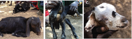
A. Goat suspected to have PPR infection in a depressed state, B. Diarrhoea and soiled anal region and hind legs, C. Showing muco-purulent ocular nasal discharges, matting of eye lids and encrusted.
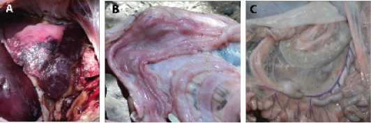
A. Inflamed lung showing hepatized apical lobe in goat, B. Hyperaeic intestinal mucosa showing hemorrhagic points, C. Swollen mesenteric lymph node (arrow) and congested mesenteric veins.
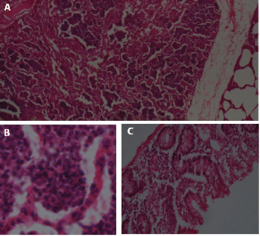
A. Lung of goat showing collapsed alveoli, B. infiltration with mononuclear cell in the alveoli and multinucleated syncitia, C. Large intestine showing proliferation of goblet cells and infiltration of inflammatory cells.
References
- • Banyard, A. C., Parida, S., Batten, C., Oura, C., Kwiatek, O., Libeau, G. 2010: Global distribution of peste des petits ruminants virus and prospects for improved diagnosis and control. Journal of General Virology.91, 2885–2897.
- • Batten, C.A., Banyard, A.C., King, D.P., Henstock, M.R., Edwards, L., Sanders, A., Buczkowski, H., Oura, C. A. L., Barrett, T. 2011: A real-time PCR assay for the specific detection of peste des petits ruminants virus. J. Virol. Methods, 171 (2), 401–404.
- • Bundza, A., Afshar, A., Dukes, T.W., Myers, D.J., Dulac Susi, G., Becker, A.W.E. 1988: Experimental PPR (goat plague) in Goats and sheep. Canadian Journal of Veterinary Research. 52, 46-52.
- • Diallo, A., Minet, C., Le Goff, C., Berhe, G., Albina, E., Libeau, G.,Barrett, T. 2007: The threat of peste des petits ruminants: progress in vaccine development for disease control. Vaccine 25, 5591–5597.
- • FAO, 2008:Peste des petits ruminants (PPR) in Morocco. EMPRES WATCH August 2008. Website: ftp://ftp.fao.org/docrep/fao/011/aj120e/aj120e00.pdf. Accessed on 21st May 2009.
- • FAO, 2009:Empres; Transboundary Disease Bulletin 33. ftp://ftp.fao.org/docrep/fao/011/i0919e/i0919e00.pdf. Accessed on 12th November 2009.
- • Kihu, S.M., Njagi,L.M., Njogu, G.N., Kamande, J.N.,Gitao, C.G. 2012: Peste des petits ruminants in Kenya; Pastoralist knowledge of the disease in goats in Samburu and Baringo Counties. Research opinions in animal & veterinary sciences, 2(11), 544-553.
- • Kul, O., Kabakci, N., Atmaca, H. T., Özkul, A. 2007: Natural Peste des Petits Ruminants Virus Infection: Novel Pathologic Findings Resembling Other Morbillivirus Infections. Veterinary Pathology 2007 44: 479-486.
- • Laboratoire Service International, (LSI) 2011: Validation of a real-time PCR kit, TaqVet® Peste des petits ruminants Virus kit; Report of kit development for detection of Peste des Petits Ruminants Virus Ref: PPRP/50. 6 Allée des écureuils – Parc d’activité du Bois Dieu - 69380 Lissieu - France.
- • Luna, L.G., (Ed.) 1968: Manual of Histologic Staining Methods of the Armed Forces Institute of Pathology. Armed Forces Institute of Pathology, Houston, USA pp. 258.
- • Munir, M., Zohari, S., Berg, M.,2013:Molecular Biology and Pathogenesis of Peste des Petits Ruminants Virus. 2013:151. ISSN 2211-7504, ISBN 978-3-642-31450-6, DOI 10.1007978-3-64231451-3 Springer Briefs in Animal Sciences
- • OIE, 2014: Disease outbreak summary, Kenya http://www.oie.int/wahis_2/public/wahid.php/Diseaseinformation/statusdetail/popup?diseaseid=5&country=KEN&y=2006&m=8&admin1=1590&detail=2&sdid=97395
- • QIAGEN, 2010:RNeasy® Mini Kit ,For purification of total RNA from animal cells, animal tissues, bacteria, and yeast, and for RNA cleanup. http://www.genome.duke.edu/cores/microarray/services/rna-qc/documents/RNeasy_Mini_Handbook.pdf Accessed on 30th July 2013.
- • Truong, T., Boshra, H., Embury-Hyatt, C., Nfon, C., Gerdts, V., Tikoo, S., Babiuk, L. A., Kara, P., Chetty, T., Mather, A., Wallace, D. B., Babiuk, S., 2014: Peste des Petits Ruminants Virus Tissue Tropism and Pathogenesis in Sheep and Goats following Experimental Infection. PLoS ONE 9(1): e87145.
- • Wamwayi, H. M., Rossiter, P. B., Kariuki, D. P., Wafula, J. S., Barrett, T., Anderson, J., 1995: Peste des petits ruminants antibodies in east Africa. Veterinary Record, 136: 199-200.
Services
Article usage statistics
Loading…