
A 17-year-old captive male puma (Puma concolor) died after presenting anorexia, vomiting, weight loss and lethargy. At necropsy, the right middle lobe of the liver was severely affected by a tumor, and small tumor nodules were disseminated throughout the other lobes. The numerous tumor nodules were also found in the lung, stomach, kidney, heart and diaphragm, which were growing together, suspiciously metastatic, projecting, 5 to 40 mm in diameter and tawny to white in color. Histopathologically, the tumor was composed of prominent papillary-acinar structures and the cells had a resemblance to the biliary epithelium. Immunohistochemically, the tumor cells were strongly reactive for cytokeratin and CD10 and were negative for carcinoembryonic antigen, fetoprotein and hepatocyte paraffin-1. Taken together, the tumor was diagnosed as cholangiocarcinoma. This is the first case report of a cholangiocarcinoma in the puma.
Short Communication · Volume 54, Issue 1, 28 December 2021 · Open Access
Cholangiocarcinoma with Multiple Organ Metastasis in a Captive Puma (Puma concolor)
·
Affiliations & Notes ⌄
- 1College of Veterinary Medicine and Bio-Safety Research Institute, Jeonbuk National University, Iksan 54596, Republic of Korea 2Department of Veterinary Pathology, College of Veterinary Medicine and Institute of Veterinary Science, Kangwon National University, Chuncheon 24341, Republic of Korea , * Corresponding author: yeonoh@kangwon.ac.kr
Article Info ⌄
- Journal:
- Pakistan Journal of Zoology
- Published:
- 28 December 2021

From the article
- Histopathological examination showed that tumor nodules were composed of cuboidal and columnar cells with round to oval nuclei, and mitotic figures were moderate to abundant.
- The histomorphological characteristics of the tumor were highly indicative of cholangiocarcinoma.
- The other tumor nodules in the lung, stomach, kidney and diaphragm also had very similar histological appearances to that of the liver nodules.
- This gives us the rationale to conclude that the liver nodules were the primary source of neoplastic cells that metastasized to the other organs.
Selected from the article's own text.
Abstract
Materials and methods
This case study discusses a 17-year-old male puma (Puma concolor) that died after presenting anorexia, vomiting, weight loss and lethargy. At necropsy, the right middle lobe of the liver was severely affected by a
massive tumor, and other lobes were affected by small multiple nodules as described in other literature (Fig. 1A) ; . The largest tumor had a characteristic lobular pattern with an umbilicated appearance, sometimes protruding above the capsule of the liver. The cut surface colored white to gray-white and sometimes carried cystic nodules containing yellow-brown fluid. The border of the tumors was irregular but clearly delineated from the adjacent hepatic parenchyma. Palpation of the nodules revealed a firm texture as seen in connective tissue-abundant tumors. This firm texture is one of the diagnostic indexes which distinguish a cholangiocarcinoma from a hepatocellular carcinoma that generally have a soft and friable texture . Numerous nodules that were growing together, suspiciously metastatic, projecting 5 to 40 mm in diameter and tawny to white in color, were scattered throughout the lung, stomach, kidney, heart and diaphragm (Fig. 1B, C, D). Tissue samples from the tumor nodule and representative tissue specimens were collected and fixed in
10% (w/v) neutral buffered formalin for 24-48h, processed routinely and embedded in paraffin wax. Sections (4 μm) were stained with hematoxylin and eosin (H&E) for light microscopy examination. Immunohistochemical (IHC) analyses were conducted as previously described using six primary antibodies: anti-cytokeratin pan type II (AE3; 1:50, eBioscienceTM, San Diego, CA, USA), anti-cytokeratin pan type I (AE1; 1:50, eBioscienceTM, San Diego, CA, USA), anti-CD10 (56C6; 1:20, Abcam®, Cambridge, MA, USA), anti-carcinoembryonic antigen (COL-1; 1:500, eBioscienceTM, San Diego, CA, USA), anti-α-fetoprotein (1E8; 1:50, eBioscienceTM , San Diego, CA, USA) and anti-hepatocyte paraffin-1 (OCH1E5; 1:20, NOVUS Biologicals, LLC, Denver, CO, USA). Briefly, endogenous peroxidase was quenched with 2% H2O2 solution for 2 min at 4°C and digested with proteinase K for 25 min at 37°C for antigen retrieval. After blocking with Power BlockTM (BioGenex, USA) for 30 min at room temperature, primary antibodies were applied at 4°C overnight. After thorough washing in PBS containing 0.1% Tween 20, the VECTASTAIN® Universal ABC kit (Vector, CA, USA) was employed and then counterstained with Mayor’s hematoxylin. Each antibody-treated slide had a corresponding mock-treated slide.
Results and discussion
Histopathological examination showed that tumor nodules were composed of cuboidal and columnar cells with round to oval nuclei, and mitotic figures were moderate to abundant. The border of the tumors was clearly delineated by fibrous connective tissue from the remaining apparently normal hepatic parenchyma. The lumen in some neoplastic tubules or acini contained copious amount of mucin that stained weakly basophilic by standard H&E stain (Fig. 2). Cells constituted prominent papillary-acinar structures and the epithelial components were separated by abundant fibrous connective tissue that yielded a firm consistency. The histomorphological characteristics of the tumor were highly indicative of cholangiocarcinoma. Immunohistochemically, the tumor cells were strongly reactive for the cholangiocyte marker cytokeratin pan type I rather than cytokeratin pan type II and were negative for the remaining hepatocyte markers, especially Hep Par 1, which is highly sensitive and specific for hepatocellular differentiation (Fig. 3) .
The other tumor nodules in the lung, stomach, kidney and diaphragm also had very similar histological appearances to that of the liver nodules. There have been a large number of metastatic cases of CCA into the peritoneum, lungs, lymph nodes, diaphragm, spleen, kidneys, heart, adrenals and bone marrow ; ; ; . This gives us the rationale to conclude that the liver nodules were the primary source of neoplastic cells that metastasized to the other organs. Cholangiocarcinomas in domestic cats are considered highly malignant with a high rate of metastasis to other organs . The puma belongs to the family Felidae and is presumably also susceptible to diseases and tumors affecting its domestic cousins. The exact cause of the cholangiocarcinoma is impossible to determine, but we speculate that the long life span of this animal (17 years) kept in captivity enabled enough time for the tumor to develop. Another factor that can be considered in the etiology of the tumor is the possible exposure to environmental chemical carcinogens during its long stay in captivity, especially from air pollutants in a crowded busy metropolis where the zoo is located and/or from the food given to the animal.
Conclusions
In conclusion, the captive puma (Puma concolor) had bile duct carcinoma seemingly due to the combined action of the internal as well as external etiologies, and to the best of authors’ knowledge, this is the first study to describe the CCA and its metastasis and its metastasis into the various peritoneal organs in a captive puma.
Acknowledgements
This research was supported by Technology Development Program (Project No. 1116043-1) for Bio-industry, Ministry for Agriculture, Food and Rural Affairs, Republic of Korea.
Statement of conflict of interest
The authors have declared no conflict of interests.
Figures
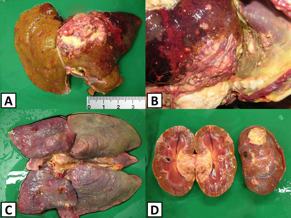
Peritoneal organs of Puma concola. Gross pathological findings exhibited yellowish tumor masses of varying sizes in the liver (A), diaphragm (B), lungs (C), and kidneys (D).
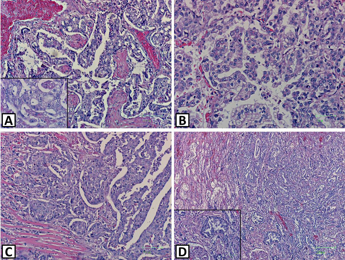
Puma concola. Papillary growth pattern of the adenocarcinoma cells in the liver (A and B), heart (C) and kidney (D). H&E. Scale bar=25 um (A inset), 50 um (B, D inset), 100 um (A,C) and 200 um (D).
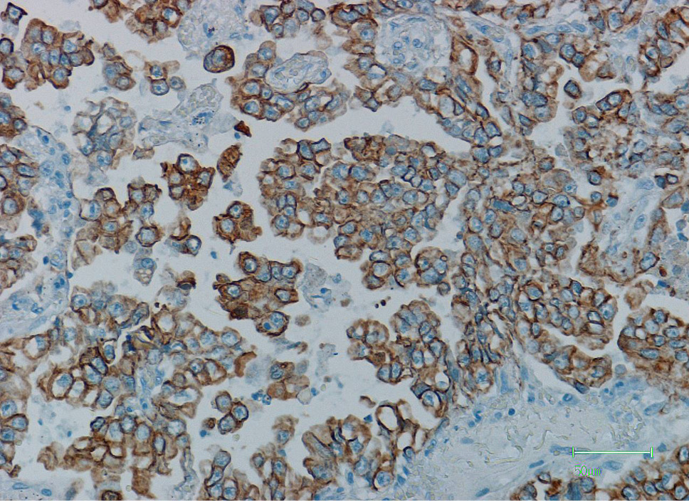
Puma concola. Cytoplasmic immunoreactivity for cytokeratin pan type I (AE I). DAB chromogen, hematoxylin counterstain. Scale bar=50 um.
References
- Allen, J.L., Martin, H.D. and Crowley, A.M., 1985. J. Am. Vet. Med. Assoc., 187: 1215.
- Anderson, L.J. and Sandison, A.T., 1968. Cancer, 21: 289-301. https://doi.org/10.1002/1097-0142(196802)21:2<289::AID-CNCR2820210219>3.0.CO;2-C[CrossRef]
- Anderson, W.I., Dougherty. E.P. and Steinberg, H., 1989. Avian Dis., 33: 594-599. https://doi.org/10.2307/1591128[CrossRef]
- Banales, J.M., Cardinale, V., Carpino, G., Marzioni, M., Andersen, J.B., Invernizzi, P., Lind, G.E., Folseraas, T., Forbes, S.J., Fouassier, L., Geier, A., Calvisi, D.F., Mertens, J.C., Trauner, M., Benedetti, A., Maroni, L., Vaquero, J., Macias, R.I., Raggi, C., Perugorria, M.J., Gaudio, E., Boberg, K.M., Marin, J.J. and Alvaro, D., 2016. Nat. Rev. Gastroenterol. Hepatol., 13: 261-280. https://doi.org/10.1038/nrgastro.2016.51[CrossRef]
- Bastianello, S.S., 1982. Onderstepoort. J. Vet. Res., 49: 195-204.
- Blechacz, B.R. and Gores, G.J., 2008. Clin. Liver Dis., 12: 131-150. https://doi.org/10.1016/j.cld.2007.11.003[CrossRef]
- Borucinska, J.D., Harshbarger, J.C. and Bogicevic, T., 2003. J. Fish Dis., 26: 43-49. https://doi.org/10.1046/j.1365-2761.2003.00435.x[CrossRef]
- Carpenter, J.L., Andrews, L.K. and Holzworth, J., 1987. In: Diseases of the cat (ed. J. Holzworth). W.B. Saunders, Philadelphia, pp. 406-596.
- Cullen, J.M. and Popp, J.A., 2002. In: Tumors in domestic animals (ed. D.J. Meuten), 4th edn. Iowa State Press, 2121 State Avenue, Ames, Iowa, pp. 495-498.
- Domínguez, M.C., Chávez, G., Trigo, F.J. and Rosales, M.L., 2001. Can. Vet. J., 42: 884-885.
- Elangbam, C.S. and Panciera, R.J., 1988. Avian Dis., 32: 594-596. https://doi.org/10.2307/1590938[CrossRef]
- García, A., Erdman, S.E., Xu, S., Feng, Y., Rogers, A.B., Schrenzel, M.D., Murphy, J.C. and Fox, J.G., 2002. Vet. Pathol., 39: 173-179. https://doi.org/10.1354/vp.39-2-173[CrossRef]
- Gibbons, P.M., Busch, M.D., Tell, L.A., Graham, J.E. and Lowenstine, L.J., 2002. Avian Dis., 46: 1062-1069. https://doi.org/10.1637/0005-2086(2002)046[1062:IPWICA]2.0.CO;2[CrossRef]
- Hou, P.C., 1964. J. Pathol. Bact., 87: 239-244. https://doi.org/10.1002/path.1700870203[CrossRef]
- Ilhan, F., Yener, Z., Vural, S.A. and Haligur, M., 2008. Bull. Vet. Inst. Pulawy, 52: 131-134.
- Lepri, E., Sforna, M., Brachelente, C. and Vitellozzi, G., 2013. J. Zoo Wildl. Med., 44: 509-512. https://doi.org/10.1638/2012-0221R1.1[CrossRef]
- Maitra, A., Murakata, L.A. and Albores-Saavedra, J., 2001. Am. J. clin. Pathol., 115: 689-694. https://doi.org/10.1309/5C2C-FP3H-GE7Q-2XJ5[CrossRef]
- Maronpot, R.R., Giles, H.D., Dykes, D.J. and Irwin, R.D., 1991. Toxicol. Pathol., 19: 561-570. https://doi.org/10.1177/019262339101900401[CrossRef]
- Martineau, D., Lemberger, K., Dallaire, A., Labelle, P., Lipscomb, T.P., Michel, P. and Mikaelian, I., 2002. Environ. Hlth. Perspect., 110: 211-262. https://doi.org/10.1289/ehp.02110285[CrossRef]
- Mikaelian, I., de Lafontaine, Y., Harshbarger, J.C., Lee, L.L. and Martineau, D., 2002. Environ. Toxicol. Chem., 21: 532-541. https://doi.org/10.1002/etc.5620210310[CrossRef]
- Miller, R.E., Boever, W.J., Thornburg, L.P. and Curtis-Velasco, M., 1985. J. Am. Vet. med. Assoc., 187: 1256-1258.
- Mischke, R., Höinghaus, R., Lütkefels, E., Buhl, K., Gerhardt, A. and Hewicker-Trautwein, M., 2003. J. Small Anim. Pract., 44: 411-414. https://doi.org/10.1111/j.1748-5827.2003.tb00178.x[CrossRef]
- Oh, Y., Ha, Y., Han, K., Seo, H.W., Kang, I., Park, C., Kim, S.C., Kim, S.H. and Chae, C., 2013. J. comp. Pathol., 148: 259-265. https://doi.org/10.1016/j.jcpa.2012.06.002[CrossRef]
- Ohta, T., Nagakawa, T., Ueda, N., Nakamura, T., Kayahara, M., Ueno, K., Miyazaki, I., Terada, T. and Nakanuma, Y., 1991. Scand. J. Gastroenterol., 26: 1107-1114. https://doi.org/10.3109/00365529109003963[CrossRef]
- Patnaik, A.K., Hurvitz, A.I., Lieberman, P.H. and Johnson, G.F., 1981. Vet. Pathol., 18: 439-444. https://doi.org/10.1177/030098588101800403[CrossRef]
- Pinkney, A.E., Harshbarger, J.C., May, E.B. and Melancon, M.J., 2001. Environ. Toxicol. Chem., 20: 1196-1205. https://doi.org/10.1002/etc.5620200608[CrossRef]
- Renner, M.S., Zaias, J. and Bossart, G.D., 2001. J. Zoo Wildl. Med., 32: 384-386.
- Schmidt, R.E. and Langham, R.F., 1967. J. Am. Vet. med. Assoc., 151: 1325-1328.
- Sironi, G. and Riccaboni, P., 1997. Vet. Rec., 3: 77-78. https://doi.org/10.1136/vr.141.3.77[CrossRef]
- Stedman, T.L., 1982. Illustrated Stedman’s medical dictionary, 24th Edn, Waverly Press, Inc. Mt. Royal and Guilford Avenues., Baltimore, MD 21202, USA, pp. 268.
- Taylor, G.N., Lloyd, R.D., Mays, C.W., Angus, W., Miller, S.C., Shabestari, L. and Hahn, F.F., 1991. Hlth. Phys., 61: 337-347.
- Trigo, F.J., Thompson, H., Breeze, R.G. and Nash, A.S., 1982. J. comp. Pathol., 92: 21-39. https://doi.org/10.1016/0021-9975(82)90040-8[CrossRef]
Services
Article usage statistics
Loading…