Introduction
Tick bites, surgical wounds, fight wounds, and perforations are the main lesions where flies prefer to lay eggs. Myiasis is described as infection and infestation by fly larvae (maggot) in the organs or tissues of animals and humans. Myiasis may result in organ disfigurations, unpleasant smell, anorexia, and weight loss. In severe cases, toxemia and sepsis caused by secondary bacterial infection are possible ; . Based on the target organ, the myiasis is categorized as cutaneous, ocular, urogenital, nasal, auricular, and so on . Ocular myiasis is defined as eye or periorbital tissue infestation by the maggots. Depending on the affected area of the eye, ocular myiasis is categorized into three groups: Ophthalmomyiasis, orbital and palpebral. Ophthalmomyiasis is also classified as ophthalmomyiasis externa (no eye penetration) and ophthalmomyiasis interna (penetration to inner parts of the eye occurs) .
Calliphoridae (blow flies), Sarcophagidae (flesh flies), and Cuterebridae families (Diptera: Cyclorrhapha) are the causative agents of most myiasis cases . According to the relationship between the host and the parasite, myiasis is divided into two types: Obligatory and facultative . In obligatory myiasis, the parasite can not complete its life cycle without the host while in facultative myiasis, a host is not necessary for the parasite to complete its life cycle. Facultative myiasis (accidental) is commonly due to the oral entrance of larvae with foods .
Sarcophagidae (a large family with more than 2600 species) flies are ovoviviparous, depositing maggots instead of eggs on carcass, feces, and open wounds. Larval stages of Wohlfahrtia magnifica (Diptera: Sarcophagidae), as an obligatory myiasis agent, parasitize many warm-blooded vertebrates, which has been reported from both humans and animals such as dogs, camels, horses, leopards, and pigs, resulting in severe myiasis of mucosal membranes and wound lesions . The myiasis caused by W. magnifica has been reported from different parts of Europe, Asia, and Africa . Wohlfahrtia sp. has been reported from various parts of Iran and different cases of human and animal myiasis due to this species have been reported previously ; ; ; ; ; ; ; . Around 61% of the animal traumatic myiasis in southern Iran were due to W. magnifica maggots and this species has been confirmed as the main traumatic myiasis agent of animals in Iran . However, no reports of orbital myiasis are reported in dogs in this country. In the present report, a case of bilateral ocular myiasis due to W. magnifica in an ownerless dog is described (Karaj, Iran), and a successful treatment method has been introduced.
Materials and Methods
Case presentation
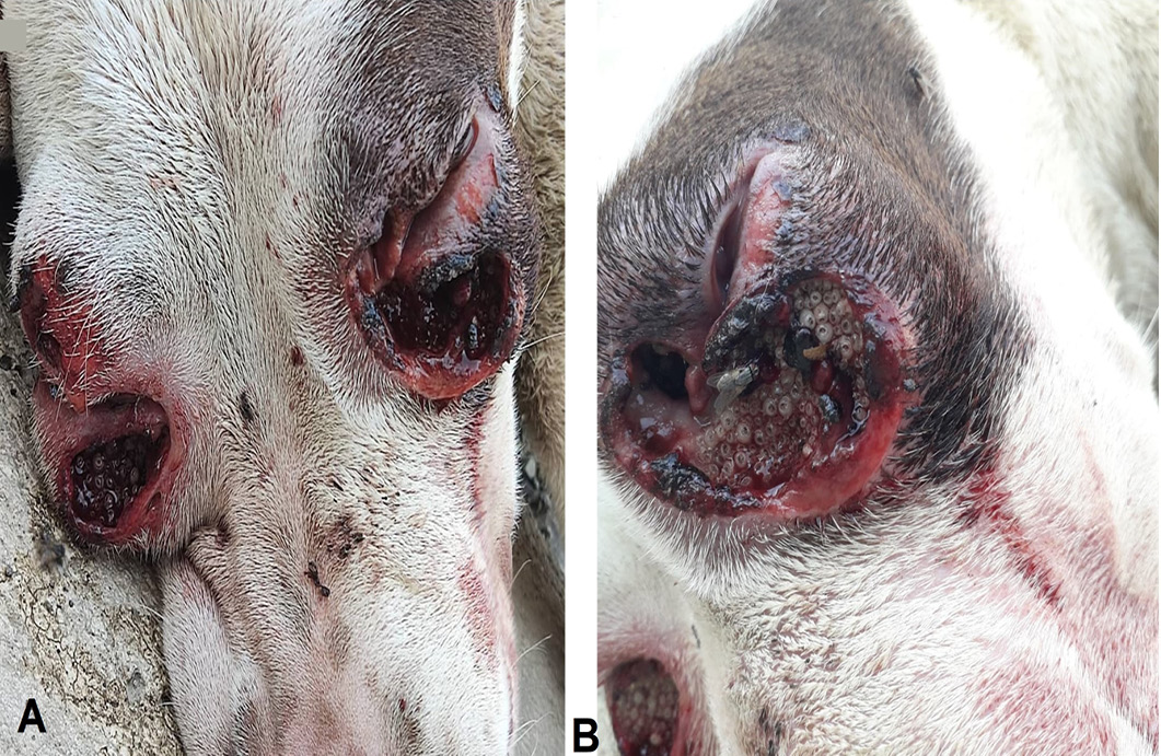
In August 2021, an adult female Pitbull, weighing 15kg, was attended to a veterinary clinic, Karaj, Iran. The dog was referred to the veterinarian with severe anorexia and ocular myiasis. The lesions were seen in both eyes, mainly in the ocular bulbs, lower eyelids, and orbital cavities (Figure 1). A close-up examination revealed the infestation with maggots, unpleasant smell, edema, erythema, ulcers, and necrotic tissues. The orbital walls of both eyes were almost not visible because of massive larvae over the eyes. The body temperature was 40.5°C representing fever.
After removing all larvae under complete anesthesia with Ketamine/ Xylazine, the wounds were washed with Povidone-iodine diluted in normal saline. Then, Permethrin 5% (Gilaranco Pharm Co., Iran) was applied to the wounds in order to reduce the number of live larvae and re-infestation. Oxytetracycline spray 2.5% (25mg/mL: 3.84% w/w) was applied directly to the wounded tissues and continued until the end of the second week (once daily). Systemic Antibiotic therapy with Penicillin/Streptomycin 1+1 (Nasrpharma CO., Iran) was also used to reduce the chance of toxemia, for 5 days (10,000 units/kilogram, once daily). To reduce the inflammation, Dexamethasone was prescribed once a day for three days (0.1 mg/kg, IM). Because the dog had permanently lost its sight, food and water were given to the animal manually for three weeks.
Ethical approval
As the study was conducted with a clinical case, ethical committee approval was not required. The treatment complies with the guidelines laid down by the ethical rights of animals following the country’s law and regulations.
Case follow-up
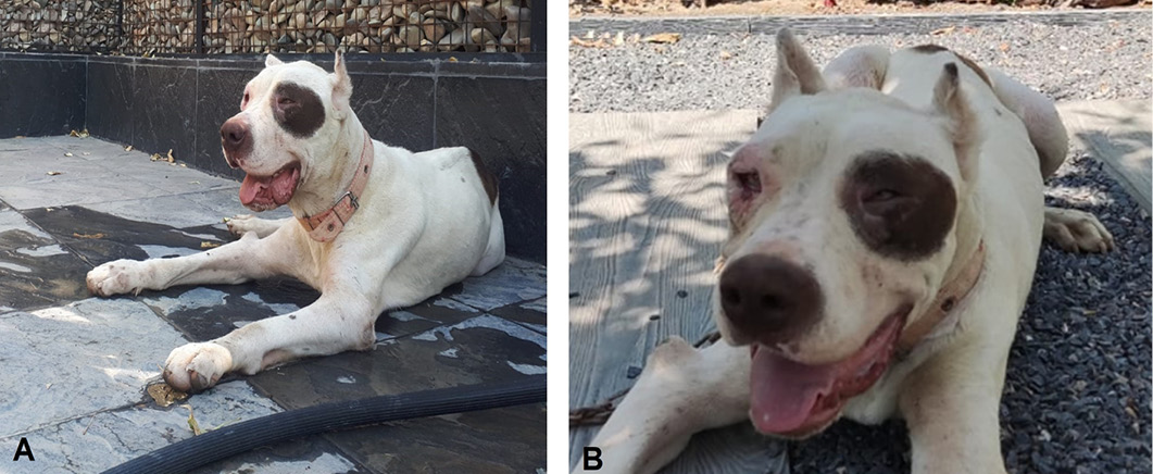
After a week of treatment, there was a progressive improvement in the animal’s condition. Ophthalmic examination of both eyes revealed that the animal was not blind. As a result, permanent loss of sight which was observed at the beginning of animal presentation might be due to edema, pain, and larvae aggregating around both eyes. Examination on day 20 showed that the animal regained its vision completely. examination on day 20 showed that the animal regained its vision completely (Figure 2).
Identification of larvae
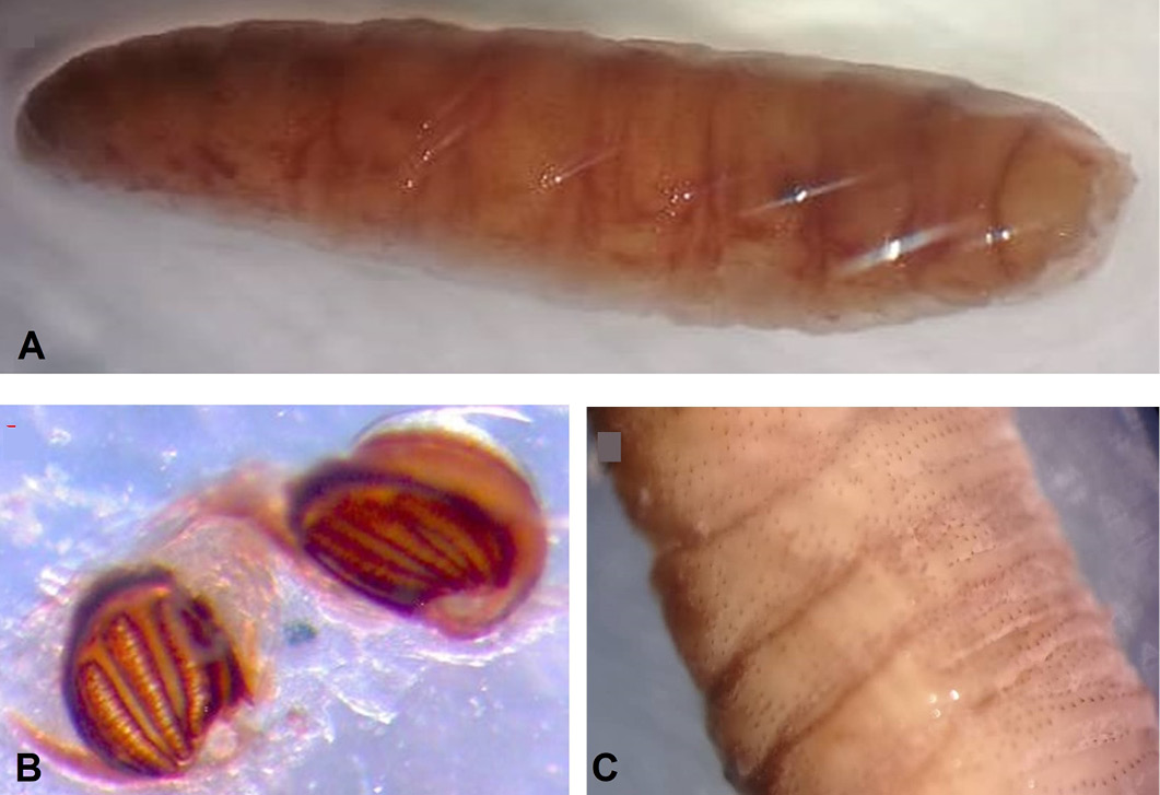
larvae were removed from the wound using forceps and then kept in ethanol 70%. The collected larvae (20) were identified using relevant keys ; ; using stereomicroscope (Olympus CX41, Tokyo, Japan). Larvae were cylindrical, grey in color, 14 to 18 mm in length, and 3 to 5 mm in width. They also had scattered stout spines on the body surface. The larvae of W. magnifica were characterized by hidden spiracles in the cavity of the posterior part. The posterior spiracle had a fairy chitinized button area with wide openings on the peritreme as well as three inner slits (Figure 3).
Results and Discussion
Tissue damage and economic losses (skin damages, reduction in growth rate, milk and meat production, and death due to toxemia) are subsequent outcomes of myiasis . The cases of myiasis have not been recorded regularly in the health system of Iran , but it seems to be prevalent in various parts especially in the areas of condensed cattle keeping industries as an occupational disease .
As far as we know, no report is present in Iran in terms of ocular myiasis in animals. However, many cases of myiasis in different organs due to W. magnifica have been reported: infestation of dogs with W. magnifica in different organs including mouth, paw, skin and, prepuce , Infestation with W. magnifica in referred dogs with a prevalence of 1.95% in Razavi Khorasan province , genital myiasis caused by W. magnifica in camel herds in the southwest of Iran , and gingival myiasis in a goat caused by W. magnifica from Mashhad, northeast of Iran . In a study conducted among slaughtered animals in Kashan, 4 species of dipterous larvae including W. magnifica were detected on sheep . Some human cases of W. magnifica myiasis have also been reported in Iran. Larvae were isolated from male and female external urogenital system and dental cavity ; ; .
Myiasis has been reported in human and many animal species; however orbital myiasis, especially in bilateral form, is not a common case. Miranda et al. reported a case of ocular myiasis of a naturally infested female Beagle dog caused by Cochliomyia hominivorax. The animal was prescribed orally with Nitenpyram alongside analgesic and anti-inflammatory medication. The treatment was reported to be effective . A case of ocular myiasis with Lucilia sericata in a sheep from Saudi Arabia has also been reported in 2008 .
Previous studies revealed Permethrin is effective on the prevention of wound re-infestation by larvae during the treatment process. Permethrin produces protection against new larval infestation for almost 2 weeks with only limited dermal absorption (<1%) ; . Safe permethrin products are now available for food animals in sprays and pour-on formulation to treat ectoparasites. Low dermal absorption and as a result low toxicity, long duration of action, and different available formulations made Permethrin a good drug of choice. Oxytetracycline is a protein synthesis inhibitor with broad-spectrum activity. Topical application of Oxytetracycline results in significant control of primary and secondary skin infections . Our treatment included Permethrin, local, as well as systemic antibiotic therapy with Oxytetracycline and Penicillin/Streptomycin, respectively and systemic anti-informatory dexamethasone. Case follow up to day 20 showed a significant improvement in the animal’s condition including weight gain, eye vision, normal temperature, and reduced edema. Previous studies also suggested Ivermectin as a potent drug in animal myiasis cases ; . It has been reported that Ivermectin is a reliable drug in larval eradication from open wounds with a long duration of action (22 days) .
Conclusions and Recommendations
The present report revealed that mechanical removing of the larvae, subsequent washing of the wound with diluted Povidone-iodine, disinfection of the surrounding with permethrin and antibiotic therapy alongside anti-inflammatory drugs are effective in the treatment of orbital myiasis in dogs. As a result, both medical and veterinary clinicians should be aware of the possibility of the presence of myiasis in different parts of patients’ bodies as well as proper treatment.
Conflict of interest
The authors have declared no conflict of interest.