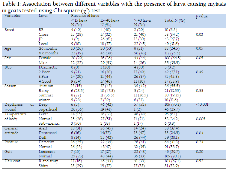
Overall prevalence of our study was 5.21% among 4338 cases which is comparable to Giangaspero et al. (2011), Alahmed (2004) who reported 3% out of 3129 in Italy, 2% out of 3712 in Riyadh Region respectively. However, Radfar and Hajmohammadi (2012), Shoorijeh et al. (2011), Gebremedhin EZ (2011), Arslan et al. (2009), Kara et al. (2005), Abo–Shehada et al. (2003) and Dorchies et al. (2000) found higher prevalence rate of myiasis. In the study, 226 cases were myiasis where goat 69%, cattle 22%, dog 7% and sheep 2% which is compared to Sergio et al. (2007) stating cattle and goat 46.4%, followed by dogs 15.3%, humans 14.7%, pigs 6%, horses 4% and sheep 1%. Apparent deviation is reflected the differences in the level of management, housing, cleanliness and as well as genetic variation in disease resistance breeds. Cross breed goat were commonly infested with myiasis (34.2%) than local (31.6%), Jamnapari (27.7%) and Black Bengal (6.5%) due to genetic variation and environment of tropics which is agreed by Kara et al. (2005) and Farkas et al. (1997); however, Cramer et al. (2002) found adult, light and short haired pure male dog breeds were mostly infested. In our study, females were significantly (p ≤ 0.05) infested with myiasis (64.5%) than male (35.5%) which had a similar finding with Radfar and Hajmohammadi (2012), but their relationship was insignificant. However, Orfanou et al. (2011), Abd El–Rahman (2010), Kara et al. (2005), Farkas et al. (1997) reported more cases in male. Shoorijeh et al. (2011) and Abo–Shehada et al. (2003) found same type of infection rate in both sexes. Poor to fatty animals were infested with myiasis, which is agreement with Gebremedhin (2011). Above 6 months old goats were more prone to myiasis (75.5%) (p ≤ 0.05) which is close to Rahman et al. (2009) where myiasis mostly occurs in cattle of over 2 years. However, different agreement by Kara et al. (2005) implicit infestation which is decreased with the age of cattle and Abo–Shehada et al. (2003) stating all age groups were equally infested. Paredes–Esquivel et al. (2012) found prevalence in lambs younger than 4 months was significantly affected (p ≤ 0.05) which was insignificant in adult sheep (p=0.081).
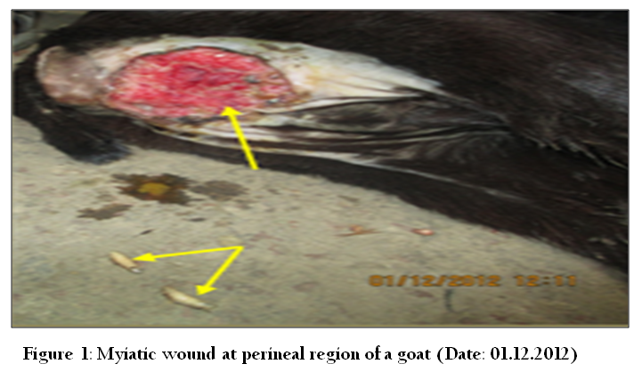
Figure 1: Myiatic wound at perineal region of a goat (Date: 01.12.2012)
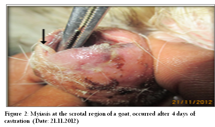
Figure 2: Myiasis at the scrotal region of a goat, occurred after 4 days of castration (Date: 21.11.2012)
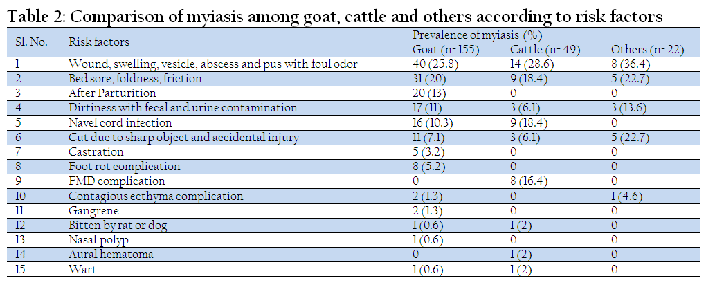
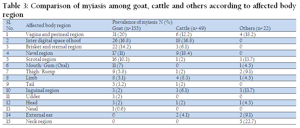
Wounds, swelling, vesicle, abscess and pus with foul odor have been identified mostly as major predisposing factors. Septic wounds of different parts of body were more prominent than others (Goat 55.5% and Cattle 69.4%). Particularly deep wound significantly harboring huge maggots compared to superficial wounds (p ≤ 0.05) which is an agreement with Farkas and Hall (1998). Among other body parts, myiasis frequently occurs at vagina and perineal region (Goat 20%, cattle 12.2%); inter digital space (Goat 16.8%, Cattle 36.8%) which coincide with Rahman et al. (2009). Moreover, Giangaspero et al. (2011) found myiatic wound on vulva and prepuce of sheep; Gaglio et al. (2011) reported three cases of genital myiasis of a goat, ram and dog in Italy; Duro et al. (2007) stated umbilical myiasis in animal; Farkas et al. (1997) found wound myiasis on external genital organs (87%) of sheep in Hungary. Our study exposed other common sites as brisket (goat 14.2% and cattle 6.1%), navel (goat 11% and cattle 18.4%), scrotal (goat 10.3% and cattle 2%), tail, inguinal, udder, thigh/rump, limb, gum, nasal, horn, ear and neck which is similar findings with Cramer et al. (2002) in dog of southern zone of Rio de Janeiro municipality, Farkas et al. (2009) in dog of Al Hoceima, northern Morocco, Beth Knapp–Tyner, (2010) in deer fawn.
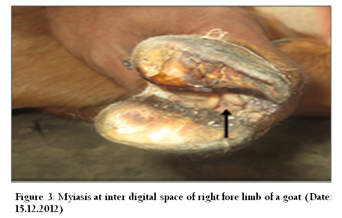
Figure 3: Myiasis at inter digital space of right fore limb of a goat (Date: 15.12.2012)
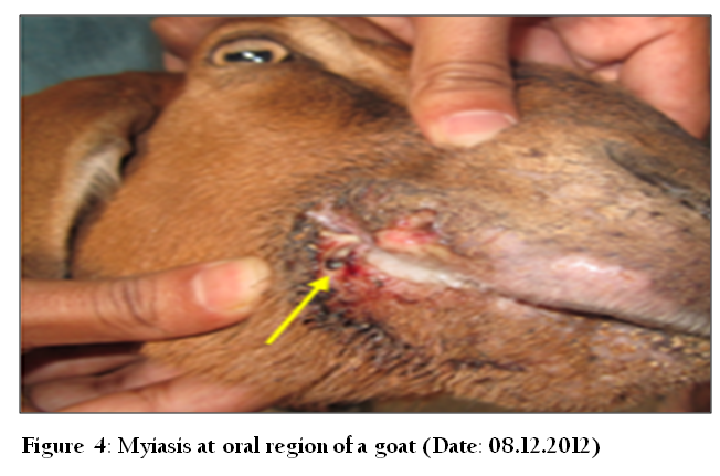
Figure 4: Myiasis at oral region of a goat (Date: 08.12.2012)
On the observation, dirtiness with fecal and urine contamination, poor housing infrastructure, floor with rough surface (brick, concrete) leads to bed sore and wound which is agreement with Bhola et al. (2012). Phillips (2009) confirmed sheep were predisposed to fly strike (cutaneous myiasis) where fleece was contaminated with feces or urine. Accidental injury and traumatic wound (nail, barbed wire, glass etc.) is another important risk factor causing myiasis. Trombetta et al. (2009) reported traumatic myiasis habitually in cattle, dogs and cats whereas Farkas and Hall (1998) found traumatic myiasis infestation >10% of animals at sheep, cattle and horses in Hungary. Additionally, Dik et al. (2012) found 22 traumatic myiasis in animals in Turkey; Ipek DN and Ipek P (2012) observed a facultative traumatic myiasis in domestic rabbit; Giangaspero et al. (2011) examined 10 traumatic myiasis in Italy. Scholtz et al. (2011) reported as presence or absence of dermatophilosis was the main predisposing factor for blowfly strike in sheep. Amazingly gangrene, wart, polyp are fascinating risk factors of myiasis.
Our study revealed that myiasis was found highest in autumn (55.5%) than cold (11.6%) due to prevailing warm and humid climate of tropic where Radfar and Hajmohammadi (2012) reported prevalence varied from 6.8% to 41.8% in August, 2007 to February, 2008 in South–eastern part of Iran; Paredes–Esquivel et al. (2012) recorded significant differences in prevalence in winter and autumn where fly activity held between May to June in the island of Majorca (Spain). In addition, Shoorijeh et al. (2011) found prevalence ranged from 6.6% in spring to 17.9% in winter in South Iran where Abd El–Rahman (2010) analyzed infestation rate in camel was significantly greater in colder (68.8%) than warmer (31%) in Western Libya. Similarly, Orfanou et al. (2011) found six cases from May to July and three cases from August to October in 163 dogs. Alem et al. (2010) described prevalence ranged from 77.7% to 98.8% from November to March in sheep and goats in Central Oromia; Arslan et al. (2009) reported prevalence of nasal myiasis was 54.3% in spring, 41% in summer, 28% in rainfall, 38.9% in winter and statistically significant differences among seasons (p ≤ 0.05) at north–eastern part of Turkey; Alahmed (2004) stated highest percentages of myiasis during Mar–May (60%) and Sept–Nov (31.5%) where temperature and relative humidity are optimum and infestation incidences were low (5% and 1.5% respectively) at dry hot season (Jun–August) and cold season (Dec–Feb) in Riyadh Region; Dorchies et al. (2000) found prevalence from 14.3% to 65% in Feb–Oct in sheep and 6.25% to 47.1% in Sept–Apr in goat in France; Farkas and Hall (1998) described myiasis season lasted from March to November where most cases were available in July and August at sheep, cattle and horses in Hungary; Amin et al. (1997) revealed high infestation rate in summer, followed by spring then autumn. However, Cramer et al. (2002) said no month of the year presented higher occurrence of myiasis cases.