
This study aimed to investigate the bacteriostatic effect of royal jelly (RJ) on methicillin-resistant Staphylococcus (MRSA) and to elucidate the underlying mechanism. MRSA cultures were exposed to varying concentrations of royal jelly (RJ), and MRSA absorbance at 600 nm was measured every 10 hours to assess the impact of RJ on MRSA proliferation. SDS-PAGE electrophoresis and Coomassie blue staining were employed to assess the impact of RJ treatment on MRSA protein expression. A 5% concentration of RJ exhibited substantial inhibition of MRSA proliferation (P < 0.01), with a concentration-dependent increase in inhibitory potency. SDS-PAGE electrophoresis and Coomassie Blue staining results suggested a potential link between royal jelly and its impact on MRSA protein expression, contributing to its inhibitory effect. RJ can disrupt the normal MRSA protein expression and impede MRSA proliferation, though it doesn’t completely eradicate MRSA.
Short Communication · Volume 57, Issue 3, 12 May 2025 · Open Access
Bacteriostatic Effect of Royal Jelly on Methicillin-Resistant Staphylococcus aureus
· · · · ·
Affiliations & Notes ⌄
- 1Department of Medical Laboratory, Fenyang College Shanxi Medical University, Fenyang, 032200, China. 2Public Health Emergency Center, Chinese Center for Disease Control and Prevention, Beijing, 102206, China. 3Department of Clinical Medicine, Fenyang College Shanxi Medical University, Fenyang, 032200, China Songhao Tian and Jinhua Li are the co-first authors. * Corresponding author: tshibm@163.com
Article Info ⌄
- Journal:
- Pakistan Journal of Zoology
- Published:
- 12 May 2025

From the article
- The emergence of these problems brings great challenges to the treatment of MRSA infection, so it is urgent to find new methods for the treatment of MRSA infection.
- The results of Coomassie brilliant blue dye showed that MRSA treated with RJ could affect the expression of MRSA protein.
- RJ has a certain inhibitory effect on MRSA, which may be related to its inhibition of MRSA protein expression.
- It is found that the effective component of inhibiting MRSA in RJ is of great significance to the prevention and treatment of MRSA.
Selected from the article's own text.
Abstract
Materials and methods
Fresh RJ, provided by Shanxi Agricultural University, was kept in the refrigerator at -20℃. The strain used in the test was MRSA (ATCC43300) purchased from Beijing Baioubowei Biotechnology Co., Ltd. The liquid medium was nutritious broth medium and the solid medium was nutritious Agar medium, which was purchased from Hangzhou Binhe microbial Reagent Co., Ltd and Qingdao Binde Biotechnology Co., Ltd. Gram staining kit was purchased from Anhui Chaohu Hongci Medical Equipment Co., Ltd.
For identification of test strains, the colony growth and hemolysis were observed when the blood agar plate was cultured at 37℃ for 24 h. The tested strains were stained with Gram staining kit and identified by PCR amplification of drug resistance gene mec A. The primer sequence is shown in Table I. The amplification conditions were as follows: pre-denaturation at 94℃ for 5 min; followed by 25 cycles at 94℃ for 30 s, 46℃ for 30 min, and 72℃ for 2 min; extension at 72℃ for 10 min.
For preparation of bacterial suspension, the strain was inoculated on solid medium, cultured for 18 h, single colony was inoculated on liquid medium and incubated in shaker at 37℃ for 24 h. The bacterial culture was diluted with PBS to produce the bacterial liquid with the concentration of 108 CFU/ml.
For bacteriostatic test liquid medium and different amounts of RJ were added to the centrifuge tube, and the same amount of MRSA was inoculated to ensure the same amount of MRSA in each tube. The final concentration of RJ in the centrifuge tube is 0%, 5%, and 10%. The bacteria in all centrifuge tubes were cultured in 200 rpm shaker and incubator at 37℃. Bacterial proliferation was assessed by measuring the absorbance at 600 nm, following the approach outlined by Ibarlucea et al. (2017).The absorbance of the culture medium at 600 nm was measured every 10 h. The experiment was repeated three times.
The soluble proteins of MRSA bacteria were rdn on SDS-PAGE according to the method of Lihong et al. (2010), MRSA was cultured in the medium containing 0%, 5%, and 10% RJ for 50 h. The supernatant was discarded after 1000 rpm centrifugation and the bacteria were retained. RIPA lysate was used to split precipitation before being centrifuged for 10 min at 13800 rpm. The supernatant was collected and the sample buffer was added. Samples were then boiled for 10 min and separated by 10% SDS-PAGE. After electrophoresis, the gel was dyed with 0.25% Coomassie brilliant blue dye and decolorized with eluent until the band was clearly visible.
One-way analysis of variance (ANOVA) was utilized to compare various groups, with data analysis performed using SPSS 20.0. Statistically significant results were defined by P-values below 0.05.
Results
Blood agar plate culture showed that yellow grape bead-like colonies were formed on the culture medium, and a clear hemolytic ring was formed around the colonies (Fig. 1A). It was purple after Gram staining, indicating that the strain was gram-positive bacterium (Fig. 1B). After PCR, 16S rRNA and mec A gene bands were amplified by agarose gel electrophoresis, and the position of the bands was consistent with the expected length (Fig. 1C). Based on the above results, the strain were determined as MRSA.
Figure 2 shown the effects of different concentrations of RJ on MRSA in the first 30 h, the absorbance of 0% RJ group at 600 nm was significantly higher than that of the other two groups. The addition of 5% and 10% RJ to the culture medium could significantly decrease the absorbance value at 600nm (P<0.01). The results showed that RJ has a strong inhibitory effect on MRSA.
Table I. mec A and 16s rRNA primer sequences.
| Gene | Primer | Sequence (5’-3’) | Size(bp) |
| mec A | mec A-F | CGGGATCCAAAGATAAAGAAATTAATAATACTA | 1948 bp |
| mec A-R | GGGGTACCTTATTCATCTATATCGTATTTTTTA | ||
| 16s rRNA | 16s rRNA-F | AAAGATGGCATCATCATTCAAC | 1372 bp |
| 16s rRNA-R | TACCGTCATTATCTTCCCCAAA |
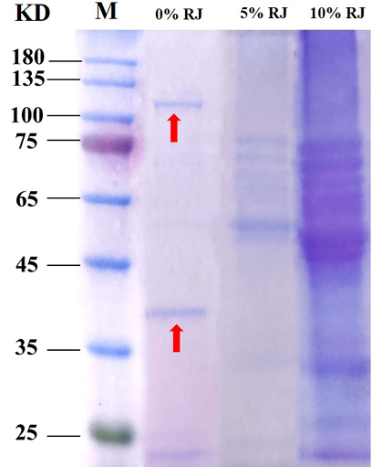
Figure 3 shown the effect of RJ on the expression of MRSA protein. It was found that after 5% RJ and 10% RJ processing, the two bands between 35 kD-45 kD and 100 kD-135 kD disappeared (red arrow marked position). The results showed that RJ could inhibit the expression of MRSA protein.
Discussion
The problem of bacterial drug resistance is becoming more and more serious. In addition to methicillin, MRSA has been found to be resistant to a variety of antibiotics, such as β-lactam, quinolone, sulfamethoxazole, erythromycin, clindamycin, linazolone, and daptomycin . At present, vancomycin is the first choice for the treatment of MRSA infection, but with the emergence of vancomycin-mediated Staphylococcus aureus (VISA) and heterogeneous vancomycin-mediated S. aureus (hVISA), the efficacy of vancomycin in the treatment of MRSA decreases . In recent years, due to the unreasonable use of vancomycin in the treatment of MRSA infection, the sensitivity of vancomycin weakens year by year, resulting in the emergence of vancomycin-resistant S. aureus (VRSA) . Lack of monitoring of vancomycin dosage leads to side effects such as red man syndrome and nephrotoxicity caused by overuse of vancomycin ; . The emergence of these problems brings great challenges to the treatment of MRSA infection, so it is urgent to find new methods for the treatment of MRSA infection.
RJ is a nutritional secretion produced by worker bees. It has been found that the components of RJ include many antimicrobial peptides, such as royalactin, apisimin, jeleines I, II, III, IV, apalbumin α and 10-HAD . Antimicrobial peptides are considered to be natural antibiotics, which can kill broad-spectrum pathogens and drug-resistant bacteria, and have a very low risk of drug resistance . Antimicrobial peptides provide new methods and ideas for solving the problem of bacterial drug resistance . A variety of antimicrobial peptides in RJ can resist gram-positive bacteria and prevent skin from being infected by pathogenic bacteria . Eshraghi and Seifollahi (2003) found that RJ has an inhibitory effect on six kinds of bacteria, including S. aureus. In vitro studies have found that RJ can effectively inhibit MRSA and has a certain effect on MRSA infection caused by spinal implantation in rabbits . This study found that RJ can inhibit the proliferation of MRSA in vitro, but was unable to eradicate MRSA completely. The results are basically consistent with the previous experimental results based on in vivo studies . The results of Coomassie brilliant blue dye showed that MRSA treated with RJ could affect the expression of MRSA protein. RJ is a mixture of water (more than 60%), protein, lipids, sugars, vitamins, minerals and free amino acids. The composition of RJ is affected by many factors, such as season, type of collector, flower variety, geographical environment, and so on . The complexity of its components determines its various biological functions. This study found that RJ is not sufficient to kill MRSA, but can affect the expression of MRSA protein. RJ was found to totally eliminate MRSA and enhance wound healing in animals . This conclusion is not consistent with the results of this study. The antibacterial activity of RJ and rape honey combination was greater than that of rape honey . The antibacterial activity of RJ may be attributed to the components contained in it. Its anti-MRSA effect needs to be further verified in clinical practice. Consequently, it is imperative to isolate and purify the active compounds within RJ that are effective against MRSA, in order to generate novel approaches for MRSA infection treatment.
Conclusions
RJ has a certain inhibitory effect on MRSA, which may be related to its inhibition of MRSA protein expression. But RJ was unable to kill MRSA completely. It is found that the effective component of inhibiting MRSA in RJ is of great significance to the prevention and treatment of MRSA.
Funding
This work was supported by the Fenyang College of Shanxi Medical University 2024 Doctoral Research Initiation Grant Program (2024BS09) and the Science and Technology Innovation Program for Higher Education Institutions in Shanxi Province (2023L474).
Statement of conflict of interest
The authors have declared no conflict of interest.
Figures
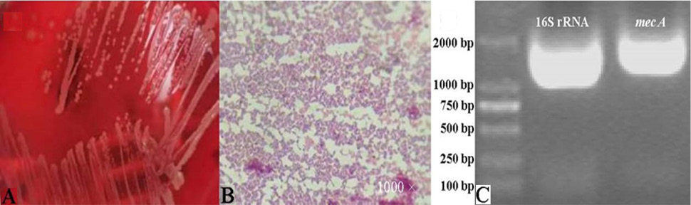
Results of strain identification (A) Blood Agar culture colony (B) Gram staining (C) Results of agarose gel electrophoresis of 16s rRNA and mec A genes.
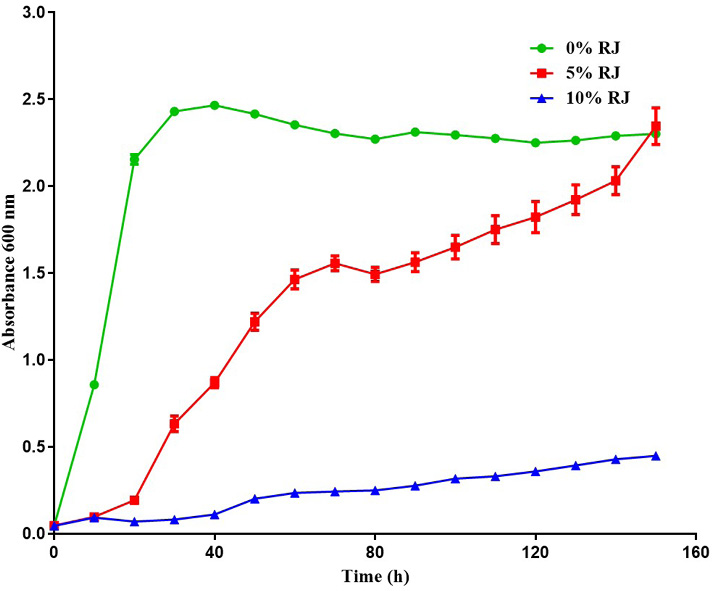
Effects of different concentrations of RJ on the proliferation of MRSA, **P<0.01 vs. 0% RJ group.
References
- Alkharsah, K.R., Rehman, S., Alkhamis, F., Alnimr, A., Diab, A. and Al-Ali, A.K., 2018. Annls Clin. Microbiol. Antimicrob., 17: 7. https://doi.org/10.1186/s12941-018-0260-2[CrossRef]
- Bărnuţiu, L.I., Al, L., Mărghitaş, D.S. and Dezmirean, C.M., 2011. Lucrari Stiintifice Zoot. Bioteh., 44: 67-72.
- Bhise, K., Sau, S., Kebriaei, R., Rice, S.A., Stamper, K.C., Alsaab, H.O., Rybak, M.J. and Iyer, A.K., 2018. Materials, 11: 1245. https://doi.org/10.3390/ma11071245[CrossRef]
- Boukraâ, L., 2015. Antiinfect. Agents Med. Chem., 13: 36-41. https://doi.org/10.2174/2211352513666150318233855[CrossRef]
- Boukraâ, L. and Sulaiman, S.A., 2009. Recent Patents Anti-infect. Drug Discov., 4: 206-213. https://doi.org/10.2174/157489109789318505[CrossRef]
- Chi, X., Liu, Z., Wang, H., Wang, Y., Wei, W. and Xu, B., 2021. J. Fd. Biochem., 45: e13701. https://doi.org/10.1111/jfbc.13701[CrossRef]
- Dadashi, M., Nasiri, M.J., Fallah, F., Owlia, P., Hajikhani, B., Emaneini, M. and Mirpour, M., 2018. J. Glob. Antimicrob. Resist., 12: 96-103. https://doi.org/10.1016/j.jgar.2017.09.006[CrossRef]
- Dinkov, D., Stratev, D., Balkanska, R., Sergelidis, D. and Vashin, I., 2016. J. Bact. Virol., 46: 36-43. https://doi.org/10.4167/jbv.2016.46.1.36[CrossRef]
- El-Gayar, M.H., Aboshanab, K.M., Aboulwafa, M.M. and Hassouna, N.A., 2016. Wound Med., 13: 18-27. https://doi.org/10.1016/j.wndm.2016.05.004[CrossRef]
- El-Gayar, M.H., Ishak, R.A.H., Esmat, A., Aboulwafa, M.M. and Aboshanab, K.M., 2022. AMB Express, 12: 37. https://doi.org/10.1186/s13568-022-01378-x[CrossRef]
- Eshraghi, S. and Seifollahi, F., 2003. Iran J. Publ. Hlth., 32: 25-30. https://doi.org/10.1023/A:1026235131969[CrossRef]
- Fratini, F., Cilia, G., Mancini, S. and Felicioli, A., 2016. Microbiol. Res., 192: 130-141. https://doi.org/10.1016/j.micres.2016.06.007[CrossRef]
- Gunaldi, O., Dağlıoğlu, Y., Tuğcu, B., Kızılyıldırım, S., Postalcı, L., Ofluoğlu, E. and Köksal, F., 2014. Turk. Neurosurg., 24: 249-252.
- Guo, J., Wang, Z., Chen, Y., Cao, J., Tian, W., Ma, B. and Dong, Y., 2021. J. Funct. Fds., 82: https://doi.org/10.1016/j.jff.2021.104514[CrossRef]
- Ibarlucea, B., Rim, T., Baek, C.K., de Visser, J., Baraban, L. and Cuniberti, G., 2017. Lab. Chip., 17: 4283-4293. https://doi.org/10.1039/C7LC00807D[CrossRef]
- Lai, C.C., Chen, C.C., Lu, Y.C., Chuang, Y.C. and Tang, H.J., 2018. Infect. Drug Resist., 11: 681-687. https://doi.org/10.2147/IDR.S159455[CrossRef]
- Lee, A.S., de Lencastre, H., Garau, J., Kluytmans, J., Malhotra-Kumar, S., Peschel, A. and Harbarth, S., 2018. Nat. Rev. Dis. Primers, 4: 18033. https://doi.org/10.1038/nrdp.2018.33[CrossRef]
- Li, S., Tao, L., Yu, X., Zheng, H., Wu, J. and Hu, F., 2021. J. Agric. Fd. Chem., 69: 14415-14427. https://doi.org/10.1021/acs.jafc.1c05942[CrossRef]
- Lihong, Q., Yan, T. and Jing, X., 2010. Microbiol. China, 37: 1628-1638.
- Meaney, C.J., Hynicka, L.M. and Tsoukleris, M.G., 2014. Pharmacother. J. Hum. Pharmacol. Drug Ther., 34: 653-661. https://doi.org/10.1002/phar.1423[CrossRef]
- Nagahama, Y., VanBeek, M.J. and Greenlee, J.D.W., 2018. J. clin. Neurosci., S0967586817313723.
- Otręba, M., Marek, Ł., Tyczyńska, N., Stojko, J. and Rzepecka-Stojko, A., 2021. Life, 11: https://doi.org/10.3390/life11121311[CrossRef]
- Park, H.G., Kim, B.Y., Park, M.J., Deng, Y., Choi, Y.S., Lee, K.S. and Jin, B.R., 2019. J. Asia-Pac. Ent., 22: 737-741. https://doi.org/10.1016/j.aspen.2019.06.005[CrossRef]
- Ramazoni, M., Siregar, M. and Jamil, K.F., 2018. IOP Conf. Ser. Earth Environ., 125: 012096. https://doi.org/10.1088/1755-1315/125/1/012096[CrossRef]
- Seven, İ., Şimşek, Ü.G., Gökçe, Z., Tatli Seven, P., Arslan, A. and Yilmaz, Ö., 2014. Turk. J. Vet. Anim. Sci., 38: 271-277. https://doi.org/10.3906/vet-1303-62[CrossRef]
- Uthaibutra, V., Kaewkod, T., Prapawilai, P., Pandith, H. and Tragoolpua, Y., 2023. Molecules, 28. https://doi.org/10.3390/molecules28030996[CrossRef]
- Zasloff, M., 2002. Nature, 415: 389-395. https://doi.org/10.1038/415389a[CrossRef]
- Zhen, X., Lundborg, C.S., Zhang, M., Sun, X., Li, Y., Hu, X., Gu, S., Gu, Y., Wei, J. and Dong, H., 2020. Sci. Rep., 10: https://doi.org/10.1038/s41598-020-60825-6[CrossRef]
Services
Article usage statistics
Loading…