
It was attempted to molecularly characterize breast cancer patients of district Bannu, KP, Pakistan for any possible polymorphism in the estrogen receptor (ESR1) gene. Blood samples in this regard were collected from clinically diagnosed eighteen breast cancer patients, all possessing invasive ductal carcinoma. DNA was amplified through PCR for 2 different sequences of ESR1 gene. Twelve normal individuals of different ages were also characterized. Amplified products were digested with Xbal and Pvull restriction enzymes and were electrophoresed to visualize single nucleotide polymorphisms (SNPs); rs9340799 (G>A) and rs2234693 (C>T) in ESR1 gene. The amplified product of 524bp of rs9340799 sequence of ESR1 gene was treated with Xbal restriction enzyme while amplified product of 451bp of rs2234693 sequence was treated with PvuII restriction enzyme. Nine patients were found homozygous and 8 were heterozygous for minor allele while 1 patient was carrying major allele. For rs2234693, all the patients were carrying major allele. All the normal individuals were lacking the above stated 2 SNPs. There is thus strong correlation between SNPs of ESR1 gene and breast cancer and hence, can be used as a significant marker in the determination of breast cancer.
Short Communication · Volume 54, Issue 6, 23 September 2022 · Open Access
Association of rs 9340799 and rs 224693 SNPs in Estrogen Receptor Gene with Breast Cancer
· · · ·
Affiliations & Notes ⌄
- 1Institute of Biological Sciences, Gomal University, D. I. Khan, Pakistan 2Institute of Numerical Sciences, Gomal University, D. I. Khan, Pakistan 3Faculty of Pharmacy, Gomal University, D. I. Khan, Pakistan * Corresponding author: sjabbarkhan@yahoo.com
Article Info ⌄
- Journal:
- Pakistan Journal of Zoology
- Published:
- 23 September 2022

From the article
- ESR1 gene has two important single nucleotide polymorphisms (SNPs); rs9340799 (G>A) and rs2234693 (C>T).
- The amplified product of 524bp (Fig. 1A) (rs9340799 sequence) of ESR1 gene was treated with Xbal restriction enzyme.
- Hence, ESR1 gene polymorphism has a significant role in breast cancer predisposition.
- SNPs in ESR1 gene have been linked with breast cancer development and can be used as one of the significant markers to determination of breast cancer .
Selected from the article's own text.
Abstract
Materials and methods
Blood samples of clinically diagnosed 18 breast cancer patients and 12 normal individuals were collected from district Bannu. All the individuals belong to Pashtun ethnic group (Table I) and were molecularly characterized for SNPs in ESR1 gene. Detailed information of each patient was recorded on study-designed pro forma. The study was approved by ethical committee of Gomal University, D. I. Khan, KP, Pakistan.
DNA was extracted by salting out method . Four ml of blood was washed with T.E (10 mM Tris HCl pH 8.0, 2 mM EDTA) after thawing. The pellet was resuspended in 4.5 ml buffer containing 10 mM Tris HCl of pH 8.2, 2mM EDTA and 400 mM NaCl. 240µg proteinase K and 80 µl of 10% SDS were added for digesting protein. It was then incubated overnight at 37°C. Proteins were precipitated with 0.4 ml of 6M NaCl by shaking vigorously for 45 sec and centrifuged at 3000 rpm for 10 min. The supernatant was transferred to another sterilized tube and DNA was precipitated with 4ml of isopropanol . DNA was dissolved in 0.35 ml TE after washing with 70% ethanol, and then heated at 70 ºC for 2 h. DNA was quantitatively measured with NanoDrop spectrophotometer (Thermo Scientific NanoDrop, 2000).
For PCR reaction to perform, 150 ng genomic DNA, 25 mM of each dNTPs, 1 unit of Taq DNA polymerase, 10 mM each of the forward and reverse primers, 1.5 mM MgCl2 and 1x Taq reaction buffer were used in 20µl reaction volume in 2 different reactions. The reaction was carried out through 30 cycles that consisted of 3 min denaturation at 95°C, 30 sec annealing at 65°C and one minute and 30 sec extension at 72°C. During the first cycle, denaturation was done at 95°C for 5 min while the final extension was done at 72°C for 10 min. Gel electrophoresis of the PCR product along with 100bp ladder was done on 2% agarose gel containing ethidium bromide for visualization.
Ten µl of amplified products were digested with Xbal and Pvull restriction enzymes under the conditions recommended by manufacturer (New England Bio labs, USA). The digested products were electrophoresed on 3% agarose gel.
Table I. Information of corresponding patient regarding age, type of breast cancer, grade, and date of diagnosis.
| S. No | Breast cancer type | Grade | Patient age (Yr) | Breast involved | rs9340799 (G>A) | rs 2234693 (C>T) |
| 1 | IDC | III | 43 | Left | Homozygous | Normal |
| 2 | IDC | III | 41 | Left | Heterozygous | Normal |
| 3 | IDC | III | 38 | Left | Homozygous | Normal |
| 4 | IDC | III | 44 | Left | Normal | Normal |
| 5 | IDC | III | 35 | Left | Heterozygous | Normal |
| 6 | IDC | III | 40 | Right | Homozygous | Normal |
| 7 | IDC | III | 30 | Right | Heterozygous | Normal |
| 8 | IDC | III | 45 | Left | Homozygous | Normal |
| 9 | IDC | III | 30 | Right | Homozygous | Normal |
| 10 | IDC | III | 26 | Left | Homozygous | Normal |
| 11 | IDC | II | 60 | Right | Heterozygous | Normal |
| 12 | IDC | II | 34 | Right | Heterozygous | Normal |
| 13 | IDC | II | 35 | Left | Heterozygous | Normal |
| 14 | IDC | II | 32 | Right | Homozygous | Normal |
| 15 | IDC | II | 24 | Left | Homozygous | Normal |
| 16 | IDC | II | 23 | Right | Heterozygous | Normal |
| 17 | IDC | II | 36 | Left | Heterozygous | Normal |
| 18 | IDC | II | 47 | Right | Homozygous | Normal |
IDC, invasive ductal carcinoma.
Results
Clinically diagnosed 18 breast cancer patients and 12 normal individuals, all belonging to Pashtun ethnic group were molecularly characterized for polymorphisms in ESR1 gene (Table I). ESR1 gene has two important single nucleotide polymorphisms (SNPs); rs9340799 (G>A) and rs2234693 (C>T). Such polymorphism if present has an impact on breast cancer predisposition. Two restriction enzymes i-e Xbal and PvuII were used for identification
of SNPs sequences in ESR1 gene. The amplified product of 524bp (Fig. 1A) (rs9340799 sequence) of ESR1 gene was treated with Xbal restriction enzyme. Samples 1, 3, 6, (Table I, Fig. 1B) and 8, 9, 10, 14, 15, 18 (not shown in the Fig. 1) were completely digested on treatment with Xbal that resulted in homozygous condition for minor allele “A”. Moreover, the samples No. 2, 5, 7 (Table I, Fig. 1) and 11, 12, 13, 16, 17 (not shown in the Fig. 1) were incompletely digested on treatment with the same enzyme that resulted in heterozygous condition for minor allele “A” while, sample No 4 (Fig. 1) was homozygous for major allele “G” after treatment with Xbal restriction. Thus, sample No 4 was wild (normal) type for rs9340799 sequence. Similarly, the amplified product of 451bp (rs2234693 sequence) of ESR1 (Fig. 1C) gene from all the breast cancer patients was also treated with PvuII restriction enzyme. In this regard, all the 18 samples were not digested even for longer exposure (Fig. 1D) by PvuII enzyme, meaning that the samples were wild (normal) type for rs2234693 sequence. Likewise, all the 12 normal individuals possessed neither rs9340799 (G>A) nor rs2234693 (C>T) polymorphism.
Discussion
Breast cancer is one of the most familiar malignancies that cause severe health problems in women globally ; . Inheritance, postmenopausal use of hormones, early age of menarche, delayed first pregnancy, breastfeeding for brief period, low parity, and a long gap between births are some of the factors concerned with the breast cancer predisposition . Additionally, hormones are one of the key risk factors . Hormones control growth and function of epithelial cells of the breast. Estrogen plays a major role in breast cancer predisposition as its over-expression may induce the cells to build mutations ; . Single-nucleotide polymorphisms (SNPs) are the most common type of genetic variations in the human genome. Large numbers of estrogen-related genes have been reported with SNPs as the probable risk factors of breast cancer in the women already have gone through menopause . Estrogen receptor alpha gene (ESR1), encoding ERα protein is more important in initiation, development and therapeutics of breast cancer. Specific SNPs in ESR1 gene are involved directly or indirectly in the change of its function, that have an impact on the risk of breast cancer ; . The two important SNPs, (rs2234693 and rs9340799), detected with PvuII and Xbal, respectively have been shown as important markers in certain types of cancer . We, here for the first time in Pakistan attempted to molecularly characterize breast cancer patients for possible polymorphism in two SNPs sequences, the rs9340799 (G>A) and rs2234693 (C>T) in ESR1 gene. No polymorphism was found for rs2234693 (C>T) of ESR1 gene of all the collected blood samples, when treated with Pvull restriction enzyme. All the 18 patients were found homozygous (normal) for major allele “G”. For rs9340799 (G>A) SNP, of the eighteen patients, 9 patients were found homozygous and 8 patients were heterozygous for minor allele “A” of ESR1 gene, while 1 patient was found normal for the said polymorphism. Interestingly, neither rs9340799 (G>A) nor rs2234693 (C>T) mutation was found in normal individuals of the same locality. This was an important finding for the patients of breast cancer in Bannu district of KP, Pakistan. Except for one patient having the age of 60 years, all the patients were below 50 years of age. Observing the molecular characterization of breast cancer with emphasis on breast involved, stage, age status, and status of ESR1 gene (homozygous or heterozygous condition), it is worth-mentioning that classification characteristics were found insignificant except age groups (p>5%). Empirically, the researchers came with findings when comparing breast cancer tissues involved and breast-cancer stage that classification criteria did appear independently at 5% level of significance. The Pearsonian coefficient for statistical comparison appeared with value 0.17 that was far greater than 5%. Similarly, on comparing breast involved and gene status, once again, very large coefficient appeared with value 0.64, and, in the same picture, cancer grade and gene status were found statistically independent (p>5%). Hence one can visibly decide that breast cancer is statistically independent of breast side and ESR1 gene status. Independent and collective effects of these different polymorphisms in ESR1 gene potentially reveal exposure to estrogen and as a result, an increase in breast cancer susceptibility. Hence, ESR1 gene polymorphism has a significant role in breast cancer predisposition.
Conclusion
SNPs in ESR1 gene have been linked with breast cancer development and can be used as one of the significant markers to determination of breast cancer
Statement of conflict of interest
The authors have declared no conflict of interest.
Figures
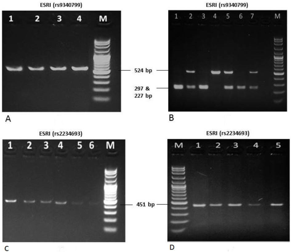
Amplified product of ESR1 gene (rs 9340799) of clinically diagnosed breast cancer patients of Pashtun ethnic group. A, Lanes 1,2,3,4 show amplified product of 524bp. Lane M is 100bp ladder as a marker. B, Digestion of amplified PCR product (524bp) with Xbal restriction enzyme. Lanes 1, 3 and 6 show the samples completely digested Xbal enzyme, producing 2 bands of 297 and 227bp (pretty close to each other that appears as single band). This confirms the presence of G>A polymorphisms and are homozygous for minor allele “A”. Lanes 2, 5 and 7 show the samples incompletely digested by the Xbal restriction enzyme and are thus heterozygous for minor allele “A”, giving 3 bands of 524, 297 and 227bp. Lanes 4 show sample not digested by Xbal enzyme and thus showing a single band of 524bp. This sample is homozygous for major allele “G”. M is a 100bp ladder. Lane 8 is blank. C, amplified product of ESR1 gene (rs2234693) of clinically diagnosed breast cancer patients of Pashtun ethnic group. D, Digestion of amplified PCR product using PvuII restriction enzyme. Lanes 1 to 8, samples are not digested by PvuII restriction enzyme, showing a single band of 451b. Thus all the patients lack rs2234693 (C>T) polymorphism. M is a 100bp ladder used as a marker.
References
- Agarwal, G., Pradeep, P.V., Aggarwal, V., Yip, C.H., and Cheung, P.S., 2007. World. J. Surg., 1: 1031-1040. https://doi.org/10.1007/s00268-005-0585-9[CrossRef]
- Ahmed, R., Shaikh, H., and Hasan, S.H., 1997. J. Pak. med. Assoc., 47: 114–116.
- Bhurgri, Y., 2004. Asia. Pac. J. Cancer, 5: 77–82.
- Boral, D., Vishnoi, M., Liu, N., Yin, W., Sprouse, M.L., Hong, D.S., Tan, T.Z., Thiery, J.P., Chang, J.C., and Marchetti, D., 2017. Nat. Commun., 8: 196. https://doi.org/10.1038/s41467-017-00196-1[CrossRef]
- Easton, D.F., Pooley, K.A., Dunning, A.M., Pharoah, P.D., Thompson, D., and Ballinger, D.G., 2009. J. natl. Cancer Inst., 101: 1012–1018.
- Ferlay, J., Parkin, D.M., and Steliarova-Foucher, E., 2010. Eur. J. Cancer, 46: 765–781. https://doi.org/10.1016/j.ejca.2009.12.014[CrossRef]
- Ferlay, J., Steliarova-Foucher, E., Lortet-Tieulent, J., Rosso, S., Coebergh, J.W., Comber, H., Forman, D., and Bray, F., 2013. Eur. J. Cancer, 49: 1374–1403. https://doi.org/10.1016/j.ejca.2012.12.027[CrossRef]
- Figueroa, J.D., and Brinton, L.A., 2012. J. natl. Cancer Inst., 104: 641–642. https://doi.org/10.1093/jnci/djs193[CrossRef]
- Hashmi, M., 1997. J. Pak. med. Assoc., 47: 75–78. https://doi.org/10.1093/occmed/47.2.75[CrossRef]
- Kanchan, R.K., Siddiqui, J.A., Mahapatra, S., Batra, S.K., and Nasser, M.W., 2020. Adv. Ther. Mol. Cancer, 19: 29. https://doi.org/10.1186/s12943-020-1140-x[CrossRef]
- Lalloo, F., and Evans, D.G., 2012. Clin. Genet., 82: 105–114. https://doi.org/10.1111/j.1399-0004.2012.01859.x[CrossRef]
- Maddams, J.B., Utley, M., and Moller, H., 2010. Br. J. Cancer, 107: 1195-1202. https://doi.org/10.1038/bjc.2012.366[CrossRef]
- Malik, I.A., Mubarik, A., Luqman, M., Ullah, K., Ahmad, M., Alam, S.M., and Mughal, T., 1992. J. environ. Pathol. Toxicol. Oncol., 11: 353.
- Miller, S.A., Dykes, D.D., and Polesky, H.F., 1998. Nucl. Acids Res., 16: 1215. https://doi.org/10.1093/nar/16.3.1215[CrossRef]
- Miyoshi, Y., and Noguchi, S., 2003. Biomed. Pharmacother., 57: 471–481. https://doi.org/10.1016/j.biopha.2003.09.008[CrossRef]
- Sereno, M., Hasko, J., Molnar, K., Media, S.J., Reisz, Z., Malho, R., Videiro, M., Tiszalvicz, L., Booth, S.A., Wilhelm, I., Krizbai, I.A., and Brito, M.A., 2020. Mol. Oncol., 14: 520-538. https://doi.org/10.1002/1878-0261.12632[CrossRef]
- Shin, H.R., Carlos, M.C., and Varghese, C., 2012. Jpn. J. clin. Oncol., 42: 867-881. https://doi.org/10.1093/jjco/hys077[CrossRef]
- Siegel, R., Naishadham, D., and Jemal, A., 2013. Cancer J. Clin., 63: 11–30. https://doi.org/10.3322/caac.21166[CrossRef]
- Stratton, M.R., and Rahman, N., 2008. Nat. Genet., 40: 17–22. https://doi.org/10.1038/ng.2007.53[CrossRef]
- Usmani, K., Khanum, A., Afzal, H., and Ahmad, N., 1996. J. environ. Pathol. Toxicol. Oncol., 15: 251–253.
- Yager, J.D., 2000. J. natl. Cancer Inst., 27: 67–73. https://doi.org/10.1093/oxfordjournals.jncimonographs.a024245[CrossRef]
- Yager, J.D., and Davidson, N.E., 2006. N. Engl. J. Med., 354: 270–282. https://doi.org/10.1056/NEJMra050776[CrossRef]
Services
Article usage statistics
Loading…