
The polycystic ovarian syndrome (PCOS) is an endocrine disease in reproductively aged women and is charactered bydyslipidemia, hyper-androgenism, acne, hirsutism, menstrual irregularities, and ovarian dysfunction resulting in infertility. A balanced body mass index, physical fitness, biochemical and endocrine ratios play vital role in proper functioning of reproductive system and guard against PCOS development. This study was conducted to investigate the role of socio-demographic variables, lipid and hormonal level/ratios in obese and non-obese PCOS individuals. For this purpose, a total of 300 subjects were recruited from Southern Punjab consisting of obese cases (n=61), non-obese cases (n=89), obese controls (n=35) and non-obese controls(n=115). Analysis of the socio-demographic variables in PCOS showed significant association with geographical area, diet, monthly income, smoking status, education, medication, infertilitycomplications and family history in obese and non-obese cases compared to controls. Statistical analysis showed a significant difference in BMI, lipid profile (serum level of HDL, LDL, CHOL/HDL, HDL/LDL and LDL/HDL ratios) and endocrine profile (estradiol and insulin levels) between obese cases and obese controls. We have also observed a significantly lowerserum HDL, estradiol, levels and HDL/LDL ratio compared to control groups. While CHOL level was significantly higher in obese cases than obese controls, the serum TG/HDL ratio, LH, FSH, testosterone and progesterone levels were significantly higher in non-obese cases as compared to non-obese controls. Taken together, we showed that obesity is not an exclusive factor involved in the development of PCOS. The occurrence and prevalence of PCOS are attributed to multiple alternative factors which warrant future studies comprising of larger cohorts and at the national levels.
Research Article · Volume 56, Issue 4, 4 June 2024 · Open Access
Association of Demographic, Lipid and Hormonal Profile with Polycystic Ovarian Syndrome in Pakistani Women
· · · ·
Affiliations & Notes ⌄
- 1Department of Zoology, The Women University Multan, Multan, Pakistan. 2MINAR Cancer Hospital, Multan. * Corresponding author: drasia@wum.edu.pk
Article Info ⌄
- Journal:
- Pakistan Journal of Zoology
- Published:
- 4 June 2024

From the article
- In regard to residential area and the percentage of non-obese cases, there was a high and significantly different compared to non-obese controls (p=0.023) (Table I).
- Regarding diet status, the current study showed significant difference between obese and non-obese cases and controls.
- Obese cases were significantly younger individual compared toobese controls.
- While CHOL level was significantly high in obese cases than obese controls, the serum TG/HDL ratio, LH, FSH, testosterone and progesterone levels were significantly higher in non-obese cases as compared to non-obese controls.
Selected from the article's own text.
Abstract
INTRODUCTION
T he polycystic ovarian syndrome (PCOS) is the most frequently occurring endocrine, multi-factorial and polygenic disorder in the women of reproductive age ; ; ; ; ; . Compared to healthy woman, PCOS-carrier females are prone to additional health issues including obesity, anxiety, depression, infertility, loss of sexuality and loss of feminism which ultimately attribute to unhealthy life style Moran et al. (2010). The PCOS is characterized by heterogeneous disease with hyperandrogenism, insulin resistance (IR), cystic ovary, anovulation and infertility . Owing to hormonal imbalance, female with PCOS display dyslipidemia, insulin resistance (IR), oxidative stress, metabolic abnormality and high level of androgen which result in infertility .
The disproportional body mass index (BMI)is considered an important factor in the prevalence of PCOS (Annagur et al., 2014). The PCOS-carrier females, especially at the pubertal age,usually show elevated BMI. Females with BMI >29.9 are more likely to develop PCOS compared to females with BMI ≤29.9 . Age also plays critical role in the aetiology of PCOS where ovarian volume gradually reduces over age . Interestingly, the PCOS occurrence is more frequentin urban areas than rural areas and it has been observed that the females of urban areas have a strong association towards PCOS development ; . Obesity is yet another factor for the development of PCOS and it has been reported that 50% of women suffering from PCOS are obese. However, obesity is not an exclusive factor for PCOS development as it has also been recorded in non-obese individuals . Obesity is also responsible for IR and hyperinsulinemia in PCOS patients ; . Obesity poses adverse effect on health in well-off societies and results in fecundity (subfertility and infertility) especially in European population . Liepa et al. (2008) has reported that PCOS females are usually overweight and obese. Lifestyle modifications such as weight loss and intake of fibrous-rich diet with decreased fats and carbohydrates contents reduce the chances to develop PCOS. Farshchi et al. (2007) have investigated the impact of calories intake and concluded that the meal should be divided into several portions per day and accompanied should be a low intake of drinks and snacks. Weight loss or insulin sensitivity drugs are considered useful; however, these are beneficial when used with proper diet and physical activity . Financial and economic burdens are significantly associated with PCOS . It has been reported that higher prevalence of an ovulatory PCOS is associated with low-income status compared to high income (Di-Fede et al., 2009). Additionally, it has also observed a correlation between education and family monthly income. Education has a pivotal role in the aetiology of ovulatory phenotypes. Di-Fede et al. (2009) have suggested that illiteracy rate was found to be significantly high in females carrying PCOS syndrome. However, Tabassum et al. (2021) have showed that highly qualified individuals were higher in number carrying PCOS.
Smoking has been shown to carry devastating effects on PCOS . Sowers et al. (2001) have suggested that smoking worsens the androgen hormone level and thus have aggravated sign and symptoms, endocrine and metabolic profile with less conception and fertility rate . Additionally, infertility has also been significantly associated with PCOS development ; Atjiosan, 2020; . Atjiosan (2020) have suggested that approximately 75 percent females carrying PCOS show infertility.
Marital status of the women plays a very important part in the pathogenicity of PCOS. It has been revealed that PCOS is more frequent in married than unmarried individuals . Both lifestyle modifications regarding health, and patient care should be provided to the PCOS females to alleviate the impact on livelihood ; . It is also considered that this syndrome may be genetic contributed and association of first-degree relatives (mother and sister) with PCOS cases are significant compared to the healthy females (Kahsar-Miller et al., 2001). Enlarged cystic ovaries with thickened, smooth, pale and capsule as well as family history are said to be the best diagnosis of this syndrome but it is not confirmed yet .
There are emerging evidences that PCOS may be accompanied with dyslipidemia, oxidative stress, infertility, hyperinsulinemia and hyperandrogenism in reproductively aged females . The dyslipidemia is involved in the PCOS development whereas BMI, cholesterol, LDL and insulin levels are increased while HDL is decreased in PCOS females , 2000). This is evident that the dyslipidemia appeared more commonly in PCOS women compared to healthy females . Cupisti et al. (2008) have found significantly high level of LH and low-density lipid and low level of HDL. In contrast, Mattsson et al. (1984) have found high level of triglycerides and very low-density lipid. However, a significantly decreased concentration of free cholesterol in low density lipoproteins was observed in PCOS patients. It has been noticed that abnormal lipid profile is common in PCOS patients as>70% of females carrying PCOS, the levels of LDL and TG were high and HDL level was low . Mobeen et al. (2016) have evaluated the relationship among BMI, clinical profile and LH/FSH ratiosin. PCOS patients. Fertile and infertile females showed elevated LH/FSH ratio. Additionally, it was noticed that FSH was significantly associated with obesity and infertility while BMI was significantly correlated with obesity and hirsutism. Lifestyle modification and weight loss were suggested to reduce sign and symptoms of infertility and hirsutism .
Testosterone has a high impact in the prevalence of PCOS as a significant association with PCOS been recorded. Serum testosterone level was considered to be a strong indicator in the severity of this syndrome . The PCOS is an endocrinological problem associated with abnormal function of estrogen and its receptors (ERs) expression. Owing to changes in signalling pathway of estrogen receptor, cell cycle, cell proliferation, invasion, migration and ovulation are affected. Female infertility is caused by abnormal ovulation which affects reproduction and pregnancy in women with PCOS. Estrogen regulates biological and physiological processes in the body with the help of estrogen receptors. Thus, identification of ER can be used as an effective target for the PCOS treatment .
Females with PCOS are IR as a significantly high level of insulin, fasting blood glucose and all plasma lipids were reported in PCOS patients as compared to controls Kumawat et al. (2021). They have also reported high level of insulin and IR in obese control group and even in lean PCOS group. In PCOS, high insulin level and IR were identified as significant contributors to reproductive and metabolic abnormalities . The features of metabolic syndrome (MS) are obesity, hypertension, IR, visceral fat deposition and dyslipidemia. These are most frequently occurring sequalae in PCOS patients than healthy controls ; . However, lipid ratios of blood and fatness indices can be used as an effective screening tool in the diagnosis of MS in PCOS women (Kałuzna et al., 2022). Taken together, PCOS is a complex syndrome and is influenced by multi-layered factors to determine the end point of the carriers.
This study aims to explore the socio-demographic variables in obese and non-obese individual for the prevalence of PCOS. In this regards, BMI, biochemical and endocrine parameters were determined in these groups in Southern Punjab, Pakistan to assess the factors and socio-demographic attributes of PCOS. The outcome will facilitate the early identification and overall impact of the PCOS in Pakistani population.
MATERIALS AND METHODS
A total of 300 females (150 PCOS cases and 150 non-PCOS controls) were accessed in Southern Punjab, Pakistan. The inclusion criteria of study subjects were PCOS women with amenorrhea, oligomenorrhea, hirsutism, acne, obesity, without taking any steroids except oral contraceptives during non-gestational period. All individuals who were consuming alcohol, were under hormonal treatment within the last 6 weeks, pregnant, hypertensive and drug-addicted were excluded from this study. The Research Ethics Committee of the Women University Multan approved the present study. Study subjects were recruited after confirmation from gynaecologists. After receiving informed consent, data of the relevant females (that came to hospitals for follow-up or medical treatment) were collected from different hospitals and clinics (MINAR, Cancer Hospital, Asma Azhar Hospital and Rubina Infertility Clinic, Multan) which provide medical facilities in Southern Punjab. Demographic data were recorded in especially designed questionnaire. For the analysis of lipid and hormonal profile, blood samples were collected in the fasting state and serum was separated.
The age for study subjects (cases and controls) ranged from 18-35 years. On the basis of BMI, cases and controls were further categorized as obese (BMI≥30) and non-obese (BMI<30). All participants were divided into four groups: Group I: obese cases (n=61), Group II: non-obese cases (n=89), Group III: obese controls (n=35) and Group IV: non-obese controls (n=115). Hormonal analyses such as LH, FSH, testosterone, progesterone and oestradiol levels were measured by enzyme immune assay (EIA) using commercially available kits (Catalogue numbers KA2280, KA0213, KA0309, KA0299 and KA0297 Abnova, Taiwan), respectively. Serum insulin level was analysed by enzyme linked immunosorbent assay (ELISA) (Insulin kit Cat. No. IN374S, Calbiotech, California). Enzymatic in vitro assay was used in order to determine lipid profile including CHOL, TG, HDL and LDL, which were estimated using commercially available kits (Cobas lipid panel, Cat No. 06380115119, Roche diagnostics GmbH). Lipid and hormonal profile ratios were evaluated on Microsoft Excel sheet and BMI was determined with the help of following formula (BMI=weight in kilograms/height in meter2). Statistical analyses were performed by Chi-square and independent Student’s t-test using SPSS software (version-20). A p-value ≤0.05 was considered to be statistically significant.
RESULTS
The data of socio-demographic variables in obese and non-obese cases and their respective controls are shown in Table I. Geographical area/residential area included urban and rural area are highlighted in Figure 1. These data revealed a significant difference between obese and non-obese cases and controls (p=0.047). In regard to residential area and the percentage of non-obese cases, there was a high and significantly different compared to non-obese controls (p=0.023) (Table I). Income of study population was categorized into three group (very low =Rs. 10,000 to 20,000, low >20,000 to 40,000 and medium to high > 40,000 (Figure 1). Statistical analysis among income groups showed a significant difference between cases and control (p=0.004). Moreover, significant difference was also observed between obese cases and obese controls (p=0.014), non-obese cases and obese controls (p=0.003), and obese and non-obese cases (p=0.009) (Fig. 1A).
Regarding smoking status of population comprised of active smokers who smoked (inhaled and exhaled), passive smokers who breathed in second hand smoke (only inhaled) and non-smokers were those not exposed to smoking. Analysis of smoking status showed significant differences between obese and non-obese cases and controls (p<0.001). Statistical analysis of smoking by chi square test showed a significant difference in obese cases vs obese controls, non-obese cases vs non-obese and obese controls (p=0.039, p<0.001 and p=0.001), respectively (Fig. 1B).
Regarding education status, the study population was comprised of illiterate (no education) and literate (primary to graduation). Percentage of literate obese cases was high than illiterate. Significant difference was observed in obese cases vs obese controls, obese cases vs non-obese controls (p=0.028 and p=0.001, respectively), non-obese cases vs non-obese controls and non-obese cases vs obese controls (p=0.007 and p=0.023) regarding education status (Table I).
The diet of study population included carbohydrate rich, proteins rich, lipid rich and mixed diet. Higher percentage of obese cases was taking mixed diet than other groups. Significant difference was found between obese cases vs obese and non-obese controls (p=0.014, p=0.003, respectively) and non-obese cases vs non-obese and obese controls (p=0.002 and p=0.043, respectively) reference to diet status (Fig. 1C).
Among married women, complications associated with infertility were divided into four categories (female cases with abortion, miscarriages, having no child and live babies with no complication) and found to be significantly different (p<0.001). Percentage of non-obese cases and controls was high with group of females having no child compared to other groups. Significant difference was observed in non-obese cases vs obese cases and non-obese controls (p=0.033 and p=0.002, respectively) and between obese and non-obese controls (p=0.013) (Fig. 1D). There were two categories regarding marital status that are married and unmarried. Statistical analysis showed no significant difference among these groups (p=0.282), however, a high percentage of married non-obese PCOS was observed compared to other groups (Table I.
Family history of studied cohort consisted of women who had family history with PCOS and no family history of PCOS. Family history was found to be associated with PCOS (p<0.001). Significantly high difference was found between obese cases vs obese and non-obese controls (p=0.005 and p=0.007). Similar difference was also observed between non-obese cases vs non-obese and obese controls (p<0.001 in both). Difference was also significant between obese and non-obese controls regarding family history (Table I).
In obese cases (Group I), the age, serum HDL, estradiol level, and HDL/LDL ratios were significantly low, whereas MI, serum CHOL, LDL, insulin levels, CHOL/DL, and LDL/HDL ratios were significantly high. However, LH, FSH, testosterone levels, TG/HDL and LH/FSH ratios were non-significantly high and serum TG, progesterone levels were non-significantly low compared to obese controls (Group III). In non-obese cases (Group II) versus l non-obese controls (Group IV),a significantly high level of BMI, LDL, CHOL/HDL, TG/HDL, LDL/HDL ratios, LH, FSH, testosterone, progesterone and insulin were observed. In contrast, HDL, HDL/LDL and serum estradiol levels were significantly low, whereas serum CHOL, TG levels and LH/FSH ratio were non-significantly high. In obese cases (Group I) the age, serum HDL, estradiol, progesterone level, HDL/LDL ratio were significantly low but BMI, serum LDL, testosterone, insulin levels, CHOL/HDL, TG/HDL, LDL/HDL, LH/FSH ratios were significantly high. A non-significant difference was observed in serum level of CHOL, TG, LH and FSH compared to non-obese controls (Group IV). In non-obese cases (Group II) compared to obese controls (Group III), the BMI, HDL level and HDL/LDL ratio were significantly low whereas CHOL, LDL, CHOL/HDL, LDL/HDL, testosterone, and insulin levels were significantly high and non-significant difference in age, serum TG, LH, FSH and progesterone level and TG/HDL and LH/FSH ratios were found. However, no significant difference was observed between obese and lean cases and obese controls and non-obese controls except for BMI and serum progesterone level. The BMI was significantly higher in obese cases and controls compared to lean cases and controls, but serum progesterone level was significantly low in obese cases than non-obese cases and significantly high in obese controls versus non-obese controls (Table II).
Table I. Association of socio-demographic variables between Obese and Non-obese subjects.
| Sociodemographic variables
| Obese | Non-obese | P-value
| ||
| Cases (n=61) | Controls (n= 35) | Cases (n=89) | Controls (n=115) | ||
| Geographical area (Residential area) | |||||
| Urban | 38 (62.3%) | 22(62.9%) | 56(62.9%)*b | 53(46.1%) | 0.047 |
| Rural | 23(37.7%) | 13(37.1%) | 33(37.1%) | 62(53.9%) | |
| Education | |||||
| Literate (Primary to graduation) | 43 (70.5%)*a, **c | 16 (45.7%) | 61 (68.5%)**b, *d | 51(44.3%) | <0.001 |
| Illiterate (no education) | 18 (29.5%) | 19 (54.3%) | 28 (31.5%) | 64 (55.7%) | |
| Marital status | |||||
| Married | 47 (77.0%) | 23 (65.7%) | 72 (80.9%) | 92 (80.0%) | 0.282 |
| Unmarried | 14 (23.0%) | 12 (34.3%) | 17 (19.1%) | 23 (20.0%) | |
| Family history | |||||
| Family history with PCOS | 43(70.5%)**a, ***c | 14 (40%)**f | 59 (66.3%)***b, **d | 20 (17.4%) | <0.001 |
| Family history with no PCOS | 18(29.5%) | 21 (60.0%) | 30 (33.7%) | 95 (82.61%) | |
Note: significant =p<0.05*, highly significant =p<0.01**, p<0.001***. a, obese cases vs obese controls; b, non-obese cases vs non-obese controls; c, obese cases vs non-obese controls; d, non-obese casesvs obese controls; e, obese cases vs non-obese cases; f, obese controls vs non-obese controls.
Table II. Comparison of age, BMI, lipid profile and endocrine profile between obese and non-obese groups.
| Parameters | Obese groups | Non-obese groups | ||
| Cases (n=61) | Controls (n= 35) | Cases (n=89) | Controls (n= 115) | |
| Age | 26.56±0.69*a*c | 28.86±0.87 | 26.84±0.598 | 28.23±0.49 |
| BMI (kg/m2 ) | 34.72±0.59**a***c***e | 32.31±0.38***f | 23.52±0.397*b***d | 22.03±0.47 |
| Lipid profile | ||||
| Cholesterol (mmol/L) | 213.97±6.10**a | 188.86±8.97 | 220.74±4.59***d | 211.42±6.09 |
| Triglycerides (mmol/L) | 169.67±2.96 | 187.20±12.60 | 171.08±2.40 | 166.10±6.59 |
| HDL (mmol/L) | 50.72±0.86***a***c | 66.54±5.42 | 51.16±0.80***b***d | 77.77±3.96 |
| LDL (mmol/L) | 167.44±3.02***a***c | 98.69±7.41 | 168.12±2.21***b***d | 109.44±4.15 |
| CHOL/HDL (mmol/L) | 4.36±0.15*a**c | 3.57±0.38 | 4.40±0.13***b**d | 3.49±0.21 |
| TG/HDL(mmol/L) | 3.43±0.87**c | 3.40±0.42 | 3.46±0.073***b | 2.69±0.18 |
| HDL/LDL (mmol/L) | 0.02±0.02***a***c | 0.83±0.012 | 0.03±0.02***b***d | 0.90±0.09 |
| LDL/HDL (mmol/L) | 3.36±0.08***a***c | 1.89±0.24 | 3.33±0.07***b***d | 1.18±0.11 |
| Hormonal profile | ||||
| LH (IU/mL) | 9.23±1.24 | 6.83±0.64 | 14.44±3.64*b | 7.37±0.52 |
| FSH (IU/mL) | 8.44±2.06 | 6.11±0.39 | 13.71±4.22*b | 6.19±0.22 |
| LH/FSH (IU/mL) | 1.93±0.84*c | 1.23±0.15 | 1.61±0.13 | 1.38±0.12 |
| Estradiol (nmol/L) | 128.36±13.92***a***c | 269.96±25.22 | 136.84±11.88***b***d | 256.79±13.86 |
| Testosterone (nmol/L) | 38.59±5.35**c | 25.11±1.82 | 45.53±5.37***b*d | 25.570±0.98 |
| Progesterone (nmol/L) | 1.28±0.26*e | 1.89±0.14*f | 2.37±0.42*b | 1.57±0.07 |
| Insulin (pmol/L) | 55.15±8.07***a***c | 10.63±1.86 | 63.16±9.65***b***d | 14.28±1.37 |
For statistical details and abbreviations, see Table I.
DISCUSSION
It remained an established fact that females with PCOS are more susceptible to health issues including obesity, anxiety, depression, loss of sexuality, infertility, loss of feminism and poor quality of life compared to healthy women . Our presented results showed that percentage of obese and non-obese cases was high in individuals living in urban area as compared to rural area. A similar observation was also noticed by Bharathi et al. (2017) who have reported high percentage of obese and non-obese cases in urban area. In the current study, a significant difference was found in residential area of non-obese cases and non-obese controls. However, overall percentage of PCOS cases residing in urban area was high than rural area in agreement to a recent finding Mushtaq et al. (2022). They have reported that females residing in urban areas were more susceptible to PCOS as compared to females of rural locality.
Present study showed that women having very low monthly income were significantly associated to obese cases as compared to obese controls. Various studies have also investigated that monthly income was significantly associated with PCOS (Di-Fede et al., 2009). However, a study reported number of working obese PCOS were less than non-obese PCOS .The current study showed smoking status was significantly different in obese and non-obese cases than obese and non-obese controls. This finding is unique in its kind as no such observation has previously been documented. However, some studies have found effect of smoking on lipid profile in PCOS and controls. A study conducted by Pau et al. (2013) found that smoking increased the triglyceride level in PCOS cases than controls. They also observed that MS was more frequent in smoker PCOS than non-smoker PCOS and controls. Craig et al. (1989) haveexamined association of smoking and lipid concentration and found increased level of TG, CHOL, LDL and low level of HDL in smoker PCOS than non-smokers.
The present study showed that obese educated females are more susceptible to PCOS compared to non-obese in contrast to a previous observation (Mahmoud et al., 2015). In this study, authors have reported that obese women were less highly educated than non-obese. Regarding diet status, the current study showed significant difference between obese and non-obese cases and controls. However, percentage of obese cases were on mixed diet. Percentage of subjects with high intake of fat rich diet (lipids) was more in non-obese cases than obese. This investigation was supported by a previous study conducted by Bandini et al. (1999) and in contrast to other studies ; .
In this study, complications associated with infertility were highly significant with obese cases than controls. Multiple previous studies ; have reported similar results. In the current study, non-significant difference was recorded when marital status was compared between both groups. However, Mahmoud et al. (2015) have reported that percentage of married obese PCOS women was more than non-obese PCOS women. However, in an investigation of overall cases and control, married females with this syndrome were associated more significantly with PCOS than that of the married controls and contrary to the investigations of Tabassum et al. (2021).
Obese cases were significantly younger individual compared toobese controls. A study conducted by Silfen et al. (2003) have reported significantly more age of obese cases than obese controls whereas no difference in age was found by Yilmaz et al. (2005). Interestingly, age was non-significantly different in non-obese cases vs controls, non-obese cases vs obese controls, obese vs non-obese cases, and obese vs non-obese controls. These results in agreement with the result of a study conducted by Taher and Mhaibes (2015). Silfen et al. (2003) and Yilmaz et al. (2005) have also showed no difference in age between obese vs. non-obese cases similar to this study, but in contrast to study by Layegh et al. (2016), who reported significant difference. In age comparison,a significant difference was observed between obese cases and lean controls in contrast to result reported by Taher and Mhaibes (2015).
BMI was significantly high in obese cases than obese and non-obese controls (Yilmaz et al. (2005), however, BMI of obese controls was significantly high than non-obese cases and controls. These findings are supported by the results of Taher and Mhaibes (2015) and Shi et al. (2021). BMI was also significantly high in obese cases and obese controls versus non-obese cases and control respectively.These finding are against the observations reported by Daghestani et al. (2021). Previous studies ; ; ; have also found significantly high BMI in obese vs non-obese cases as we have reported in this study.
We have shown that CHOL and LDL serum levels were significantly high in obese cases than obese controls in agreement with result of other studies ; ; and in contrast to result of a study by Yilmaz et al. (2005). High level of CHOL and LDL may be due to obesity or diet habit. Dyslipidemia may be a problem in PCOS women but it cannot be manifestation of syndrome. The latter study reported no-significant difference in the level of CHOL and LDL. Serum CHOL and TG levels were not different in obese and non-obese cases vs non-obese controls, in contrast to findings of a study by Ibrahim et al. (2020). Similarly, no difference was observed in CHOL and TG levels between obese cases and non-obese cases. Our results were not consistent with findings of Layegh et al. (2016), and Ibrahim et al. (2020). No significant difference was found between obese cases and obese controls in serum TG level that is in agreement to findings of a study of Shi et al. (2021), but not in agreement with results reported by other studies ; ; ; Daghestani et al., 2018). This may be due to consumption of fat rich diet. No significant difference in serum TG level was observed between non-obese cases and non-obese controls similar to findings of Yilmaz et al. (2005) but deviated from result of other studies ; ; . This difference may be due to ethnic variations. Significantly high level of LDL and low level of HDL were found in obese and non-obese cases than non-obese controls similar to findings of Sayed and Bakhiet (2020). According to studies Daghestani et al. (2021) and Ibrahim et al. (2020), LDL level was significantly highin non-obese cases compared to non-obese controls. Our results are in agreement with the results of this study. High level of LDL and low level of HDL may be associated with PCOS. Serum HDL level was significantly low in obese cases than obese controls similar to finding of Yilmaz et al. (2005), and Legro et al. (2001) but in contrast to result of study by Shi et al. (2021), Daghestani et al. (2021). While serum HDL level was low significantly in non-obese PCOS than non-obese controls matched to the result of some studies ; ; ; but in contrast to results reported by Ibrahim et al. (2020). Serum HDL/LDL ratio was significantly low while LDL/HDL was significantly high in obese case and non-obese cases than obese and non-obese controls as well as in obese cases than non-obese controls, but no published data was found for comparison. Non-significantly different serum level of CHOL, HDL, TG and LDL were observed in obese cases than lean cases and in obese controls than lean controls respectively similar to the finding of Amini et al. (2014) except for the serum TG level. In contrast to this study serum TG level was significantly high in obese cases than lean cases reported by Yilmaz et al. (2005), and Amini et al. (2014). Results of other studies ; showed serum CHOL, TG and LDL levels high significantly and non-significantly high level of HDL in contrast to this study in obese cases than non-obese cases. In contrast to our results, (Daghestani et al. (2021) have found significant differencelevels in CHOL, HDL, TG and LDL between obese controls and non-obese controls. Silfen et al. (2003) have reported significantly high level of LDL and low level of HDL in adolescents in contrast to our results in obese cases than non-obese cases.
In present study the LH level was not different significantly in obese cases than obese controls in contrast to result of some studies ; but similar to the result of this study LH level was high significantly in non-obese cases vs. non-obese controls reported by Nambiar et al. (2016). High level of LH in PCOS may be due to neuroendocrine abnormality leading to high level of androgen. Serum level of FSH was not statistically different in obese cases than obese control as well as between obese cases and non-obese cases similar to finding of a study ( . Serum LH, FSH and testosterone levels were not significantly different in obese cases vs obese control. Similar results were found by Ibrahim et al. (2020) but serum LH, FSH and testosterone level significantly high in non-obese cases compared to non-obese controls contrary to findings of. Statistically no difference in the LH/FSH ratio was found in obese and non-obese cases vs obese and non-obese controls in contrast to findings of study by Shi et al. (2021).
Serum testosterone and insulin levels were significantly high and LH/FSH ratio was elevated in obese cases than non-obese controls consistent with results of Taher and Mhaibes (2015) . No significant difference was found in serum levels of LH, FSH and LH/FSH ratios between obese and non-obese cases.These results are in agreement with the observations made by Taher and Mhaibes (2015). Serum LH, FSH, estradiol, testosterone, insulin level and LH/FSH ratio was non significantly different in obese vs non-obese cases, but progesterone level was significantly low in contrast to the result of Mahmoud et al. (2015) that reported significant difference in LH, FSH, testosterone, insulin level and non-significant difference in progesterone, but result of his study showed non-significant difference in serum level of estrogen similar to result of this study. Silfen et al. (2003) showed significantly high level of progesterone in obese cases than non-obese cases in contrast to result of this study. Silfen et al. (2003) found significant low level of LH in obese cases than non-obese in contrast to our study but similar to this study no difference in FSH and testosterone level in obese cases than non-obese cases was reported. Findings of Yilmaz et al. (2005), Ibrahim and Abdelsalam (2015), and Sachdeva et al. (2019) were also in agreement to our study that LH and FSH levels were non significantly different between obese cases than non-obese cases but Yilmaz et al. (2005) showed significantly high level of testosterone and LH/FSH ratio in contrast to result of present study. Layegh et al. (2016) showed non-significant difference in LH/FSH ratio, serum estradiol and testosterone level that is similar to result of present study in obese cases than non-obese cases. Statistically no significant difference was observed in serum level of LH, FSH, testosterone, insulin and LH/FSH ratio in obese than non-obese controls, consistent with results of Taher and Mhaibes (2015) .
Serum level of estradiol non-significantly whereas progesterone level was significantly high between obese and non-obese controls. In the current study, there was significantly low level of estradiol in obese and non-obese cases than obese and non-obese controls, respectively. These results offer novel insights as currently no such observations are reported in the literature. However, Mahmoud et al. (2015), Layegh et al. (2016) have showed non-significant difference in estradiol level between obese cases than non-obese cases similar to result of this study. Serum level of testosterone non-significantly high in obese cases than obese controls but significantly high in non-obese cases compared to non-obese controls in contrast to findings of Dongargaonkar et al. (2020). Researchers of this study havefound that testosterone level was significantly high in obese cases than obese controls and non-significantly high in non-obese cases than non-obese controls. In the current study, significantly high level of testosterone was observed in obese cases than non-obese controls and non-obese cases than obese controls. However, testosterone level was not different in obese versus non-obese cases and obese vs lean controls.
Serum insulin level was significantly high in obese cases than obese control similar to previous findings ; and in contrast to other studies (Daghestani et al. (2021). Serum insulin level was also significantly high in non-obese cases than non-obese controls also similar to the findings of Dongargaonkar et al. (2020). Similarly, serum insulin level was significantly high in non-obese cases than obese controls in contrast to findings of Taher and Mhaibes (2015) which means that PCOS in both obese and non-obese cases was associated with insulin resistance. In this case-control study insulin level was not statistically different between obese cases and non-obese cases similar to result of studies conducted by Layegh et al. (2016); Kumawat et al. (2021) but in contrast to result of Silfen et al. (2003).It has also been reported that a significant difference exists between obese cases (with BMI >23) than non-obese cases ; ; . Similar to findings of Silfen et al. (2003) and Taher and Mhaibes (2015), no significant difference was observed in insulin level between obese and non-obese control in this study but in contrast to report of study by Daghestani et al. (2021), who found significant difference.
CONCLUSION
Based on presented results, it is plausible to conclude that BMI, serum LDL, insulin levels and LDL/HDL ratio were significantly high in obese cases and non-obese compared to obese and non-obese controls. However, serum HDL, estradiol levels and HDL/LDL ratio were significantly lower compared to control groups. While CHOL level was significantly high in obese cases than obese controls, the serum TG/HDL ratio, LH, FSH, testosterone and progesterone levels were significantly higher in non-obese cases as compared to non-obese controls. Therefore, it’s evident that obesity is not the main factor involved in the development of PCOS and there might be alternative factors playing role in the PCOS development which warrant future studies.
Acknowledgements
This manuscript is based on the PhD research work of FA. The authors would like to thank hospitals of Multan including MINAR Cancer Hospital, Asma Azhar Hospital and Rubina Infertility Clinic, Multan for providing facility to approach volunteers and patients for collection of data for this study. The authors are highly obliged to Department of Zoology, The Women University Multan, Multan and MINAR Cancer Hospital, Multan, Pakistan for providing facilities to conduct this study.
Funding
The study received no external funds.
Ethical statement
Ethical approval of the study was obtained from University Research Ethics Committee under the approval letter number WUM/UREC/00011 dated 29-10-2020.
Statement of conflict of interest
The authors have declared no conflict of interest.
Figures
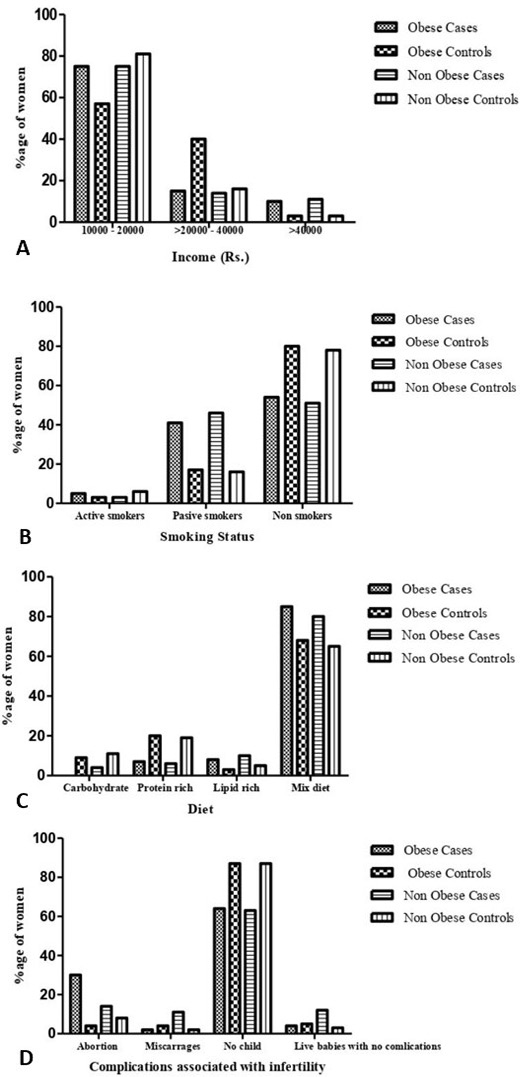
Frequency of obese and non-obese subjects in different income groups (A), regarding smoking status (B), diet (C) and regarding compilations an associated with infertility (D).
References
- Aljefri, Y.E., Alahmadi, R.A., Alajmi, R.S., Alkhamisi, T.A., Maaddawi, H.A., Alraddadi, A.A. and Alamri, A.M., 2021. Cutaneous manifestations and hormonal changes among polycystic ovary syndrome patients at a tertiary care center. Cureus, 13: e20593. https://doi.org/10.7759/cureus.20593[CrossRef]
- Amini, L., Sadeghi, M.R., Oskuie, F., Kamali, K., and Maleki, H., 2014. Lipid profile in women with polycystic ovary syndrome. Crescent J. Med. Biol. Sci., 1: 147-150.
- Annagür, B.B., Tazegül, A. and Akbaba, N., 2014. Body image, self-esteem and depressive symptomatology in women with polycystic ovary syndrome. Noro Psikiyatr. Ars., 51: 129. https://doi.org/10.4274/npa.y6778[CrossRef]
- Atijosan, A.B., 2020. Torturing the helpless: A review of PCOS induced infertility from a gender perspective. J. Gend. Power, 14: 157-168. https://doi.org/10.2478/jgp-2020-0019[CrossRef]
- Azziz, R., Marin, C., Hoq, L., Badamgarav, E. and Song, P., 2005. Health care-related economic burden of the polycystic ovary syndrome during the reproductive life span. J. Clin. Endocr. Metab., 90: 4650-4658. https://doi.org/10.1210/jc.2005-0628[CrossRef]
- Bandini, L.G., Vu, D., Must, A., Cyr, H., Goldberg, A. and Dietz, W.H., 1999. Comparison of high-calorie, low-nutrient-dense food consumption among obese and non-obese adolescents. Obes. Res., 7: 438-443. https://doi.org/10.1002/j.1550-8528.1999.tb00431.x[CrossRef]
- Begum, G.S., Shariff, A., Ayman, G., Mohammad, B., Housam, R., and Khaled, N., 2017. Assessment of risk factors for development of polycystic ovarian syndrome. Diabetes, 1: 164-167.
- Bharathi, R.V., Swetha, S., Neerajaa, J., Madhavica, J.V., Janani, D.M., Rekha, S.N. and Usha, B., 2017. An epidemiological survey: Effect of predisposing factors for PCOS in Indian urban and rural population. Middle East Fertil. Soc. J., 22: 313-316. https://doi.org/10.1016/j.mefs.2017.05.007[CrossRef]
- Bolúmar, F., Olsen, J., Rebagliato, M., Sáez-Lloret, I., Bisanti, L., and European Study Group on Infertility and Subfecundity, 2000. Body mass index and delayed conception: A European multicenter study on infertility and subfecundity. Am. J. Epidemiol., 151: 1072-1079. https://doi.org/10.1093/oxfordjournals.aje.a010150[CrossRef]
- Boyle, J.A., Cunningham, J., OˈDea, K., Dunbar, T. and Norman, R.J., 2012. Prevalence of polycystic ovary syndrome in a sample of Indigenous women in Darwin, Australia. Med. J. Aust., 196: 62-66. https://doi.org/10.5694/mja11.10553[CrossRef]
- Cooper, H.E., Spellacy, W.N., Prem, K.A. and Cohen, W.D., 1968. Hereditary factors in the Stein-Leventhal syndrome. Am. J. Obst. Gynecol., 100: 371-387. https://doi.org/10.1016/S0002-9378(15)33704-2[CrossRef]
- Craig, W.Y., Palomaki, G.E., and Haddow, J.E., 1989. Cigarette smoking and serum lipid and lipoprotein concentrations: An analysis of published data. Br. med. J., 298: 784-788. https://doi.org/10.1136/bmj.298.6676.784[CrossRef]
- Cupisti, S., Kajaia, N., Dittrich, R., Duezenli, H., Beckmann, M.W., and Mueller, A., 2008. Body mass index and ovarian function are associated with endocrine and metabolic abnormalities in women with hyperandrogenic syndrome. Eur. J. Endocrinol., 158: 711-720. https://doi.org/10.1530/EJE-07-0515[CrossRef]
- Daghestani, M.H., Daghestani, M., Daghistani, M., El-Mazny, A., Bjørklund, G., Chirumbolo, S., Al Saggaf, S.H. and Warsy, A., 2018. A study of ghrelin and leptin levels and their relationship to metabolic profiles in obese and lean Saudi women with polycystic ovary syndrome (PCOS). Lipids Hlth. Dis., 17: 1-9. https://doi.org/10.1186/s12944-018-0839-9[CrossRef]
- Daghestani, M.H., Omair, M., Daghestani, M., Abdel-Razeq, S.S., Kaya, N. and Warsy, A., 2021. Influence of b2 adrenergic receptor polymorphism (rs1042713 and rs1042714) on anthropometric, hormonal and lipid profiles in polycystic ovarian syndrome. J. Med. Biochem., 40: 74-85. https://doi.org/10.5937%2Fjomb0-26183
- Di Fede, G., Mansueto, P., Longo, R.A., Rini, G., and Carmina, E., 2009. Influence of sociocultural factors on the ovulatory status of polycystic ovary syndrome. Fertil. Steril., 91: 1853-1856. https://doi.org/10.1016/j.fertnstert.2008.02.161[CrossRef]
- Dongargaonkar, P.G., Aher, G.S. and Gavali, U.G., 2020. Non-obese and obese women with polycystic ovary syndrome-their metabolic characteristics and insulin resistance. Age, 23: 25. https://doi.org/10.36348/sijog.2020.v03i03.002[CrossRef]
- Farshchi, H., Rane, A., Love, A. and Kennedy, R.L., 2007. Diet and nutrition in polycystic ovary syndrome (PCOS): pointers for nutritional management. Obstet. Gynecol., 27: 762-773. https://doi.org/10.1080/01443610701667338[CrossRef]
- Gambineri, A., Pelusi, C., Vicennati, V., Pagotto, U. and Pasquali, R., 2002. Obesity and the polycystic ovary syndrome. Indian J. Ophthalmol., 26: 883-896. https://doi.org/10.1038/sj.ijo.0801994[CrossRef]
- Hahn, S., Tan, S., Sack, S., Kimmig, R., Quadbeck, B., Mann, K., and Janssen, O.E., 2007. Prevalence of the metabolic syndrome in German women with polycystic ovary syndrome. Exp. Clin. Endocrinol. Diabetes, 115: 130-135. https://doi.org/10.1055/s-2007-967093[CrossRef]
- Hsu, M.I., 2013. Changes in the PCOS phenotype with age. Steroids, 78: 761-766. https://doi.org/10.1016/j.steroids.2013.04.005[CrossRef]
- Ibrahim, T.A.E.S., Ali, A.E.S. and Radwan, M.E.H., 2020. Lipid profile in women with polycystic ovary syndrome. Egypt. J. Hosp. Med., 78: 272-277. https://doi.org/10.21608/ejhm.2020.70969[CrossRef]
- Ibrahim, W. and Abdelsalam, K.E.A., 2015. Levels of FSH, LH, SHBG, total testosterone, and LH/FSH ratio in Sudanese patients with polycystic ovary syndrome in relation to body mass index. Int. J. Curr. Res., 7: 11919-11922.
- Ishak, A., Kadir, A.A., Hussain, N.H.N. and Ismail, S.B., 2012. Prevalence and characteristics of metabolic syndrome among polycystic ovarian syndrome patients in Malaysia. Int. J. Collaborat. Res. Intern.Med. Publ. Hlth., 4: 0-0.
- Kahsar-Miller, M.D., Nixon, C., Boots, L.R., Go, R.C. and Azziz, R., 2001. Prevalence of polycystic ovary syndrome (PCOS) in first-degree relatives of patients with PCOS. Fertil. Steril., 75: 53-58. https://doi.org/10.1016/S0015-0282(00)01662-9[CrossRef]
- Kałużna, M., Czlapka-Matyasik, M., Kompf, P., Moczko, J., Wachowiak-Ochmańska, K., Janicki, A. and Ziemnicka, K., 2022. Lipid ratios and obesity indices are effective predictors of metabolic syndrome in women with polycystic ovary syndrome. Ther. Adv. Endocrinol. Metabol., 13: 20420188211066699. https://doi.org/10.1177/20420188211066699[CrossRef]
- Karli, P., and Ayan, D., 2018. Evaluation of hormonal profile in polycystic ovary syndrome. Int. J. Innov. Res. Med. Sci., 2: 5-8.
- Kiranmayee, D., Kavya, K., Himabindu, Y., Sriharibabu, M., Madhuri, G.L.J. and Venu, S., 2017. Correlations between anthropometry and lipid profile in women with PCOS. J. Hum. Reprod. Sci., 10: 167. https://doi.org/10.4103/jhrs.JHRS_108_16[CrossRef]
- Kousta, E., White, D.M., Cela, E., McCarthy, M.I. and Franks, S., 1999. The prevalence of polycystic ovaries in women with infertility. Hum. Reprod., 14: 2720-2723. https://doi.org/10.1093/humrep/14.11.2720[CrossRef]
- Kumawat, M., Choudhary, P. and Aggarwal, S., 2021. Association of serum leptin with anthropometric indices of obesity, blood lipids, steroidal hormones, and insulin resistance in polycystic ovarian syndrome. J. Hum. Reprod. Sci., 14: 228. https://doi.org/10.4103/jhrs.jhrs_35_21[CrossRef]
- Lamb, J.D., Johnstone, E.B., Rousseau, J.A., Jones, C.L., Pasch, L.A., Cedars, M.I. and Huddleston, H.G., 2011. Physical activity in women with polycystic ovary syndrome: Prevalence, predictors, and positive health associations. Am. J. Obst. Gynecol., 204: 352-e1. https://doi.org/10.1016/j.ajog.2010.12.006[CrossRef]
- Layegh, P., Mousavi, Z., Tehrani, D.F., Parizadeh, S.M.R. and Khajedaluee, M., 2016. Insulin resistance and endocrine-metabolic abnormalities in polycystic ovarian syndrome: Comparison between obese and non-obese PCOS patients. Int. J. Reprod. Biomed., 14: 263-270. https://doi.org/10.29252/ijrm.14.4.263[CrossRef]
- Legro, R. S., Kunselman, A. R. and Dunaif, A., 2001. Prevalence and predictors of dyslipidemia in women with polycystic ovary syndrome. Am. J. Med., 111: 607-613. https://doi.org/10.1016/S0002-9343(01)00948-2[CrossRef]
- Li, J., Wu, Q., Wu, X.K., Zhou, Z.M., Fu, P., Chen, X.H., Yan, Y., Wang, X., Yang, Z.W., Li, W.L. and Stener-Victorin, E., 2018. Effect of exposure to second-hand smoke from husbands on biochemical hyperandrogenism, metabolic syndrome and conception rates in women with polycystic ovary syndrome undergoing ovulation induction. Mol. Hum. Reprod., 33: 617-625. https://doi.org/10.1093/humrep/dey027[CrossRef]
- Liepa, G.U., Sengupta, A. and Karsies, D., 2008. Polycystic ovary syndrome (PCOS) and other androgen excess related conditions: Can changes in dietary intake make a difference? Nutr. Clin. Pract., 23: 63-71. https://doi.org/10.1177/011542650802300116[CrossRef]
- Liu, Q., Xie, Y.J., Qu, L.H., Zhang, M.X. and Mo, Z.C., 2019. Dyslipidemia involvement in the development of polycystic ovary syndrome. Taiwan J. Obstet. Gynecol., 58: 447-453. https://doi.org/10.1016/j.tjog.2019.05.003[CrossRef]
- Mahmoud, M.I., Habeeb, F. and Kasim, K., 2015. Reproductive and biochemical changes in obese and non-obese polycystic ovary syndrome women. Alex. J. Med., 51: 5-9. https://doi.org/10.1016/j.ajme.2014.03.002[CrossRef]
- March, W.A., Moore, V.M., Willson, K.J., Phillips, D.I., Norman, R.J. and Davies, M.J., 2009. The prevalence of polycystic ovary syndrome in a community sample assessed under contrasting diagnostic criteria. Mol. Hum. Reprod., 25: 544-551. https://doi.org/10.1093/humrep/dep399[CrossRef]
- Mattsson, L.Å., Cullberg, G., Hamberger, L., Samsioe, G. and Silfverstolpe, G., 1984. Lipid metabolism in women with polycystic ovary syndrome: Possible implications for an increased risk of coronary heart disease. Fertil. Steril., 42: 579-584. https://doi.org/10.1016/S0015-0282(16)48142-2[CrossRef]
- Messinis, I.E., Messini, C.I., Anifandis, G., and Dafopoulos, K., 2015. Polycystic ovaries and obesity. Best Pract. Res. Clin. Obstet. Gynaecol., 29: 479-488. https://doi.org/10.1016/j.bpobgyn.2014.11.001[CrossRef]
- Mihailidis, J., Dermesropian, R., Taxel, P., Luthra, P., and Grant-Kels, J.M., 2017. Endocrine evaluation of hirsutism. Int. J. Womens Dermatol., 3: S6-S10. https://doi.org/10.1016/j.ijwd.2017.02.007[CrossRef]
- Mobeen, H., Hamid, H., Shafiq, A., Adil, M., Kashif, M. and Yousuf, N.W., 2016. LH/FSH, BMI and clinical profile in polycystic ovarian syndrome: A correlative study. Int. J. Biochem. Res. Rev., 9: 1. https://doi.org/10.9734/IJBCRR/2016/21676[CrossRef]
- Moran, L., Gibson-Helm, M., Teede, H. and Deeks, A., 2010. Polycystic ovary syndrome: A biopsychosocial understanding in young women to improve knowledge and treatment options. J. Psychosom. Obstet. Gynaecol., 31(1), 24-31. https://doi.org/10.3109/01674820903477593[CrossRef]
- Mushtaq, A., Bibi, A. and Kausar, N., 2022. Increased risk of infertility, marital maladjustment and psychological morbidity in PCOS patients of southern Punjab, Pakistan. Pakistan J. Zool., 55: 1-8. https://doi.org/10.17582/journal.pjz/20210919130943[Full text][CrossRef]
- Nambiar, V., Vijesh, V.V., Lakshmanan, P., Sukumaran, S. and Suganthi, R., 2016. Association of adiponectin and resistin gene polymorphisms in South Indian women with polycystic ovary syndrome. Eur. J. Obstet. Gynecol. Reprod. Biol., 200: 82-88. https://doi.org/10.1016/j.ejogrb.2016.02.031[CrossRef]
- Patel S., 2018. Polycystic ovary syndrome (PCOS), an inflammatory, systemic, lifestyle endocrinopathy. J. Steroid Biochem. Mol. Biol., 182: 27-36. https://doi.org/10.1016/j.jsbmb.2018.04.008[CrossRef]
- Pau, C.T., Keefe, C.C. and Welt, C.K., 2013. Cigarette smoking, nicotine levels and increased risk for metabolic syndrome in women with polycystic ovary syndrome. Gynecol. Endocrinol., 29: 551-555. https://doi.org/10.3109/09513590.2013.788634[CrossRef]
- Ramezani, T.F., Rashidi, H., Bahri, K.M., Tohidi, M., and Azizi, F., 2014. The prevalence of metabolic disorders in various phenotypes of polycystic ovary syndrome: A community based study in Southwest of Iran. Reprod. Biol. Endocrinol., 12: 1-6. http://www.rbej.com/content/12/1/89, https://doi.org/10.1186/1477-7827-12-89[CrossRef]
- Sachdeva, G., Gainder, S., Suri, V., Sachdeva, N. and Chopra, S., 2019. Obese and non-obese polycystic ovarian syndrome: Comparison of clinical, metabolic, hormonal parameters, and their differential response to clomiphene. Indian J. Endocrinol. Metab., 23: 257. https://doi.org/10.4103/ijem.IJEM_637_18[CrossRef]
- Sayed, A., and Bakhiet, A., 2020. Anti-müllerian hormone and lipid profile in sudanese obese women diagnosed with polycystic ovary syndrome. Sudan J. Sci. Technol., 21: 100-105.
- Shi, W., Zhao, Q., Zhao, X., Xing, C. and He, B., 2021. Analysis of endocrine and metabolic indexes in non-obese patients with polycystic ovary syndrome and its compare with obese patients. Diabetes Metab. Syndr. Obes., 14: 4275. https://doi.org/10.2147/DMSO.S329108[CrossRef]
- Silfen, M.E., Denburg, M.R., Manibo, A.M., Lobo, R.A., Jaffe, R., Ferin, M., Levine, L.S. and Oberfield, S.E., 2003. Early endocrine, metabolic, and sonographic characteristics of polycystic ovary syndrome (PCOS): Comparison between nonobese and obese adolescents. J. Clin. Endocrinol. Metab., 88: 4682-4688. https://doi.org/10.1210/jc.2003-030617[CrossRef]
- Sirmans, S.M. and Pate, K.A., 2014. Epidemiology, diagnosis, and management of polycystic ovary syndrome. Clin. Epidemiol., 6: 1. https://doi.org/10.2147/CLEP.S37559[CrossRef]
- Song, Y., Ye, W., Ye, H., Xie, T., Shen, W. and Zhou, L., 2019. Serum testosterone acts as a prognostic indicator in polycystic ovary syndrome-associated kidney injury. Physiol. Rep., 7: e14219. https://doi.org/10.14814/phy2.14219[CrossRef]
- Sowers, M.F., Beebe, J.L., McConnell, D., Randolph, J. and Jannausch, M., 2001. Testosterone concentrations in women aged 25–50 years: Associations with lifestyle, body composition, and ovarian status. Am. J. Epidemiol., 153: 256-264. https://doi.org/10.1093/aje/153.3.256[CrossRef]
- Tabassum, F., Jyoti, C., Sinha, H.H., Dhar, K. and Akhtar, M.S., 2021. Impact of polycystic ovary syndrome on quality of life of women in correlation to age, basal metabolic index, education and marriage. PLoS One, 16: e0247486. https://doi.org/10.1371/journal.pone.0247486[CrossRef]
- Taher, M.A. and Mhaibes, S.H., 2015. Assessment of some trace elements in obese and non-obese polycystic ovary syndrome (PCOS). Int. J. Sci. Res., 6: 1333-1341.
- Talbott, E.O., Guzick, D.S., Sutton-Tyrrell, K., McHugh-Pemu, K.P., Zborowski, J.V., Remsberg, K.E. and Kuller, L.H., 2000. Evidence for association between polycystic ovary syndrome and premature carotid atherosclerosis in middle-aged women. Arterioscler. Thromb. Vasc. Biol., 20: 2414-2421.
- Talbott, E., Guzick, D., Clerici, A., Berga, S., Detre, K., Weimer, K. and Kuller, L., 1995. Coronary heart disease risk factors in women with polycystic ovary syndrome. Arterioscler. Thromb. Vasc. Biol., 15: 821-826. https://doi.org/10.1161/01.ATV.15.7.821[CrossRef]
- Wild, R.A., Rizzo, M., Clifton, S., and Carmina, E., 2011. Lipid levels in polycystic ovary syndrome: Systematic review and meta-analysis. Fertil. Steril., 95: 1073-1079. https://doi.org/10.1016/j.fertnstert.2010.12.027[CrossRef]
- Xiang, S.K., Hua, F., Tang, Y., Jiang, X.H., Zhuang, Q. and Qian, F.J., 2012. Relationship between serum lipoprotein ratios and insulin resistance in polycystic ovary syndrome. Int. J. Endocrinol., 2012. Article ID 173281. https://doi.org/10.1155/2012/173281[CrossRef]
- Xu, X.L., Deng, S.L., Lian, Z.X. and Yu, K., 2021. Estrogen receptors in polycystic ovary syndrome. Cells, 10: 459. https://doi.org/10.3390/cells10020459[CrossRef]
- Yilmaz, M., Biri, A., Bukan, N., Karakoç, A., Sancak, B., Törüner, F. and Paşaoğlu, H., 2005. Levels of lipoprotein and homocysteine in non-obese and obese patients with polycystic ovary syndrome. Gynaecol. Endocrinol., 20: 258-263. https://doi.org/10.1080/09513590400027265[CrossRef]
Services
Article usage statistics
Loading…