
This study was conducted to find the antiviral potential of ivermectin against Peste des Petits Ruminants Virus (PPRV) in vitro. Firstly, cell cytotoxicity of ivermectin in the Vero cells was determined using MTT assay. The antiviral activity of ivermectin was determined using viral inhibition assays and median tissue culture infectious dose (TCID50). The results showed that 2.5 µM concentration of the ivermectin is non-toxic to the Vero cells. At this concentration, PPRV titre was significantly reduced (p < 0.001) by two log to 103.0 TCID50/0.1 mL as compared to virus control 105.5 TCID50/0.1 mL. Moreover, the ivermectin exposure after the viral attachment and entry steps able to reduce virus titre more as compare to viral attachment and entry steps that predicts the possible mechanism of the drug. Therefore, our study first time demonstrated the antiviral potential of ivermectin against PPRV in vitro and its future potential for its use as anti-PPRV therapeutics.
Short Communication · Volume 53, Issue 4, 8 June 2021 · Open Access
Antiviral Potential of Ivermectin against Peste des Petits Ruminants Virus (PPRV)
· · · · ·
Affiliations & Notes ⌄
- 1Institute of Microbiology, University of Veterinary and Animal Sciences, Lahore 2Institute of Biochemistry and Biotechnology, University of Veterinary and Animal Sciences, Lahore , * Corresponding author: sohail.raza@uvas.edu.pk
Article Info ⌄
- Journal:
- Pakistan Journal of Zoology
- Published:
- 8 June 2021

From the article
- The results of TCID50 shows that the titre of virus was reduced significantly in the drug-treated cells and ivermectin have potential to show antiviral activity against PPR replication.
- To determine the effect of the drug on PPRV binding and entry into the cells, mean virus titre determined by TCID50 of the samples of mock-treated and drug-treated cells was compared which was 105.4 TCID50/ 0.1 mL and 104.3 TCID50/0.1 mL, respectively (Fig. 1D).
- This indicates that ivermectin showed little effect on the entry of PPRV into the cells.
- To determine the effect of the drug on post-entry stages of PPRV replication, the ivermectin was added after virus attachment and entry stage.
Selected from the article's own text.
Abstract
Materials and methods
Vero cells (ATCC® CCL-81) and PPRV strain Nigeria 75/1 (KY628761.1) were obtained from Quality Operations Laboratory, the University of Veterinary and Animal Sciences. The propagation and titre determination of virus was performed on Vero cells. Cells were grown in Dulbecco’s Modified Eagle Medium (Caisson Laboratories, USA) with 10% fetal bovine serum (Capricorn Scientific) at 37°C in 5% CO2.
The virus titre was calculated by median tissue
culture infectious dose (TCID50). Briefly, 100 μL of virus suspension which was serially 10 fold diluted was added to each well of 96 well microtiter plate having confluent Vero cells. Virus control was also run alongside. Results of TCID50 were read when the virus control wells showed cytopathic effect (CPE’s). All the experiments were performed in triplicate.
For cytotoxicity assay of Ivermectin, Vero cells were added into the 96 well plates until the cells became 100% confluent. The cells were exposed with 1.25 μM, 2.5 μM, 5 μM and 10 μM of ivermectin for 24 h. The cytotoxicity assay was performed by MTT assay kit (Abcam, Cambridge, UK) using the manufacturer’s instructions. Ivermectin (Abcam, Cambridge, UK) stock solution was prepared in Dimethyl Sulfoxide (DMSO) at 50 mM and stored in aliquots at -20°C for up to 1 month.
For viral inhibition assay, Vero cells were infected with 105.9 TCID50 PPRV in the presence of 2.5 μM concentration of ivermectin and incubated at 37°C for 2 h, after which the unabsorbed viruses were washed with PBS. Fresh maintenance medium was added along with 2.5 μM concentration of ivermectin and kept in the wells for 7 days. As a control, cells were infected with 105.9 TCID50 PPRV in the absence of the drug. After 7 days samples were collected for virus titration.
For binding and entry assay, confluent Vero cells were infected with 105.9 TCID50 of PPRV in the presence of 2.5 μM concentration of ivermectin and incubated at 37°C for 2 h. The control well was infected with 105.9 TCID50 of the virus in the absence of the drug. After incubation, the drug was removed and washed with PBS three times to remove the unabsorbed virus. Fresh media without drug was added to allow the growth of virus for 7 days.
For determining the effect of Ivermectin on replication of PPRV Vero cells were infected with 105.9 TCID50 PPRV for 2 h to allow the virus entry into cells. After 2 h, washing with PBS was done to remove the unabsorbed viruses. The cells were then treated with 2.5μM Ivermectin for 7 days. The control well was infected with the virus, without adding drug.
All the experiments were performed three times individually and data presented as means ± standards deviation (SD). The results were analysed by Graph pad prism software (version 6.0), Student t-test was applied to the results to compare means of the TCID50 value of the test group with the control. Statistical significance represented by asterisks is marked correspondingly in Figure 1 (*p < 0.05, **p < 0.01, ***p < 0.001)
Results and discussion
The cell cytotoxicity assay procedure indicated that the cells were 100% viable at a drug concentration of 1.25 μM and 2.5 μM while the cells were less than 50% viable at 5 μM concentration (Fig. 1A). This shows that ivermectin was safe to the Vero cells when used at 2.5μM concentration, as there was no change in the morphology of cells as compared with the mock-treated cells. The non-toxic concentrations of ivermectin as reported on Hela cells, BHK-21 cells MDBK cells were found to be 50 μM, 3 μM, 25 μM, respectively. This change in the values of non-toxic concentration could be due to use of different cells and the environment in which the experiments were carried out.
Effect of ivermectin on PPRV replication was determined by comparing the mean viral titres of virus control and drug-treated cells for the whole length of infection which was 105.5 TCID50/0.1 mL and 103.0 TCID50/0.1 mL, respectively (Fig. 1B, C). The results of TCID50 shows that the titre of virus was reduced significantly in the drug-treated cells and ivermectin have potential to show antiviral activity against PPR replication. Moreover, ivermectin able to inhibit virus titre by 4 logs in both PRV and Bovine Herpesvirus-1 (BoHV-1) while there was 49.63 % reduction in dengue virus production after ivermectin treatment . These results suggest a broad antiviral activity of ivermectin against both RNA and DNA viruses.
To determine the effect of the drug on PPRV binding and entry into the cells, mean virus titre determined by TCID50 of the samples of mock-treated and drug-treated cells was compared which was 105.4 TCID50/ 0.1 mL and 104.3 TCID50/0.1 mL, respectively (Fig. 1D). This indicates that ivermectin showed little effect on the entry of PPRV into the cells. Similarly, no effect of ivermectin was detected on BoHV-1 and PRV binding/attachment and entry steps . Moreover, ivermectin does not involve in the inhibition of flaviviruses at early steps of virus replication . Our data and the previous data largely predicts that ivermectin does not affect the early stages of viral replication
To determine the effect of the drug on post-entry stages of PPRV replication, the ivermectin was added after virus attachment and entry stage. The mean viral titre of the virus control and the cells treated with drug was compared which was 105.3 TCID50/0.1 mL and 102.9 TCID50/0.1 mL, respectively (Fig. 1E). This shows that ivermectin largely inhibited the replication steps of PPRV. Similarly, ivermectin inhibits post-entry stages of virus replication in BoHV-1 , PRV and Flaviviruses by inhibiting importin alpha/beta mediated transport of the viral proteins. These results predict that ivermectin may affect the PPRV replication at the post-entry stages of the virus replication cycle that involves the cellular importins. This predicts the function of ivermectin during PPRV replication.
Statement of conflict of interest
The authors declare no conflict of interest.
Figures
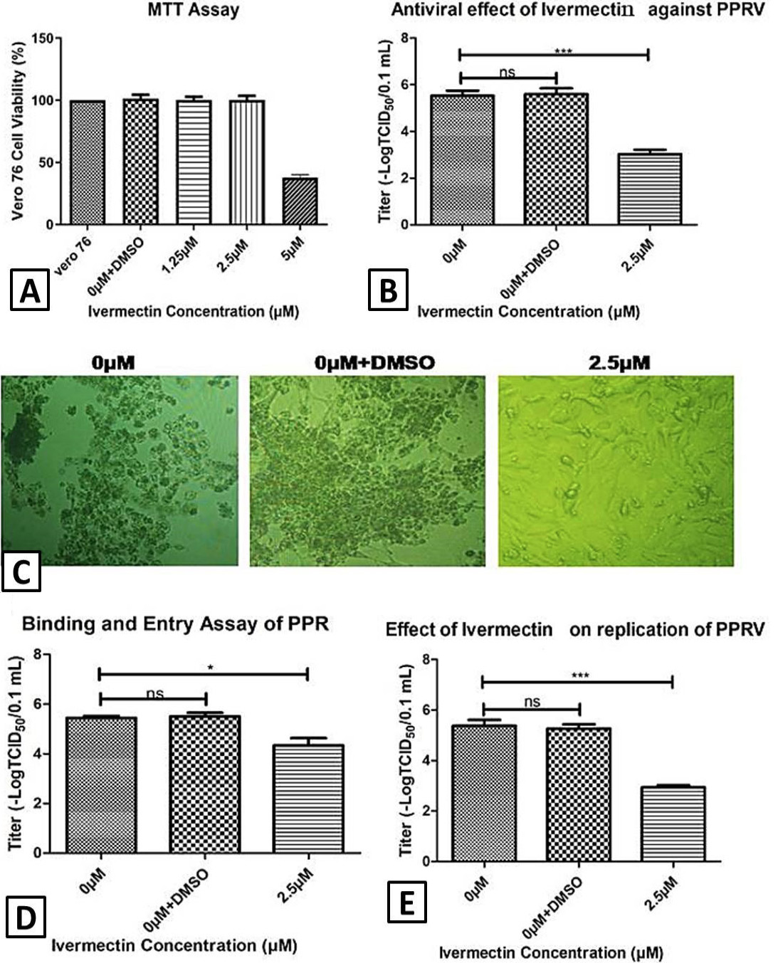
Antiviral activity of Ivermectin against PPRV. A, confluent Vero cells were exposed with different concentrations of ivermectin and cell cytotoxcic concentrations were determined 48 hrs post drug exposure using MTT assay. B, antiviral activity of ivermectin was determined using TCID50 after drug exposure during the course of infection. C, cellular morphology at 400x magnification under microscope in control cells and drug treated cells after infection. D, effect of ivermectin on viral attachment and penetration. E, effect of ivermectin on post entry stages of viral replication (0µM, virus control without drug; 0µM+DMSO, DMSO control; 2.5µM, ivermectin 2.5µM).
References
- Abubakar, M., Khan, H.A., Arshed, M.J., Hussain, M. and Ali, Q., 2011. Small Rumin. Res., 96: 1-10. https://doi.org/10.1016/j.smallrumres.2010.10.006[CrossRef]
- Anees, M., Shabbir, M.Z., Muhammad, K., Nazir, J., Shabbir, M.A., Wensman, J.J. and Munir, M., 2013. BMC Vet. Res., 9: 60. https://doi.org/10.1186/1746-6148-9-60[CrossRef]
- Caly, L., Druce, J.D., Catton, M.G., Jans, D.A. and Wagstaff, K.M., 2020. Antiviral Res., 178: 104787. https://doi.org/10.1016/j.antiviral.2020.104787[CrossRef]
- Irshad, H., Ullah, A., Ahsan, A., Zahur, A.B., Basra, M.J., Azhar, M.S., Afzal, M. and Jahangir, M., 2019. Pak. Vet. J., 39: 606-608.
- Khan, H.A., Siddique, M., Abubakar, M., Arshad, M.J. and Hussain, M., 2008. Small Rumin. Res., 79: 152-157. https://doi.org/10.1016/j.smallrumres.2008.07.021[CrossRef]
- Libeau, G., Diallo, A. and Parida, S., 2014. Anim. Front., 4: 14-20. https://doi.org/10.2527/af.2014-0003[CrossRef]
- Lv, C., Liu, W., Wang, B., Dang, R., Qiu, L., Ren, J., Yan, C., Yang, Z. and Wang, X., 2018. Antiviral Res., 159: 55-62. https://doi.org/10.1016/j.antiviral.2018.09.010[CrossRef]
- Mastrangelo, E., Pezzullo, M., De Burghgraeve, T., Kaptein, S., Pastorino, B., Dallmeier, K., de Lamballerie, X., Neyts, J., Hanson, A.M., Frick, D.N., Bolognesi, M. and Milani, M., 2012. J. Antimicrob. Chemother., 67: 1884-1894. https://doi.org/10.1093/jac/dks147[CrossRef]
- OIE, 2015. Global strategy for the control and eradication of PPR. World Organization for Animal Health (OIE).
- Raza, S., Shahin, F., Zhai, W., Li, H., Alvisi, G., Yang, K., Chen, X., Chen, Y., Chen, J. and Hu, C., 2020. Microorganisms, 8: 409. https://doi.org/10.3390/microorganisms8030409[CrossRef]
Services
Article usage statistics
Loading…