
Stress granules (SGs), a type of RNA foci, were formed in the cytoplasm of eukaryotic cells upon some unfavorable environmental stress. Previously, we found that Tudor domain containing 1 (SND1)-containing SGs actively communicate with the nuclear and cytosolic pool of HeLa cells. Here, we are interested to investigate the dynamic distribution of three nuclear proteins, including heterogeneous nuclear ribonucleoprotein A1 (hnRNP A1), Hu Antigen R (HuR) and T cell intracellular antigen 1 (TIA1), in the SG aggregation and nucleus/cytoplasm localization under stress condition. We found that hnRNP A1, HuR and TIA1-containing SGs were aggregated in the cytoplasm of HeLa cells, and accompanies the alteration of nucleus/cytoplasm localization during arsenite induced-oxidative stress. Increased hnRNP A1 fluorescence signal within cytoplasm was detected from 3% of normal cells to the 28% of stressed cells; in contrast, 87% of cells with strong hnRNP A1 signal within nucleus reduced to 50% during stress. In addition, transport receptor importin-β pathway seems to be involved in the nuclear import of hnRNP A1, rather than HuR and TIA1. However, the slightly enhanced cytoplasmic accumulation of hnRNP A1 can not influence the formation of hnRNP A1 granules during oxidative stress. Timely and effective dynamic distribution of specific stress-associated proteins in the section of nucleus, cytoplasm, and SG structure is more likely to contribute to the minimization of the detrimental condition-induced cellular damage.
Research Article · Volume 51, Issue 5, 12 June 2019 · Open Access
Analysis of the Correlation between Stress Granule Assembly and Nucleus/Cytoplasm Localization of hnRNP A1, HuR and TIA1 During Arsenite-Induced Oxidative Stress
· · · · · · ·
Affiliations & Notes ⌄
- 1School of Nursing, Tianjin Medical University, Tianjin, 300070, China 2Department of Biochemistry and Molecular Biology, Department of Immunology, School of Basic Medical Sciences, Tianjin Medical University, Tianjin, 300070, China 3Laboratory of Molecular Immunology, Research Center of Basic Medical Science, Tianjin Medical University, Tianjin, 300070, China 4Tianjin Key Laboratory of Cellular and Molecular Immunology, Tianjin Medical University, Tianjin, 300070, China 5Key Laboratory of Educat
Article Info ⌄
- Journal:
- Pakistan Journal of Zoology
- Published:
- 12 June 2019

From the article
- Cytoplasmic SG formation was linked to nucleus/cytoplasm localization of hnRNP A1, HuR and TIA1 during arsenite induced-oxidative stress.
- Here, we found that the arsenite-induced oxidative stress induces the increased cytoplasmic signal, but the decreased nuclear signal of hnRNP A1.
- Taken together, our study focuses on the association between stress granule formation and nucleus/cytoplasm location of hnRNP A1, HuR and TIA1.
- In response to unfavorable environmental stress, timely and effective dynamic distribution of specific stress-associated proteins in the section of nucleus, cytoplasm, and SGs is more likely to contribute to the minimization of the detrimental condition-induced cellular damage.
Selected from the article's own text.
Abstract
INTRODUCTION
Eukaryotes tend to inhibit the global translation and modulate the synthesis, refolding, modification, turnover, kinetics of protein, in order to maintain the homeostasis of intracellular compartments and improve cell survival ; . The assembly of stress granules (SGs), a type of RNA- containing particle structure without membranous border in the cytoplasm, was considered as one conserved cell protective approach, targeting the transient harmful environmental assaults, such as oxidative stress, heat shock and virus infection ; ; . SGs harbor translationally arrested translation preinitiation complexes (PICs), and contain higher concentration of RNA transcripts and protein components, including eukaryotic translation initiation factor 2α (eIF2α), GAP SH3 domain-binding protein 1 (G3BP1), staphylococcal nuclease and Tudor domain containing 1 (SND1), heterogeneous nuclear ribonucleoprotein A1 (hnRNP A1), Hu Antigen R (HuR) and T cell intracellular antigen 1 (TIA1) ; ; .
Several factors, including cell characteristics, stress type, time and intensity of stimulus, and post-translational modification of protein components, influence the accumulated size/number, and duration of SG structure ; ; . Pathogenesis and therapeutic strategies of cancer and some other clinical diseases, such as ischemia, amyotrophic lateral sclerosis (ALS) and Alzheimer’s disease (AD), were also linked to aberrant SG biology ; ; ; ; .
The assembly and disassembly of SGs in mammalian cells were reported to be related to several biological processes or cellular activities, such as autophagy, apoptosis, mitochondrial stress and antiviral immune response ; ; ; . It is meaningful to investigate the dynamic distribution of stress-associated protein in the SG aggregation and nucleus/cytoplasm localization during stress. Previously, we found that SND1-containing SGs actively communicate with the nuclear and cytosolic pool of HeLa cells . Here, in order to further investigate the correlation between highly dynamic SG formation and nucleo-cytoplasmic transport in HeLa cells exposed to arsenite-induced oxidative stress, we focused on three SG-associated protein components, namely hnRNP A1, HuR and TIA1, which are primarily nuclear.
Materials and Methods
Cell culture and drug treatment
Dulbecco’s minimal essential medium (DMEM, Invitrogen Life Technologies) with 10% fetal bovine serum (FBS) was used for the culture of HeLa cells.
Oxidative stress was induced to the treatment of 0.5 mM sodium arsenite. In addition, 10 μM Importazole (IPZ, Millipore/Merck, Germany) was utilized for the alteration of nucleus/cytoplasm location of targeting proteins
Plasmids and cell transfection
Two expression plasmids of RFP-hnRNPA1 and RFP-HuR were kindly provided by Prof. John Goodier (University of Pennsylvania School of Medicine, USA). Plasmid encoding GFP-TIA1 were kindly provided by Prof. Tom C. Hobman (McGill University, Montreal, Canada). Lipofectamine 2000 (Invitrogen, Barcelona, Spain) was used to transfect the above plasmids into HeLa cells, according to the manufacturer’s instructions.
Imaging collection and nucleus/cytoplasm location
4’,6-diamidino-2-phenylindole (DAPI, Sigma-Aldrich, USA) was used for staining the nucleus region of HeLa cells. The image data was collected via an inverted research microscope Leica. The ratio of cells with the nucleus-localized signal, cytoplasm-localized signal, or full cell-localized signal of hnRNP A1, HuR, and TIA1, per all cells was calculated, respectivly.
Granule quantification and statistical analysis
A total of 50~100 HeLa cells were randomly scored in each experiment. Calculation of the percentage of targeting granule positive cells was performed. SG number in selected cells was analyzed, and cell ratio of four different granule number ranges, namely 1~10, 11~20, 21~30 and >30 per cell, were calculated, respetively. Independent-Sample Student’s T Test was performed via SPSS 16.0 software. P value less than 0.05 means the existence of statistical significant difference.
Results
Cytoplasmic SG formation was linked to nucleus/cytoplasm localization of hnRNP A1, HuR and TIA1 during arsenite induced-oxidative stress
In order to study the relationship between the nucleus/cytoplasm localization and cytoplasmic SG formation, three nucleocytoplasmic shuttling protein components, including hnRNP A1, HuR and TIA1, were targeted. HeLa cells were cultured and transiently transfected with plasmids of RFP-hnRNP A1, RFP-HuR, GFP-TIA1, rspectively. The RFP and GFP vectors were included as control. Treatment of 0.5 mM sodium arsenite for 1 hour was used to induce the oxidative stress of HeLa cell. As shown in Figure 1A, RFP-tagged hnRNP A1 protein was mainly located in the nucleus section of cells under normal condition. Upon the oxidative stress, we observed the formation of hnRNP A1 potitive (hnRNP A1+) SGs in the majority of HeLa cells (Fig. 1A, Fig. 2A, P<0.05) and the alteration of nucleus/cytoplasm localization signal (Fig. 2B). Increased hnRNP A1 fluorescence signal within cytoplasm was detected from 3% of normal cells to the 28% of stressed cells; in contrast, 87% of cells with strong hnRNP A1 signal within nucleus reduced to 50% during stress (Fig. 2B).
Furthermore, we also found that HuR-containing SGs were aggregated in the cytoplasm of HeLa cells exposed to oxidative stress (Fig. 1B; Fig. 2C, P<0.05), as expected. Similarly, 11% with strong cytoplasmic HuR signal under normal condition increased to 18% under stress condition, whereas 56% with strong nuclear HuR signal reduced to 49% upon the oxidative stress (Fig. 2D).
The same trend was also observed for TIA1 protein (Fig. 1D; Fig. 2E, P<0.05; Fig. 2F). However, this phenomenon was not detectable for the vector control of GFP (Fig. 1C) and RFP (Fig. 1E). These suggested that the cytoplasmic aggragation of hnRNP A1, HuR and TIA1-containing SGs was linked to the nucleus/cytoplasm localization during arsenite induced-oxidative stress.
The effect of IPZ on the nucleus/cytoplasm localization, SG formation of hnRNP A1, HuR and TIA1 under stress condition
Next, we aim at analyzing whether altered nucleus/cytoplasm localization can influence the SG formation during stress. HeLa cells were cultured in the presence (+) or absence (-) of 10 μM Importazole (IPZ), a small molecule inhibitor of the transport receptor importin-β . As shown in Figure 3A and Figure 4A, only a slight trend for the alteration of nucleus/cytoplasm localization was observed for hnRNP A1, after the treatment of IPZ. 50% with strong nuclear hnRNPA1 signal under normal condition reduced to 41%; whereas 28% with strong cytoplasmic hnRNPA1 signal increased to 34% (Fig. 4A). Moreover, there is no difference for SG size (Fig. 3A) and the portion of cells with hnRNP A1 positive (hnRNP A1+) SGs (Fig. 4B) in HeLa cells treated with IPZ or not. We also failed to observe the statistical significant difference for the cells with different granule numbers, including 1~10, 11~20, 21~30, >31, between the IPZ (-) and IPZ (+) group (Fig. 4C). These suggested that IPZ-mediated slight alteration of nucleus/cytoplasm
localization does not significantly affect the formation of hnRNP A1-containing SGs in HeLa cells during the arsenite-induced oxidative stress.
With regards to HuR protein, we are surprised to observe an increased nuclear aggregation in stressed HeLa cells (Fig. 3B and Fig. 4D). Even though no difference for the HuR positive (HuR+) SG formation (Fig. 4E) and HuR+ SG size (Fig. 4B), decreased portions of HeLa cells with the granule number of 11~20, 21~30, was observed in the group of IPZ (+), compared with IPZ (-) group (Fig. 4F, P<0.05). In addition, there are similar change for the nucleus/cytoplasm localization (Fig. 4G), the portions of HeLa cells with TIA1 positive (TIA1+) SGs (Fig. 4H), TIA1+ SG size (Fig. 3D) and different granule numbers (Fig. 4I). No change was observed in the control group of RFP (Fig. 4C) and GFP (Fig. 4E). Overall, these data suggested that transport receptor importin-β pathway seems to be involved in the nuclear import of hnRNP A1, rather than HuR and TIA1. The slightly enhanced cytoplasmic accumulation of hnRNP A1 can not influence the formation of hnRNP A1-containing SGs during oxidative stress.
Discussion
Nucleocytoplasmic shuttling system, including nuclear localization signal (NLS), nuclear export signal (NES) and importin-α/ importin-β pathway, is essential for the nuclear transport of many proteins among eukaryotes (Cardarelli, 2017). The environmental stress was linked to the dynamic flux of proteins between the nuclear and cytoplasmic compartment. For instance, some stress conditions, such as starvation and heat shock, inhibit the importin α/ importin-β-mediated nuclear import of the small GTPase Gsp1p . In addition, nuclear transport factors, such as Importin-α1, Importin-α4 and Importin-α5, were reported to be recruited to SG structure . Meanwhile, a number of nuclear protein components are involved in the formation of cytoplasmic SGs ; . In the present study, we quantitatively analyzed the correlation between SG assembly and nucleus/cytoplasm localization of nuclear hnRNP A1, HuR and TIA1 proteins during arsenite-induced oxidative stress.
hnRNP A1, a rapid nucleo-cytoplasmic shuttling protein and alternative splicing factor, binds nascent RNA polymerase II manuscripts in cytoplasm , and functions in the pre-mRNA processing, RNA metabolism, cellular apoptosis, stress adaption, post-transcription modulation, microRNAs processing and telomere maintenance . hnRNP A1 was found to be localized into SG structure during stress, and its recruitment into SGs depends on the existance of methylarginine residues within the arginine-glycine-glycine (RGG)-motif region ; . In addition, RanGTP-binding nuclear transport receptor transportin1 was invloved in the nuclear reimport of hnRNP A1 . Guil et al. (2006) reported that Mnk1/2-mediated hnRNP A1 phosphorylation during stress fails to bind sufficiently transportin 1, which is helpful for the cytoplasmic aggregation of hnRNP A1 into SGs. Here, we found that the arsenite-induced oxidative stress induces the increased cytoplasmic signal, but the decreased nuclear signal of hnRNP A1. The inhibition of nuclear import or enhanced nuclear export may contribute to the cytoplasmic accumulation of hnRNP A1, when HeLa cells undergo the stress of sodium arsenite. Similarly, the treatment of osmotic shock can induce the cytoplasmic accumulation of hnRNP A1 in NIH 3T3 cells through the serine-specific phosphorylation of a C-terminal F-peptide, and the activation of the mitogen-activated protein kinase kinase 3/6-p38 signaling pathway . Previous FRAP results showed that hnRNP A1 protein moves continuously in and out of SG structure . The high kinetic behaviour allows the cells to respond quickly to the adverse stress, by way of dynamically influencing and equilibrating the subcellular distribution of hnRNP A1.
We also utilized a small molecule inhibitor of the transport receptor importin β (IPZ) to analyze the effect of altered nucleus/cytoplasm localization on the assembly of hnRNP A1 granules. We observed the slightly increased cytoplasmic hnRNP A1 signal after the treatment of10 μM IPZ, under the normal (data not shown) and stress conditions. However, we failed to observe the change of hnRNP A1 granule formation. It is pissible that the increased amount of cytoplasmic hnRNP A1 protein was not up to the level of affecting the SG assembly. Previously, we found that the inhibition of nuclear export of SND1 protein could influence the efficient aggreation of SND1 granule in the cytoplasm of HeLa cells . The effect of stress-responsive nucleus/cytoplasm localization on the SG formation may be protein-specific.
TIA-1 recognizes adenine/uridine rich elements (AREs) within 3’-untranslated regions (3’-UTR) of RNA, and the prion-like aggregation of TIA1 was required for the formation of mammalian SGs under stress conditions ; ; . TIA1 exhibited the similiar cellular kinetics with hnRNP A1 . Here, we observed the oxidative stress-induced slightly decreased nuclear accumulation of TIA1, however, IPZ treatment fails to influence the nucleus/cytoplasm localization of TIA1. HuR, a classical member of embryonic lethal abnormal vision (ELAV) family, recognizes AU-rich elements (AREs) sequences of targeting mRNAs, and is involved in the regulation of RNA metabolism . HuR shuttles between nucleus and cytoplasm, and heat shock inhibits the interaction between HuR and cytoplasmic mRNA . HuR locolized into SGs as well , and RanGTP-binding nuclear transport receptor Transportin 2 was shown as nuclear import factor of HuR . Even though a slightly increased cytoplasmic signal of HuR was oberved under the oxidative stress, IPZ treatment did not alter the nuclear import of HuR but decrease the portions of HeLa cells with the granule number of 11~20, 21~30, which involved some unknown reasons.
Conclusion
Taken together, our study focuses on the association between stress granule formation and nucleus/cytoplasm location of hnRNP A1, HuR and TIA1. Cytoplasmic SG aggregation behavior is linked to the alteration of nucleus/cytoplasm localization of hnRNP A1. In response to unfavorable environmental stress, timely and effective dynamic distribution of specific stress-associated proteins in the section of nucleus, cytoplasm, and SGs is more likely to contribute to the minimization of the detrimental condition-induced cellular damage.
Acknowledgement
This work was supported by grants from the National Science Foundation for Distinguished Young Scholars of China (31125012), National Natural Science Foundation of China (31571380, 31701182, and 31670759), Innovation Team Development Plan of the Ministry of Education (IRT13085), Tianjin Enterprise Science and Technology Commissioner Project (18JCTPJC59400).
Conflicts of interest
The authors declare they have no conflicts of interest.
Figures
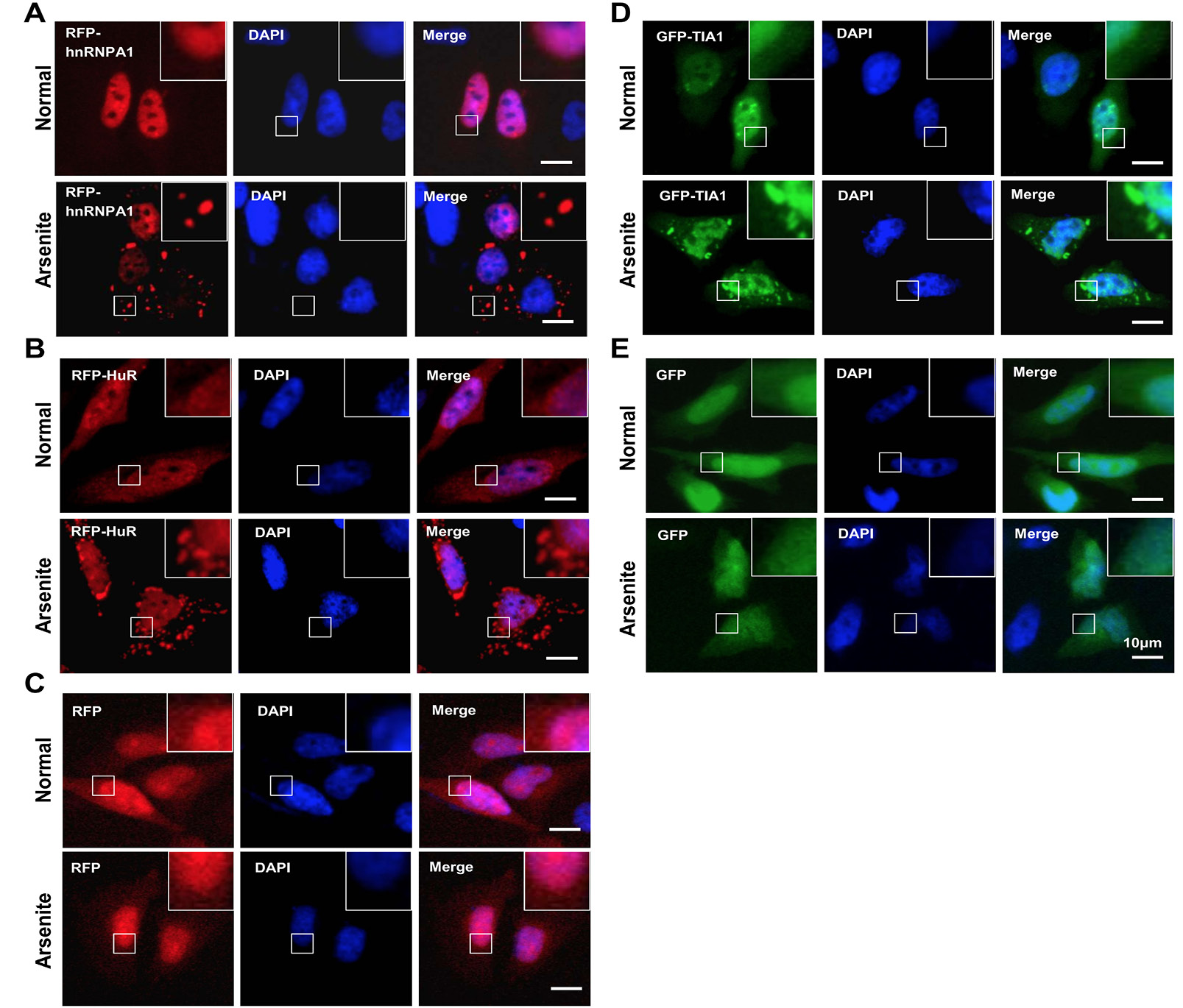
Image data for SG formation and nucleus/cytoplasm localization of hnRNP A1, HuR and TIA1 upon oxidative stress. HeLa cells were cultured and transiently transfected with plasmids of RFP-hnRNP A1 (A), RFP-HuR (B), RFP vector(C), GFP-TIA1 (D), and GFP vector (E), respectively, as indicated. After 24 hours, cells were untreated (Normal) or treated with 0.5 mM sodium arsenite for 1 hour (Arsenite). The nuclenus section was stained by DAPI. An inverted research microscope Leica was then used to collect the image data. Bar=10 μm.
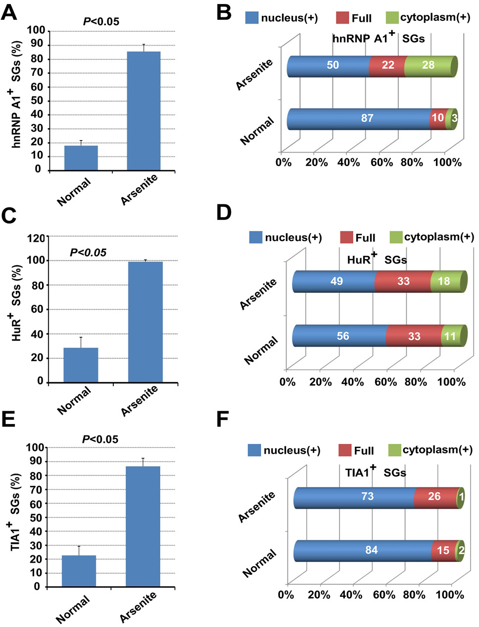
Quantification analysis for nucleus/cytoplasm localization of hnRNP A1, HuR and TIA1 upon oxidative stress. The percentage of hnRNP A1 (A), HuR (C), TIA1 (E) marked SG positive cells were measured, respectively. An independent-sample Student’s T Test was performed using SPSS 16.0. P<0.05 means significant difference. The ratio of cells with the nucleus-localized signal, cytoplasm-localized signal, or full cell-localized signal of hnRNP A1 (B), HuR (D), and TIA1 (F) in all selected cells was also calculated, respectivly.
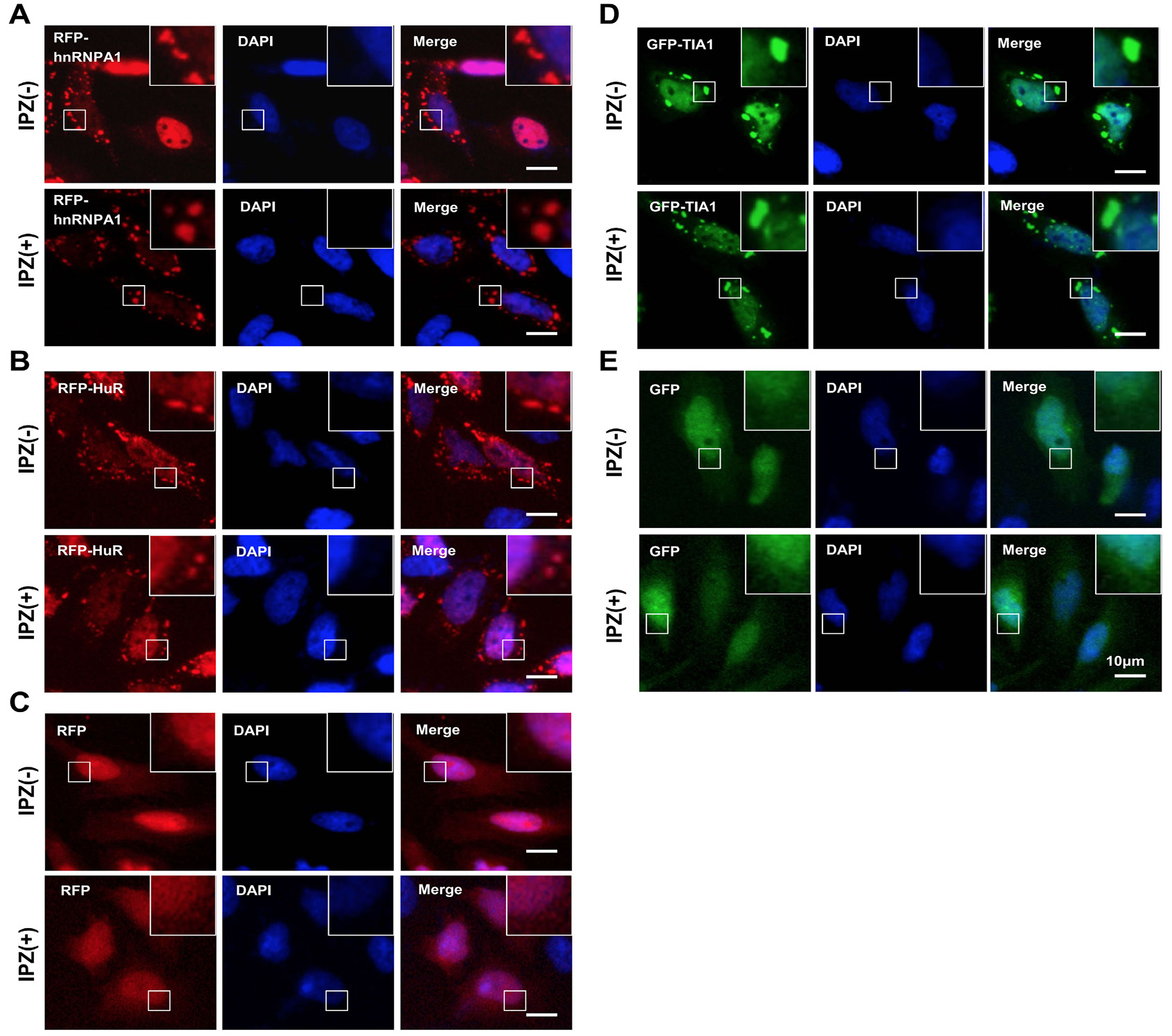
Image data for effect of IPZ on the nucleus/cytoplasm localization, SG formation of hnRNP A1, HuR and TIA1 during arsenite-induced oxidative stress. HeLa cells were cultured and transiently transfected with plasmids of RFP-hnRNP A1 (A), RFP-HuR (B), RFP (C), GFP-TIA1 (D), and GFP (E), respectively, as indicated. After the pretreated with 10 μM IPZ for 6 hours [IPZ (+)] or not [IPZ(-)], cells were treated with 0.5 mM sodium arsenite for 1 hour. The nuclenus section was stained by DAPI. An Inverted research microscope Leica was then used to collect the image data. Bar=10 μm.
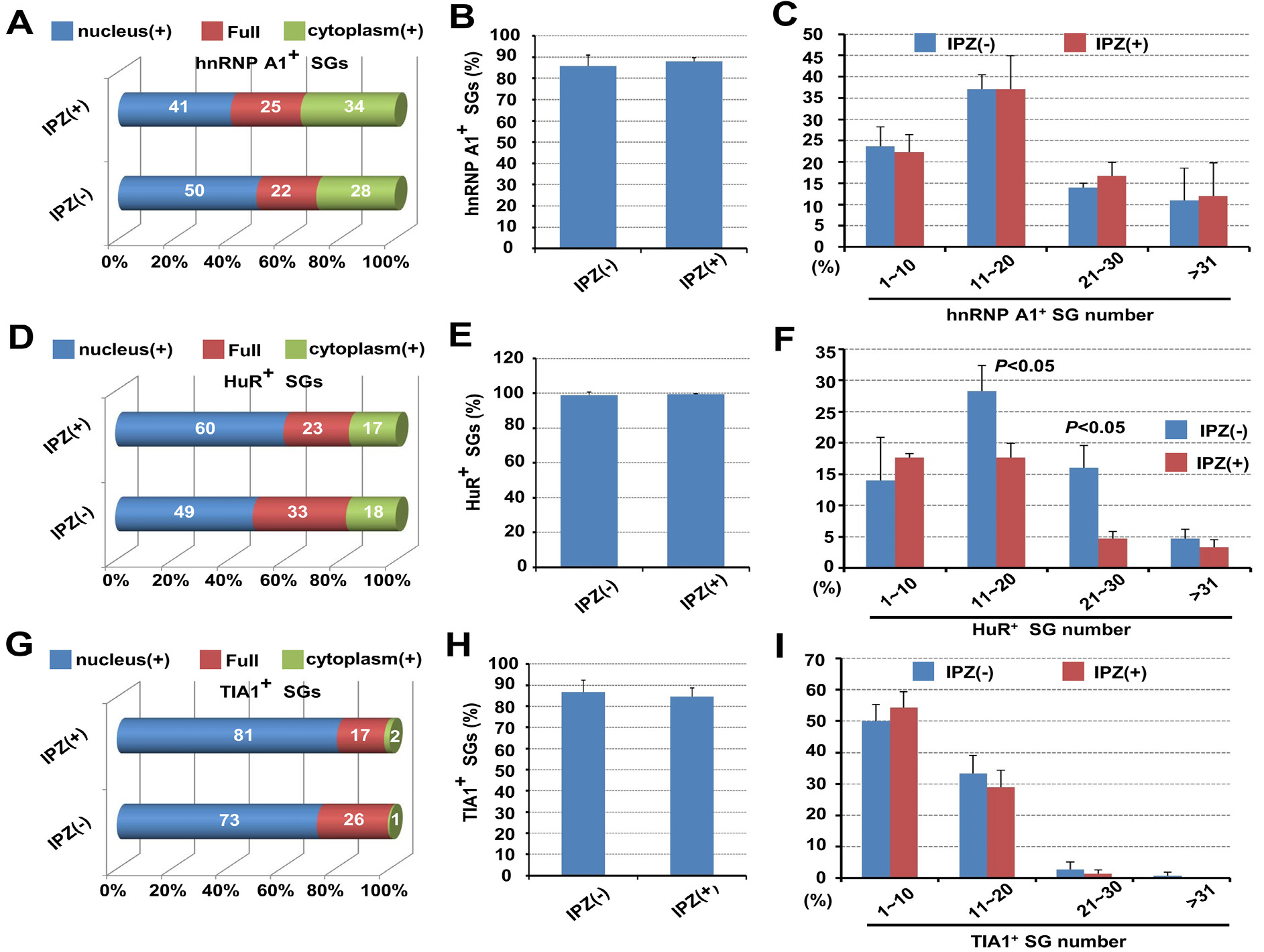
Quantification analysis for the effect of IPZ on the nucleus/cytoplasm localization, SG formation of hnRNP A1, HuR and TIA1 during arsenite-induced oxidative stress. The ratio of cells with the nucleus-localized signal, cytoplasm-localized signal, or full cell-localized signal of hnRNP A1 (A), HuR (D), and TIA1 (G) in all selected cells was calculated, respectivly. The percentage of hnRNP A1 (B), HuR (E), TIA1 (H) marked SG positive cells, and numbers of hnRNP A1 (C), HuR (F), TIA1 (I) marked granules per cell were also measured. Based on SG number per cell, there are four categories, namely 1~10, 11~20, 21~30 and >30. An independent-sample Student’s T Test was performed using SPSS 16.0. P<0.05 means significant difference.
References
- Alberti, S., Mateju, D., Mediani L. and Carra, S., 2017. Granulostasis: Protein quality control of RNP granules. Front. mol. Neurosci., 10: 84. https://doi.org/10.3389/fnmol.2017.00084[CrossRef]
- Allemand, E., Guil, S., Myers, M., Moscat, J., Caceres J.F. and Krainer, A.R., 2005. Regulation of heterogenous nuclear ribonucleoprotein A1 transport by phosphorylation in cells stressed by osmotic shock. Proc. natl. Acad. Sci. USA, 102: 3605-3610. https://doi.org/10.1073/pnas.0409889102[CrossRef]
- Anderson, P., Kedersha N. and Ivanov, P., 2015. Stress granules, P-bodies and cancer. Biochim. biophys. Acta, 1849: 861-870. https://doi.org/10.1016/j.bbagrm.2014.11.009[CrossRef]
- Buchan, J.R., 2014. mRNP granules. Assembly, function, and connections with disease. RNA Biol., 11: 1019-1030. https://doi.org/10.4161/15476286.2014.972208[CrossRef]
- Cardarelli, F., 2017. Time-resolved biophysical approaches to nucleocytoplasmic transport. Comput. Struct. Biotechnol. J., 15: 299-306. https://doi.org/10.1016/j.csbj.2017.03.005[CrossRef]
- Fu, X., Gao, X., Ge, L., Cui, X., Su, C., Yang, W., Sun, X., Zhang, W., Yao, Z., Yang X. and Yang, J., 2016. Malonate induces the assembly of cytoplasmic stress granules. FEBS Lett., 590: 22-33. https://doi.org/10.1002/1873-3468.12049[CrossRef]
- Gallouzi, I.E., Brennan, C.M., Stenberg, M.G., Swanson, M.S., Eversole, A., Maizels, N. and Steitz, J.A., 2000. HuR binding to cytoplasmic mRNA is perturbed by heat shock. Proc. natl. Acad. Sci. USA, 97: 3073-3078. https://doi.org/10.1073/pnas.97.7.3073[CrossRef]
- Gao, X., Fu, X., Song, J., Zhang, Y., Cui, X., Su, C., Ge, L., Shao, J., Xin, L., Saarikettu, J., Mei, M., Yang, X., Wei, M., Silvennoinen, O., Yao, Z., He J. and Yang, J., 2015. Poly(A)(+) mRNA-binding protein Tudor-SN regulates stress granules aggregation dynamics. FEBS J., 282: 874-890. https://doi.org/10.1111/febs.13186[CrossRef]
- Gilks, N., Kedersha, N., Ayodele, M., Shen, L., Stoecklin, G., Dember L.M. and Anderson, P., 2004. Stress granule assembly is mediated by prion-like aggregation of TIA-1. Mol. biol Cell., 15: 5383-5398. https://doi.org/10.1091/mbc.e04-08-0715[CrossRef]
- Grammatikakis, I., Abdelmohsen K. and Gorospe, M., 2017. Posttranslational control of HuR function. Wiley Interdiscip. Rev. RNA., 8: e1372. https://doi.org/10.1002/wrna.1372[CrossRef]
- Guil, S., Long J.C. and Caceres, J.F., 2006. hnRNP A1 relocalization to the stress granules reflects a role in the stress response. Mol. cell. Biol., 26: 5744-5758.
- Guttinger, S., Muhlhausser, P., Koller-Eichhorn, R., Brennecke, J. and Kutay, U., 2004. Transportin2 functions as importin and mediates nuclear import of HuR. Proc. natl. Acad. Sci. USA, 101: 2918-2923. https://doi.org/10.1073/pnas.0400342101[CrossRef]
- Jean-Philippe, J., Paz, S. and Caputi, M., 2013. hnRNP A1: The Swiss army knife of gene expression. Int. J. mol. Sci., 14: 18999-19024. https://doi.org/10.3390/ijms140918999[CrossRef]
- Kedersha, N. and Anderson, P., 2002. Stress granules: Sites of mRNA triage that regulate mRNA stability and translatability. Biochem. Soc. Trans., 30: 963-969. https://doi.org/10.1042/bst0300963[CrossRef]
- Kedersha, N., Cho, M.R. Li, W., Yacono, P.W., Chen, S., Gilks, N., Golan, D.E. and Anderson, P., 2000. Dynamic shuttling of TIA-1 accompanies the recruitment of mRNA to mammalian stress granules. J. cell. Biol., 151: 1257-1268. https://doi.org/10.1083/jcb.151.6.1257[CrossRef]
- Lloyd, R.E., 2016. Enterovirus Control of Translation and RNA Granule Stress Responses. Viruses, 8: 93 https://doi.org/10.3390/v8040093[CrossRef]
- Mahboubi, H., Seganathy, E., Kong, D. and Stochaj, U., 2013. Identification of novel stress granule components that are involved in nuclear transport. PLoS One, 8: e68356. https://doi.org/10.1371/journal.pone.0068356[CrossRef]
- Mahboubi, H. and Stochaj, U., 2017. Cytoplasmic stress granules: Dynamic modulators of cell signaling and disease. Biochim. biophys. Acta, 1863: 884-895. https://doi.org/10.1016/j.bbadis.2016.12.022[CrossRef]
- McCormick, C. and Khaperskyy, D.A., 2017. Translation inhibition and stress granules in the antiviral immune response. Nat. Rev. Immunol., 17: 647–660. https://doi.org/10.1038/nri.2017.63[CrossRef]
- Monahan, Z., Shewmaker, F. and Pandey, U.B., 2016. Stress granules at the intersection of autophagy and ALS. Brain Res., 1649(Pt B): 189-200. https://doi.org/10.1016/j.brainres.2016.05.022[CrossRef]
- Panas, M.D., Ivanov, P. and Anderson, P., 2016. Mechanistic insights into mammalian stress granule dynamics. J. cell. Biol., 215: 313-323. https://doi.org/10.1083/jcb.201609081[CrossRef]
- Pinol-Roma, S. and Dreyfuss, G., 1992. Shuttling of pre-mRNA binding proteins between nucleus and cytoplasm. Nature, 355: 730-732. https://doi.org/10.1038/355730a0[CrossRef]
- Protter, D.S. and Parker, R., 2016. Principles and properties of stress granules. Trend. Cell Biol., 26: 668-679. https://doi.org/10.1016/j.tcb.2016.05.004[CrossRef]
- Rebane, A., Aab, A. and Steitz, J.A., 2004. Transportins 1 and 2 are redundant nuclear import factors for hnRNP A1 and HuR. RNA, 10: 590-599. https://doi.org/10.1261/rna.5224304[CrossRef]
- Sfakianos, A.P., Whitmarsh A.J. and Ashe, M.P., 2016. Ribonucleoprotein bodies are phased in. Biochem. Soc. Trans., 44: 1411-1416. https://doi.org/10.1042/BST20160117[CrossRef]
- Soderholm, J.F., Bird, S.L., Kalab, P., Sampathkumar, Y., Hasegawa, K., Uehara-Bingen, M., Weis K. and Heald, R., 2011. Importazole, a small molecule inhibitor of the transport receptor importin-beta. ACS Chem. Biol., 6: 700-708. https://doi.org/10.1021/cb2000296[CrossRef]
- Stochaj, U., Rassadi R. and Chiu, J., 2000. Stress-mediated inhibition of the classical nuclear protein import pathway and nuclear accumulation of the small GTPase Gsp1p. FASEB J., 14: 2130-2132. https://doi.org/10.1096/fj.99-0751fje[CrossRef]
- Takahashi, M., Higuchi, M., Matsuki, H., Yoshita, M., Ohsawa, T., Oie M. and Fujii, M., 2013. Stress granules inhibit apoptosis by reducing reactive oxygen species production. Mol. cell. Biol., 33: 815-829. https://doi.org/10.1128/MCB.00763-12[CrossRef]
- Thomas, M.G., Loschi, M., Desbats, M.A. and Boccaccio, G.L., 2011. RNA granules: the good, the bad and the ugly. Cell. Signal., 23: 324-334. https://doi.org/10.1016/j.cellsig.2010.08.011[CrossRef]
- Wall, M.L. and Lewis, S.M., 2017. Methylarginines within the RGG-Motif Region of hnRNP A1 Affect Its IRES trans-acting factor activity and are required for hnRNP A1 stress granule localization and formation. J. mol. Biol., 429: 295-307. https://doi.org/10.1016/j.jmb.2016.12.011[CrossRef]
- Waris, S., Wilce, M.C. and Wilce, J.A., 2014. RNA recognition and stress granule formation by TIA proteins. Int. J. mol. Sci., 15: 23377-23388. https://doi.org/10.3390/ijms151223377[CrossRef]
Services
Article usage statistics
Loading…