
The arsenite resistant Bacillus cereus 3.1S, isolated from pesticide industry effluent, showed maximum growth at pH 7 at 37 °C in LB medium after 24 h of incubation. The strain tolerated As3+ up to 40 mM and also showed resistance against Pb2+ (8mM), Cd2+ (6mM), Cr6+ (6mM), and Cu2+ (10mM). The arsenite oxidase is responsible for the conversion of arsenite (As3+) into arsenate (As5+) and the predominant form of arsenite oxidase was intracellular and its optimum activity recorded was determined as 730 and 750µM/min (calculated by both Safranine O spectrophotometric and molybdene blue methods) at pH 7 and 37°C in the presence of Zn2+ as cofactor. The protein profile of B. cereus 3.1S, showed two bands of approximately 14 and 70 kDa, which had their possible role in arsenite oxidation. This was confirmed by transforming E. coli DH5α with plasmid DNA of B. cereus 3.1S. This arsenite resistant bacterial strain oxidized 76 and 86.5% As3+ from the original industrial wastewater after 3 and 6 days of incubation, respectively. This bacterially treated wastewater, when used for plant growth, revealed an improved growth of Vigna radiata as compared to the original (untreated) wastewater. This multiple metal resistant bacterium’s ability to convert toxic arsenite into relatively less toxic form may find potential application in environmental biotechnology.
Research Article · Volume 52, Issue 6, 6 September 2020 · Open Access
Multiple Metal Resistant Bacillus cereus 3.1S Isolated from Industrial Effluent has Promising Arsenite Oxidizing Potential
· · ·
Affiliations & Notes ⌄
- 1Department of Microbiology and Molecular Genetics, University of the Punjab, Lahore 2Department of Zoology, GC University, Lahore, Pakistan , * Corresponding author: rehman.mmg@pu.edu.pk
Article Info ⌄
- Journal:
- Pakistan Journal of Zoology
- Published:
- 6 September 2020

From the article
- However, the arsenite oxidase isolated from bacteria showed the substantially decreased activity at 90 oC and in case of B. cereus 3.1S it was 9%.
- Mung beans plants showed efficient growth in B. cereus 3.1S purified wastewater as compared to the growth in untreated wastewater which indicates the efficiency of bacterial arsenite oxidase.
- The arsenite oxidase, which transforms toxic As3+ into less toxic As5+, showed maximum activity at 37ºC, pH 7 and in the presence of Zn2+.
- The bacterial strain oxidized 76 and 86.5% As3+ after 3 and 6 days of incubation, respectively from the real industrial wastewater.
Selected from the article's own text.
Abstract
Introduction
Huge quantity of untreated industrial waste water is being discharged from industries such as metal processing, mining, textile, tanneries, pigment production, pharmaceuticals, pesticides, smelting, organic chemicals, alloy industries and storage batteries, rubber and plastics manufacturing industries, severely pollute the environment ; Elahi and Rehman, 2019). Heavy metals containing lumber and wood products aggravate water pollution even more day by day when they get entered into the fresh water bodies . Arsenic due to its non-degradability and persistent nature accumulates in the water bodies thus decreasing the water potential to sustain life.
Arsenic entry into the water system is the first key step of arsenicosis that seriously affects the health of human beings . Arsenic exists mainly in two forms; arsenite (As3+) and arsenate (As5+), the former is more toxic and is present in high concentration in the wastewater as compared to the later one which is less toxic and less available in the environment. Arsenic and its compounds are mutagenic, teratogenic, and carcinogenic in nature . It is a major cause of skin, liver, lung, and lymphatic cancer, and also highly toxic for the kidneys . Arsenic exacerbates human health by negatively affecting mental capabilities, decreasing the levels of red and white blood cells’ production, and also causes abdominal cramps, weakness, diarrhea, headaches, anemia ; . Moreover, it has also been linked to be a cause of type II diabetes in human beings . The continuous exposure of high concentration of arsenic compounds causes infertility and miscarriages in women as well as weak immune system in neonates. Only 0.05 mg l-1 arsenic quantity is permissible in drinking water, as recommended by the World Health Organization (WHO).
Arsenic is being removed from the contaminated sites through commonly used techniques such as isolation, physical separation, immobilization, extraction of metal, and toxicity reduction . These methods are not eco-friendly; they are also expensive due to use of chemicals in order to detoxify arsenic impurities. Microorganisms have the ability to oxidize, reduce, and adsorb arsenic. They are capable of detoxifying arsenic by changing arsenite into arsenate (oxidation), and by adding methyl group to the arsenic (methylation). Thus, arsenite oxidizing bacteria could play a vital role in the bioremediation process ; utilizing their ability to detoxify more toxic and mobile arsenite into less toxic and slowly mobile arsenate. Therefore, they are suggested to be used in environmental clean-up operations , 2018; .
In the present study, a multiple metal resistant bacterium isolated from industrial effluents has been found to have arsenite oxidizing potential. Arsenite oxidase activities, an enzyme responsible to convert arsenite into arsenate, were also evaluated at different pH, temperature, and metal ions. Impact of As3+ on the growth pattern of bacterial strain and changes in proteomics and cell physiology were also determined.
MATERIALS AND METHODS
Samples collection and isolation of arsenite resistant bacteria
Samples of industrial wastewater were collected in the sterile screw capped bottles, from different industrial sites of Lahore, Pakistan. Then, 100 µl of wastewater specimen was spread on acetate minimal salt medium (MSM) agar plates augmented with 100 µg/ml of arsenite for bacterial isolation. The minimum inhibitory concentration (MIC) was determined by preparing MSM-agar plates supplemented with increasing concentrations of sodium dihydrogen arsenite (NaH2AsO3) up to 3000 µg/ml. The 13 isolated bacteria were evaluated for their tolerance to As3+ at different concentrations and the bacterium 3.1S showing highest resistance (40 mM) was selected for further study.
Bacterial characterization
Arsenite resistant bacterial isolate was characterized morphologically and biochemically by using standard methods (Cappuccino and Sherman, 2001). The molecular characterization of the bacterium was done by isolating DNA (Supplementary Fig. S1a) according to Sambrook et al. (2001), and 16S rRNA gene amplification (Supplementary Fig. S1b) was done using universal primer pair RS1 and RS3. Fermentas purification kit (# K0513) was used to clean PCR products, and sequenced with Genetic analysis system model CEQ-800 (Beckman) Coulter Inc. Fullerton, CA, USA. The data obtained after sequencing was submitted to GenBank to get the accession number.
Heavy metal resistance
Resistance pattern of the bacterial isolate against other heavy metals e.g. as lead (PbNO3), cadmium (CdCl2), copper (CuSO4.5H2O), nickel (NiCl2.6H2O), chromium (K2Cr2O7), and arsenite (NaH2AsO3) were also checked. MSM broth medium with different concentrations of heavy metals was prepared i.e. 100, 300, 500, 1000, 1500, 2000, and 2500 µg/ml , and inoculated with log phase culture of bacterium. Culture was allowed to incubate at 37 oC for 24 h, and then growth (O.D600) was determined.
Optimization of growth conditions
The optimum cultivation parameters of the bacterium were determined i.e., temperature and pH, by cultivating it at various temperatures (25oC, 30oC, 37oC, and 42oC) and pH (5, 6, 7, 8, and 9) in 100 ml Luria broth, inoculated (100 μl) with log phase culture (inoculum O.D600nm was maintained at 0.5). Cell density was determined by taking absorbance at O.D600nm after 24 h of incubation.
Effect of arsenite on bacterial growth
Growth curves of bacterial strain were determined by growing the bacterial isolate in LB broth augmented with 100 μg/ml NaH2AsO3 (treated) and 1% glucose (control), inoculated with log phase bacterial culture (1 ml). Growth was measured by taking optical densities at O.D600nm at 0 h (immediately after inoculation), and after a regular interval of four hours up to 36 h.
Determination of arsenite oxidase activity
Qualitative method (AgNO3 assay)
Acetate minimal agar plate augmented with NaH2AsO3 (100 µg/ml) was streaked with the loop full of bacterial culture, incubated at 30°C for 48 h and was flooded with 0.1 M silver nitrate solution. Arsenate produces brownish precipitates when combined with AgNO3 and these observations were made as mentioned by Simeonova et al. (2004).
Quantitative methods
Intracellular enzyme: Acetate minimal medium containing NaH2AsO3 (100 µg/ml) was inoculated with bacterial isolate and without inoculation (control), and incubated for up to four days. Culture was centrifuged, and the pellet was washed with a 50 mM phosphate buffer (pH 7) twice before sonication. Cells were sonicated at 4ºC (on ice) for 15 sec with an interval of 1 minute, centrifuged at 14,000 xg for 10 min and supernatant thus obtained was shifted to new eppendorf. This supernatant was used to assay the intracellular enzyme activity.
Extracellular enzyme: MSM, augmented with 100 µg/ml NaH2AsO3, was prepared and inoculated with the bacterial culture and without inoculation (Control). Flasks were incubated at 28 oC for 24, 48, 72, and 96 h, and after completion of incubation, cells were harvested by centrifugation at 14000 xg for 10 min, and supernatant was separated. This supernatant was used for the estimation of extracellular enzyme activity.
Enzyme activity reaction mixture contained each sample supplemented with As3+ (100 μg/ml) along with 1 ml of 2% potassium iodate, and 1 ml of 1 M hydrochloric acid. The mixture was mixed gently until the color turned bright yellow. Then, 0.02% safranin O (0.5 ml) was added, and volume was made with distilled water up to 100 ml, and mixed gently for 2 -3 min. The pH of the solution was adjusted at 4 with the help of 2 ml acetate buffer, and the flask was shaken well. OD532nm of the reactions was measured against reagent blank .
Arsenate determination by molybdene blue
Arsenate was estimated as reported by Lenoble et al. (2003). In this, the reaction mixture contained 4 ml enzyme (intracellular or extracellular), 2 ml of reagent A (dissolving 1390.5 g of (NH4)6-Mo7O24_4H2O in 9 M H2SO4), 1 ml of reagent B (dissolving 0.5 g of ascorbic acid in 100 ml sterilized distilled water) and 50 ml of deionized water. OD870nm was determined after regular time intervals (0, 5, 10, 15, 20, 25 and 30 min). Control was also treated in the same manner and observations were made.
For the determination of arsenite oxidation potential, 4 ml of bacterial enzyme (extracellular or intracellular) was mixed with ascorbic acid solution (100 µl) and reagent A (200 µl), and deionized water was added to make the volume up to 5 ml. Reaction mixture was incubated at room temperature for 30 min and OD870 nm was measured. Arsenite oxidation potential of the bacterial strain was calculated from arsenate standard curve .
Enzyme assay
The bacterium was cultivated in MSM with and without 100µg/ml NaH2AsO3 for three days. Culture was harvested by centrifugation for 10 min at 6,000 rpm and the cells obtained were washed with 50mM phosphate buffer (pH 7) twice. The cell pellet was sonicated three times, for 15 sec at 4ºC with a time interval of 1 minute between each sonication cycle. The sonicated pellet was centrifuged for 10 min at 14,000 xg, and the supernatant obtained was shifted to a fresh Eppendorf, regarded as the soluble fraction.
Two (50 ml) sterilized flasks, labeled as “treated” and “control”, were taken. The reaction system consist of 0.1 ml bacterial sample (supernatant or resuspended pellet), along with 0.9 ml of reaction mixture containing 20 μl of 1 M phenazine methosulfate (PMS), 2 µl of 1 M 2,4-dichlorophenolindophenol (DCPIP), 2 μl of 1 M NaH2AsO3, and volume of the reaction mixture was made up to 10 ml in properly labeled flask, and incubated for 30 min at 37oC. Similar procedure was performed for the treated as well as control samples. Finally O.D600nm was determined .
Effect of temperature, pH and metal ions on enzyme activity
Optimal conditions for the maximum activity of arsenite oxidase of the B. cereus 3.1S were determined. For the optimal temperature, crude extract (100 µl) was incubated at various temperatures (30, 37, 42, 55, 70 and 90 oC) for 30 min then crude extract (100 µl) was mixed with 0.9 ml reaction mixture [20 μl of 1 M phenazine methosulfate (PMS), 2 µl of 1 M 2, 4-dichlorophenolindophenol (DCPIP), 2 μl of 1 M NaH2AsO3, and 9.76 ml of H2O]. To check the enzyme thermostability, the reaction was carried out by incubating assay mixture at 37°C for 30 min.
For pH optimization, the enzyme extract was incubated at pH values 4, 5, 6, 7, 8, and 9 in different buffers. Buffers used were acetate buffer (pH 4), sodium acetate (pH 5-6), Na2HPO4 (pH 7-8), and Tris HCl (pH 9). Then 200 μl from each buffer and 200 μl of the crude extract mixed and incubated for 60 min at 37oC. After that 200 μl from the enzyme-buffer mixture was mixed with 0.8 ml of reaction mixture [20 μl of 1 M PMS, 2 µl of 1 M DCPIP, 2 μl of 1 M NaH2AsO3, and 9.76 ml of H2O] and incubated at 37oC for 30 min.
The metal ions effect on the enzyme activity was determined. For this, 100 μl crude enzyme was mixed with metal ion solution i.e., Zn2+, Ca2+, Cu2+, Mg2+ and was incubated at 37oC for 30 min. Then 0.9 ml of reaction mixture [20 μl of 1 M PMS, 2 µl of 1 M DCPIP, 2 μl of 1 M NaH2AsO3, and 9.76 ml of H2O] was added and incubated again for 30 min at 37oC. All the enzyme activities were determined by measuring absorbance at 600 nm.
Estimation of glutathione and non-protein thiols
Under arsenite stress, the altered levels of glutathione (GSH) and other non-protein thiols (NPSHs) were measured.
SDS-polyacrylamide electrophoresis
The bacterium was cultivated in MSM broth with and without 100 µgAs3+/ml and SDS- PAGE was done according to Laemmli (1970).
Arsenite oxidase gene amplification and transformation
Amplification of arsenite oxidase gene was done by using degenerate primers as reported by Que´me´neur et al. (2008). Briefly, the reaction mixture consisted of 50 µL of distilled water containing 6 µl of genomic DNA and 5 µl of each primer (10 pmol), (Amersham Pharmacia, Piscataway, NJ, USA). The polymerase chain reaction (PCR) was performed according to Jinbo et al. (2007). Plasmid from bacterial isolate was isolated by using mini-preparation protocol according to Sambrook et al. (2001). Extracted plasmid DNA (550bp) was analyzed by gel electrophoresis and E. coli DH5α was transformed with the isolated plasnied DNA . Arsenite-oxidizing ability of transformants was checked by using the method of Simeonova et al. (2004).
Oxidation of As3+ in industrial wastewater
The bacterial strain potential to oxidize As3+ was determined by inoculating 300 ml bacterial culture (24 h old) in 1 liter of industrial wastewater. Also as control samples, autoclaved and un-autoclaved wastewater was placed under the same experimental conditions. In the wastewater, As3+ initial concentration was determined according to Noreen and Rehman (2016) and final As3+ concentration (100 µg/ml) was adjusted with NaH2AsO3. Changes in As3+ concentration were calculated with the help of a calibration curve prepared under the same experimental conditions.
Microbial treated wastewater use for plant growth
To check the effect of treated wastewater on the plant growth system, small pots were filled with autoclaved soil and seeds of Vigna radiata (mung beans) were cultivated in them. At least three experimental pots were employed; each pot differs from one another in watering scenario. Two pots served as controls; one was watered with original wastewater and the other with tap water, while the third one was watered with bacterially treated wastewater (experimental). The plants were cultivated under 1:1 light and dark for 10 days at room temperature. Finally, the growth in control, tap and untreated wastewater was compared.
Statistical analysis
Observations were made and all the experiments run in triplicate. At least three separate flasks were usually maintained for one treatment. Each time three readings were taken, their mean, and standard error of the mean were calculated.
RESULTS
As3+ resistant bacterium
Wastewater samples were taken from leather, pesticide and steel industry near Lahore, Pakistan. The temperature of wastewater samples ranged between 23-35°C, pH ranged between 5.5 to 8, and the sample color was muddy. Thirteen different colonies, appeared on MS-agar plates (100 µg As3+/ml), were selected and evaluated for their ability to tolerate maximum As3+ concentration. Only one (3.1S) showed growth on MS-agar plates containing up to 40 mM As3+ (3000 µg/ml) which also showed resistance against Pb2+ (8 mM), Cd2+ (6 mM), Cr6+ 6 (mM) and Cu2+ (10 mM). This bacterial isolate 3.1S showed maximum homology with the genus of Bacillus (Table I). The data obtained from 16S rRNA gene sequencing for Bacillus cereus (3.1S) was submitted to GenBank database under accession number of KF003020. The dendrogram on the basis of homology was also created (Fig. 1).
Table I. Morphological and biochemical characteristics of B. cereus 3.1S.
| Morphological and biochemical characteristics | B. cereus 3.1S |
| Gram staining form | +ve |
| Spore staining | +ve |
| Starch hydrolysis | -ve |
| Triple sugar iron | -ve |
| Mannitol salt agar | -ve |
| Methyl red | +ve |
| Motility | +ve |
| Indole | -ve |
| MacConkey agar | -ve |
| Catalase | +ve |
| Oxidase | -ve |
| Pigment production | -ve |
| Citrate utilization | +ve |
Figure 2 shows effect of As3+ on the bacterial growth at 37oC and pH of 7. The growth of bacterium slowed down and the maximum growth was obtained after 24 h in the presence of 100 µg As3+/ml as against 10 h in the control.
Arsenite oxidase activity in B. cereus 3.1S
Figure 3A shows production of brownish precipitates after treatment AgNo3 in culture plates where bacteria were grown in the preserve the arsenaite. On the other hand, control plates/ bright showed yellow precipitates in which no bacteria were present. B. cereus 3.1S assay results clearly demonstrated the presence of arsenite oxidase activity that converts As3+ (arsenite) into less toxic form As5+ (arsenate).
Moreover, Safranin O spectrophotometric method was used to determine any change in the concentration of As3+ present in the bacterial culture medium after a specific incubation time period. Assay results revealed that extra cellular and intracellular arsenite oxidase of 3.1S oxidized 63. 8% and 71%, respectively As3+ to As5+ after 96 h of growth. Figure 3B shows that the predominant form of the enzyme was intracellular as compared to the extracellular (OD 0.846nm).
Arsenite oxidase assay was performed after 24, 48, and 72 h of bacterial growth for soluble fraction to determine enzyme activity in µM/min/μg in different time intervals. Arsenite oxidase revealed the maximum activity after 30 min of incubation of reaction mixture of 72 h grown bacterial culture. This assay was performed to confirm that enzyme activity was increased with the increase in time of growth and reaction mixture (Fig. 3C).
Characterization of arsenite oxidase
Bacterial enzyme showed maximum activity at 37oC i.e. 66.5% (Fig. 4A) and at pH 7 i.e. 124.5% (Fig. 4B). All the tested heavy metal ions enhanced the arsenite oxidase activity but the maximum enzyme activity was determined in the presence of Zn2+ (29.6%) when compared with the control containing no metal ions (Fig. 4C).
Effect of As3+ on antioxidant molecules
It was found that As3+ stress stimulates GSH and NPSHs levels in B. cereus 3.1S (Table II). In the presence of 100 μg As3+/ml, 1597% and 86% increase in GSH and NPSHs was determined as compared to the control. This increase in antioxidants molecules indicates that bacterial cells try to maintain homeostasis by neutralizing the reactive oxygen species which are produced during metal stress.
Table II. Effect of As3+ (100 μg/ml) on the glutathione level (GSH, GSSG) and non-protein thiols (NPT) of B. cereus 3.1S.
| Control | As3+ treated | % increase | |
| GSH (mMg−1 FW) | 10.50 | 26.47 | 1597 |
| GSSG (mMg−1 FW) | 9.02 | 7.88 | - |
| Total glutathione (mMg−1 FW) | 19.52 | 34.35 | - |
| GSH/GSSG ratio | 1.16 | 3.35 | - |
| NPT | 1.21 | 2.07 | 86 |
Effect of As3+ on protein projde
A significant increase in intracellular protein content was shown by the bacterium in the presence of As3+, presumptively arsenite oxidase (Fig. 5A). The enzyme has a large subunit of 60kDa protein and smaller subunit with low molecular weight 14kDa protein in the bacterial protein sample under As3+ stress. The enzyme concentration was very low in a control sample containing no As3+ stress.
Arsenite oxidase gene is responsible for oxidation of As3+
A 550 bp product was obtained, after amplifying the arsenite oxidase gene, by using degenerate primers which presumably indicates the presence of enzyme protein in the bacterial culture under metal stress (Fig. 5B). The plasmid DNA containing arsenite oxidase gene isolated from B. cereus 3.1S (Supplementary Fig. S2) was transformed into E. coli DH5α (Supplementary Fig. S3A). E. coli strain containing plasmid DNA showed the colonies on minimal acetate medium with arsenite stress (Supplementary Fig. S3B).
Arsenite oxidizing ability of the transformants was observed by brownish precipitation appearing on agar plates when AgNO3 reacts with arsenate. Otherwise bright yellow precipitation takes place when AgNO3 reacts with arsenite. The assay results reveal arsenite oxidase activity that converts arsenite into less toxic arsenate (Supplementary Fig. S4).
Oxidation of As3+ in industrial wastewater by B. cereus 3.1S
The As3+ oxidation in industrial wastewater was clearly observed by observing change in color of the medium supplemented with bacterial culture as compared to the control containing no bacterial cells. As3+ was oxidized 76 and 86.5% by the bacterial strain after 3 and 6 days of incubation at room temperature (Fig. 6) showing the promising potential of the bacterial strain to detoxify the wastewater containing arsenic.
B. cereus 3.1S treated wastewater enhances plant growth
Vigna radiata (mung beans) seeds irrigated with B. cereus 3.1S treated wastewater germinated normally and attained good growth size within 10 days as compared to the seeds irrigated with untreated wastewater. Seeds watered with untreated industrial wastewater showed delayed germination and poor growth (Fig. 7). From this experiment it can be deduced easily that microbially-treated industrial wastewater can be used safely for irrigation purposes.
DISCUSSION
Microorganisms have evolved survival strategies to thrive in the metal increasing environment. In the current investigation, wastewater samples were collected from pesticide industry near Lahore, Pakistan and the bacterium isolated has the potential to resist As3+ contamination up to 3000 µg/ml (40 mM). This research lab is embarked upon isolating the heavy metals resistant microorganisms including arsenic resistant bacteria from the tannery and industrial effluents ; ; .
Besides As3+, B. cereus 3.1S also developed resistance against other heavy metals e.g. Cu2+, Cd2+, Cr6+, and Pb2+ at varying concentration. The arsenite oxidizing bacteria possess the property to resist other toxic metal ions . Similarly, Muller et al. (2003), Drewniak et al. (2008), Bachate et al. (2013), and Das and Barooah (2018) also reported that arsenite tolerating bacteria were able to resist other heavy metals including Se, Mn, Cr, Sb, Cd, Ni, Zn, Pb and Cu.
Silver and Phung (2005) reported that arsenite oxidase is located both on membrane and within the periplasmic space. Microorganisms produce arsenite oxidase both intra and extracellularly, which is involved in converting more toxic As3+ to less toxic As5+. The oxidation ability of B. cereus 3.1S was visualized by AgNO3 assay by producing brown precipitates. Our findings are in good agreement with other researchers ; ; . Liao et al. (2011) found that arsenite oxidase transformed 65% As3+ to As5+ within 30 min of incubation and complete As3+ oxidation was achieved within 50 min. In this study, arsenite oxidase of soluble fraction from B. cereus, 72 h grown culture, was able to transform 43% As3+ to As5+ after 30 min of incubation.
The temperature effect on the enzyme activity was evaluated by pre-incubation of enzyme at a temperature range of 30-90oC. B. cereus arsenite oxidase showed maximum activity at 37 oC as reported by Bachate et al. (2013). Many studies have been reported that the optimum temperature for bacterial arsenite oxidase activity is 30oC ; ; . However, the arsenite oxidase isolated from bacteria showed the substantially decreased activity at 90 oC and in case of B. cereus 3.1S it was 9%. Enzyme activity is markedly affected by pH. The optimum pH for arsenite oxidase activity (124.5%) from B. cereus 3.1S was 7 and it was decreased markedly over both sides of optimum pH being 119% at pH 6 and 84% at pH 8 for B. cereus. Our findings are similar with other studies that reported the same results from other bacterial species ; ; ; . Generally, pH optimum for the enzyme is in the range of 6 to 7. Enzyme activity was increased in the presence of each metal; however, the maximum activity was obtained in the presence of Zn2+.
In the current study, contents of GSH and NPSHs were increased when grown under metal stress which is indicative of the fact that this bacterium is able to tolerate arsenic induced ROS stress. The level of GSH and NPSHs was increased up to 1597 and 86% in B. cereus 3.1S as compared to the cells grown without metal stress. It has been reported that GSH has reducing potential when arsenate enters into the bacterial cell (E. coli), As5+ reacts with ATP and GSH and converts into As3+ . As3+ reduction in plants through GSH has also been documented by Singh et al. (2006). The oxidative stress resulting from the formation of ROS is combated by antioxidant molecules including GSH and NPSHs. Metallothioneins (MTs) are involved in the metal scavenging process (Fig. 7).
The enzyme presence was also confirmed by SDS-PAGE and results showed that approximately 60 and 14kDa protein bands were present in the As3+ treated sample (Fig. 5A). Exactly the similar findings were reported by other researchers ; . It has been documented that presence of As3+ induces the enzyme synthesis which is composed of two polypeptides; large catalytic subunit has molecular weight of 98 and smaller has 14kDa . The same results were reported by Ellis et al. (2001) and Yang and Rosen (2016). Koechler et al. (2010) reported that Herminiimonas arsenicoxydans contains aoxB protein with molecular mass of 92kDa.
The impact of microbial purified wastewater on the growth of V. radiata was also determined. Mung beans plants showed efficient growth in B. cereus 3.1S purified wastewater as compared to the growth in untreated wastewater which indicates the efficiency of bacterial arsenite oxidase. Noreen and Rehman (2016) reported that B. cereus and A. junii treated industrial wastewater was safe for the growth of mung beans plants.
CONCLUSIONS
In conclusion, B. cereus showed maximum growth at 37ºC and pH of 7. The multiple metal resistant bacterium tolerated As3+ up to 40 mM due to employing various mechanisms including antioxidant molecules and metallothioneins. The arsenite oxidase, which transforms toxic As3+ into less toxic As5+, showed maximum activity at 37ºC, pH 7 and in the presence of Zn2+. This inevitable role of arsenite oxidase was also confirmed by transformation. The bacterial strain oxidized 76 and 86.5% As3+ after 3 and 6 days of incubation, respectively from the real industrial wastewater. This microbially treated wastewater is safe for the growth of plants. This metal resistant bacterium with good oxidizing ability, may find potential applications in environmental biotechnology.
ACKNOWLEDGEMENT
This is to acknowledgement the support of University of the Punjab (Grant no. Env-67), Lahore, Pakistan to accomplish this research work.
Supplementary material
There is supplementary material associated with this article. Access the material online at: https://dx.doi.org/10.17582/journal.pjz/20190620050601
Statement of conflict of interest
The authors have declared no conflict of interest.
Figures
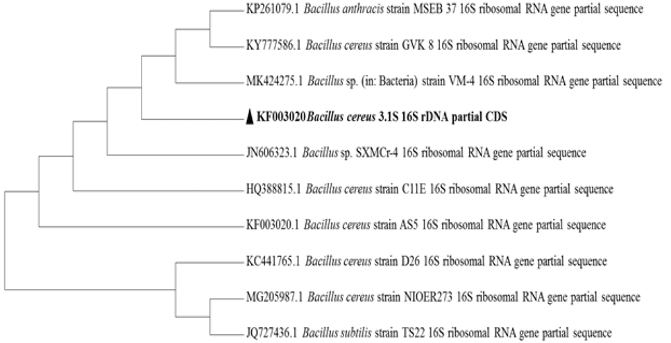
Neighbor-joining methods based tree of selected B. cereus 3.1S strains. B. anthracis strain MSEB 37 (KP261079.1), B. cereus strain GVK 8 (KY777586.1), Bacillus sp. strain VM-4 (MK424275.1), Bacillus sp. strain SXMCr-4 (JN606323.1), B. cereus strain CIIE (HQ388815.1), B. cereus strain D26 (KC441765.1), B. cereus strain NIOER273 (MG205987.1), and B. cereus strain TS22 (JQ727436.1).
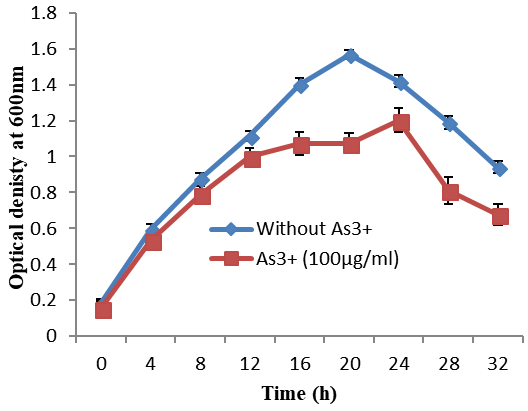
Growth pattern of B. cereus 3.1S growing in 100 μgAs3+//ml and without As3+ in minimal salt medium.
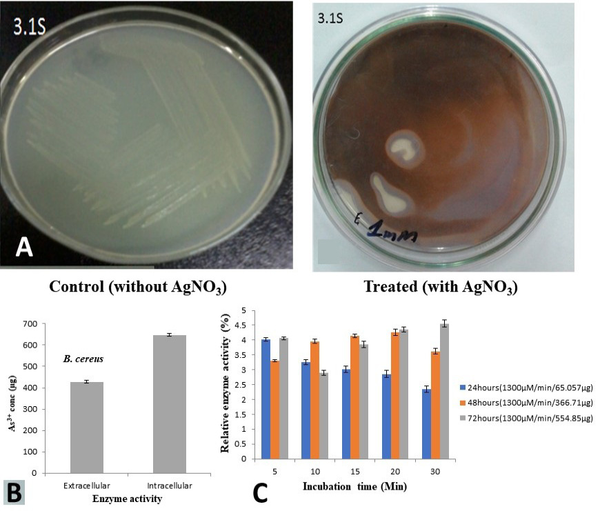
Appearance of brown precipitates on plate, containing 100 μgAs3+/ml, is showing the arsenite oxidizing ability of B. cereus 3.1S as compared to the control in AgNO3 assay (A), Concentration of As5+ transformed during extra and intra-cellular enzyme activity (B), Enzyme assay for soluble fraction of bacterial isolate 3.1S (C).
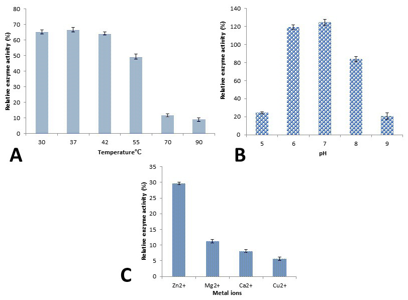
Effect of temperature (A), pH (B) and metal ions (C) on arsenite oxidase activity.
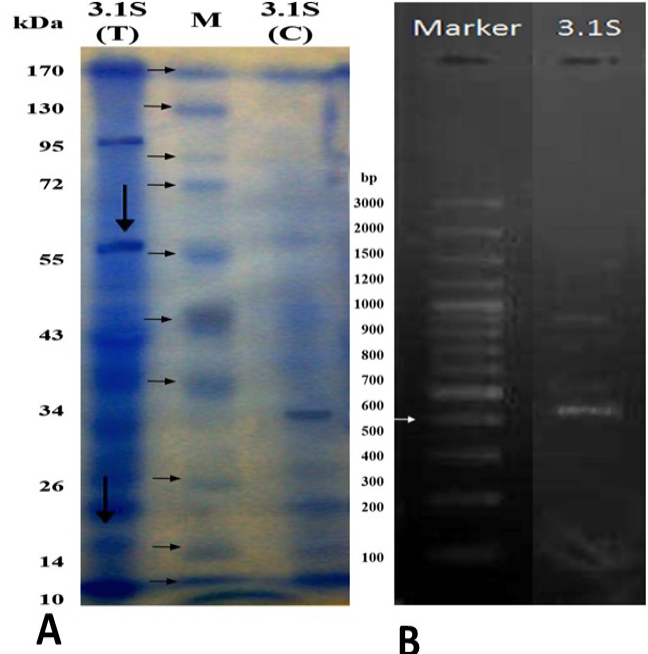
A: SDS-PAGE of bacterial isolates intracellularly with arsenite and without arsenite in the culturing medium. On right hand side is marker a line (M), next to it, control sample of 3.1S (C) and 3.1S (T). Treated sample have the arsenite in the medium and represented with (T) while control does not have arsenite in the medium and marked with (C). B: Agarose gel (1%) is showing the amplified product of arsenite oxidase gene from B. cereus 3.1S. Lane marker and 3.1S is showing DNA marker and B. cereus, respectively.
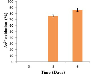
As3+ oxidation (%) by B. cereus 3.1S from industrial wastewater after 3 and 6 days of incubation at room temperature.
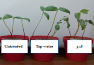
Growth of Vigna radiata (mung beans) in microbially treated wastewater (B. cereus 3.1S), tap water and untreated wastewater after 10 days of incubation under 1:1 light and dark period.
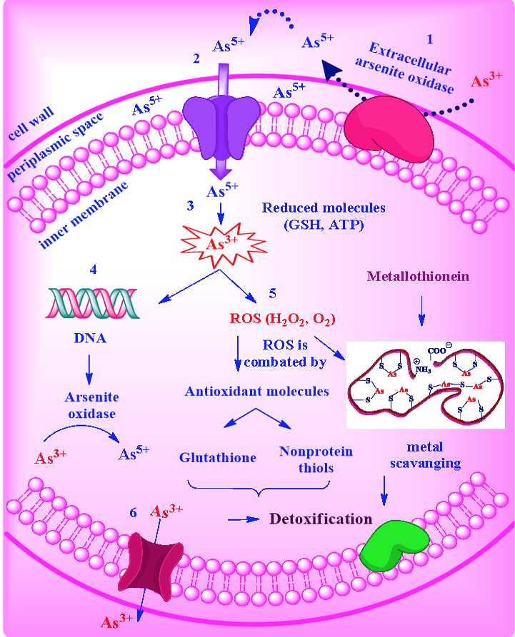
Proposed As3+ resistance and oxidation mechanism in gram positive bacterium, B. cereus 3.1S. (1) As3+ oxidizes into As5+ extracellularly by arsenite oxidase. (2) As5+ enters into cell cytoplasm. (3) As5+ reduces into As3+ by reduced molecules e.g., GSH and ATP. (4) As3+ induces DNA to make arsenite oxidase which converts As3+ into As5+. (5) As3+ is also involved in the generation of ROS and the oxidative stress resulted by the formation of ROS is combated by antioxidant molecules including glutathione and non-protein thiols. Metallothionenis are involved in metal scavenging process. (6) After certain level of accumulation, As3+ expel out through the efflux system.
References
- Bachate, S.P., Nandre, V.S., Ghatpande, N.S. and Kodam, K.M., 2013. Simultaneous reduction of Cr (VI) and oxidation of as (III) by Bacillus firmus TE7 isolated from tannery effluent. Chemosphere, 90: 2273–2278. https://doi.org/10.1016/j.chemosphere.2012.10.081[CrossRef]
- Butt, A.S. and Rehman, A., 2011. Isolation of arsenite-oxidizing bacteria from industrial effluents and their potential use in wastewater treatment. World J. Microbiol. Biotechnol., 27: 2435–2441. https://doi.org/10.1007/s11274-011-0716-4[CrossRef]
- Cappucino, J.G. and Sherman, N., 2001. Microbiology: A laboratory manual, 6th ed. Pearson Education, Benjamin Cummings, San Francisco.
- Conrads, T., Hemann, C., George, G.N., Pickering, I.J., Prince, R.C. and Hille, R., 2002. The active site of arsenite oxidase from Alcaligenes faecalis. J. Am. Chem. Soc., 124: 11276–11277. https://doi.org/10.1021/ja027684q[CrossRef]
- Das, S. and Barooah, M., 2018. Characterization of siderophore producing arsenic-resistant Staphylococcus sp. strain TA6 isolated from contaminated groundwater of Jorhat, Assam and its possible role in arsenic geocycle. BMC Microbiol., 18:104. https://doi.org/10.1186/s12866-018-1240-6[CrossRef]
- Dey, U., Chatterjee, S. and Mondal, N.K., 2016. Isolation and characterization of arsenic-resistant bacteria and possible application in bioremediation. Biotechnol. Rep., 10: 1-7. https://doi.org/10.1016/j.btre.2016.02.002[CrossRef]
- Drewniak, L., Styczek, A., Majder-Lopatka, M. and Sklodowska, A., 2008. Bacteria, hyper tolerant to arsenic in the rocks of an ancient gold mine, and their potential role in dissemination of arsenic pollution. Environ. Pollut., 156: 1069–1074. https://doi.org/10.1016/j.envpol.2008.04.019[CrossRef]
- Elahi, A. and Rehman, A., 2018. Multiple metal resistance and Cr6+ reduction by bacterium, Staphylococcus sciuri A-HS1, isolated from untreated tannery effluent. J. King Saud Uni. Sci., 31: 1005-1013. https://doi.org/10.1016/j.jksus.2018.07.016[CrossRef]
- Ellis, P.J., Conrads, R., Hille, R. and Kuhn, P., 2001. Crystal structure of the 100kDa arsenite oxidase from Alcaligenes faecalis in two crystal forms at 1.64 A and 2.03 A. Structure, 9: 125-132. https://doi.org/10.1016/S0969-2126(01)00566-4[CrossRef]
- Ghosh, D., Bhadury, P. and Routh, J., 2018. Coping with arsenic stress: Adaptations of arsenite-oxidizing bacterial membrane lipids to increasing arsenic levels. Microbiol. Open, 7: e00594. https://doi.org/10.1002/mbo3.594[CrossRef]
- Heinrich-Salmeron, A., Cordi, A., Brochier-Armanet, C., Halter, D., Pagnout, C., Abbaszadeh-Fard, E., Montaut, D., Seby, F., Bertin, P.N., Bauda, P. and Arsène-Ploetze, F., 2011. Unsuspected diversity of arsenite-oxidizing bacteria revealed by a widespread distribution of the aoxB gene in prokaryotes. Appl. environ. Microbiol., 77: 4685–4692. https://doi.org/10.1128/AEM.02884-10[CrossRef]
- Ilyas, S. and Rehman, A., 2018. Metal resistance and uptake by Trichosporon asahii and Pichia kudriavzevii isolated from industrial effluents. Arch. environ. Protect., 44: 75-82.
- Jinbo, X., Wenming, W., Haoxin, F. and Gejiao, W., 2007. Arsenite-oxidizing Agrobacterium and arsenic resistant microorganisms of a Chinese coal mine ecosystem1. State Key Laboratory of Agricultural Microbiology, Huazhong Agricultural University, Wuhan, http://www.paper.edu.cn.
- Kohnhorst, A., Allan, L., Pokethitiyoke, P. and Anyapo, S., 2002. Sustainable environmental sanitation and water services. 28th WEDC Conference, Kolkata, India.
- Koechler, S., Cleiss-Arnold, J., Proux, C., Sismeiro, O., Dillies, M.-A., Goulhen-Chollet, F., Hommais, F., Lie`vremont, D., Arse`ne-Ploetze, F., Coppe´e, J.-Y. and Bertin, P.N., 2010. Multiple controls affect arsenite oxidase gene expression in Herminii monasarsenicoxydans. BMC Microbiol., 10: 53. https://doi.org/10.1186/1471-2180-10-53[CrossRef]
- Krumova, K., Nikolovska, M. and Groudeva, V., 2008. Isolation and identification of arsenic-transforming bacteria from arsenic contaminated sites in Bulgaria. Biotechnol. Biotech. Equip., 22: 721–728. https://doi.org/10.1080/13102818.2008.10817541[CrossRef]
- Laemmli, U.K., 1970. Cleavage of structural proteins during the assembly of the head of bacteriaphage T4. Nature, 227: 680-685. https://doi.org/10.1038/227680a0[CrossRef]
- Lenoble, V., Deluchat, V., Serpaud, B. and Bollinger, J.-C., 2003. Arsenite oxidation and arsenate determination by the molybdene blue method. Talanta, 61: 267-276. https://doi.org/10.1016/S0039-9140(03)00274-1[CrossRef]
- Liao, V.H.-C., Chu, Y.-J., Su, Y.-C., Hsiao, S.-Y., Wei, C.-C., Liu, C.-W., Liao, C.-M., Shen, W.-C. and Chang, F.-J., 2011. Arsenite-oxidizing and arsenate-reducing bacteria associated with arsenic-rich groundwater in Taiwan. J. Contam. Hydrol., 123: 20-29. https://doi.org/10.1016/j.jconhyd.2010.12.003[CrossRef]
- Lim, K.T., Shukor, M.Y. and Wasoh, H., 2014. Physical, chemical, and biological methods for the removal of arsenic compounds. Biomed. Res. Int., 2014: 503784. https://doi.org/10.1155/2014/503784[CrossRef]
- Marzan, L.W., Hossain, M., Mina, S.A., Akter, Y. and Chowdhury, A.M.M.A., 2017. Isolation and biochemical characterization of heavy-metal resistant bacteria from tannery effluent in Chittagong city, Bangladesh: Bioremediation viewpoint. Egypt J. aquat. Res., 43: 65-74. https://doi.org/10.1016/j.ejar.2016.11.002[CrossRef]
- Mead, M.N., 2005. Arsenic: In search of an antidote to a global poison. Environ. Health Perspect., 113: A378–A386.
- Mohammadi, T., Moheb, A., Sadrzadeh, M. and Razmi, A., 2005. Modeling of metal ions removal from wastewater by electrodialysis. Sep. Purif. Technol., 41: 73–82. https://doi.org/10.1016/j.seppur.2004.04.007[CrossRef]
- Muller, D., Lievremont, D., Simeonova, D.D., Hubert, J.C. and Lett, M.C., 2003. Arsenite oxidase aox genes from a metal-resistant betaproteobacterium. Bacteriology, 185: 135-141. https://doi.org/10.1128/JB.185.1.135-141.2003[CrossRef]
- Mu, Y., Zhou, X., Liu, L., Zhou, X.-K., Zeng, X.-C. and Li, W.-J., 2019. Pseudaminobacter arsenicus sp. nov., an arsenic-resistant bacterium isolated from arsenic-rich aquifers. Int. J. Syst. Evol. Microbiol., 69: 791-797. https://doi.org/10.1099/ijsem.0.003238[CrossRef]
- Noreen, A. and Rehman, A., 2016. Arsenite oxidizing multiple metal resistant bacteria isolated from industrial effluent: Their potential use in wastewater treatment. World J. Microbiol. Biotechnol., 32: 133. https://doi.org/10.1007/s11274-016-2079-3[CrossRef]
- Oremland, R.S. and Stolz, J.F., 2005. Arsenic, microbes and contaminated aquifers. Trends Microbiol., 13: 45–49. https://doi.org/10.1016/j.tim.2004.12.002[CrossRef]
- Pasha, C. and Narayana, B., 2008. Determination of arsenic in environmental and biological samples using Toluidine blue or Safranine O by simple spectrophotometric method. Bull. environ. Contam. Toxicol., 81: 47–51. https://doi.org/10.1007/s00128-008-9454-1[CrossRef]
- Pattanapipitpaisal, P., Brown, N.L. and Macaskie, L.E., 2001. Chromate reduction and 16S rRNA identification of bacteria isolated from a Cr (VI)-contaminated site. Appl. Microbiol. Biotechnol., 57: 257–261. https://doi.org/10.1007/s002530100758[CrossRef]
- Que´me´neur, M., Heinrich-Salmeron, A., Muller, D., Lie‘vremont, D., Jauzein, M., Bertin, P.N., Garrido, F. and Joulian, C., 2008. Diversity surveys and evolutionary relationships of aoxB genes in aerobic arsenite-oxidizing bacteria. Appl. environ. Microbiol., 74: 4567–4573. https://doi.org/10.1128/AEM.02851-07[CrossRef]
- Raja, C.E. and Omine, R., 2012. Arsenic, boron and salt resistant Bacillus safensis MS11 isolated from Mongolia desert soil. Afr. J. Biotechnol., 11: 2267-2275. https://doi.org/10.5897/AJB11.3131[CrossRef]
- Rehman, A., Butt, A.S. and Hasnain, S., 2010. Isolation and characterization of arsenite oxidizing Pseudomonas lubricans and its potential use in bioremediation of waste water. Afr. J. Biotechnol., 9: 1493-1498. https://doi.org/10.5897/AJB09.1663[CrossRef]
- Santini, J.M. and Hoven, R.N.V., 2004. Molybdenum-containing arsenite oxidase of the chemolithoautotrophic arsenite oxidizer NT-26. J. Bact., 186: 1614–1619. https://doi.org/10.1128/JB.186.6.1614-1619.2004[CrossRef]
- Santini, J.M., Sly, L.I., Schnagl, R.D. and Macy, J.M., 2000. A new chemolithotrophic arsenite-oxidising bacterium isolated from a gold mine: Phylogenetic, physiological, and preliminary biochemical studies. Appl. environ. Microbiol., 66: 92–97. https://doi.org/10.1128/AEM.66.1.92-97.2000[CrossRef]
- Sambrook, J., MacCallum, P. and Russell, D., 2001. Molecular cloning: A laboratory manual. 3rd edition. Cold Spring Harbor Laboratory Press.
- Satyapal, G.K., Mishraa, S.K., Srivastavab, A., Ranjanc, R.K., Prakash, K., Haquea, R. and Kumar, N., 2018. Possible bioremediation of arsenic toxicity by isolating indigenous bacteria from the middle gangetic plain of Bihar, India. Biotechnol. Rep., 17: 117-125. https://doi.org/10.1016/j.btre.2018.02.002[CrossRef]
- Satyapal, G.K., Rani, S., Kumar, M. and Kumar, N., 2016. Potential role of arsenic resistant bacteria in bioremediation: Current status and future prospects. J. Microb. Biochem. Technol., 8: 256-258. https://doi.org/10.4172/1948-5948.1000294[CrossRef]
- Silver, S. and Phung, L.T., 2005. A bacterial view of the periodic table: Genes and proteins for toxic inorganic ions. J. indust. Microbiol. Biotechnol., 32: 587–605. https://doi.org/10.1007/s10295-005-0019-6[CrossRef]
- Simeonova, D.D., Lievremont, D., Lagarde, F., Muller, D., Groudeva, V. and Lett, M-C., 2004. Micro plate screening assay for detection of arsenite oxidizing and arsenate-reducing bacteria. FEMS Microbiol. Lett., 237: 249–253. https://doi.org/10.1111/j.1574-6968.2004.tb09703.x[CrossRef]
- Simeonova, D.D., Micheva, K., Muller, D.A.E., Lagarde, F., Lett, M.C. and Groudeva, V.I., 2005. Arsenite oxidation in batch reactors with alginate-immobilized ULPAsI strain. Biotechnol. Bioeng., 91: 441–446. https://doi.org/10.1002/bit.20530[CrossRef]
- Singh, N., Ma, L.Q., Srivastava, M. and Rathinasabapathi, B., 2006. Metabolic adaptations to arsenic-induced oxidative stress in Pteris vittata L. and Pteris ensiformis L. Pl. Sci., 170: 274–282.
- Stolz, J.F., Basu, P., Santini, J.M. and Oremland, R.S., 2006. Arsenic and selenium in microbial metabolism. Annu. Rev. Microbiol., 60: 107–130. https://doi.org/10.1146/annurev.micro.60.080805.142053[CrossRef]
- Tsai, S.-L., Singh, S. and Chen, W., 2009. Arsenic metabolism by microbes in nature and the impact on arsenic remediation. Curr. Opin. Biotechnol., 20: 659–667. https://doi.org/10.1016/j.copbio.2009.09.013[CrossRef]
- Tseng, C.-H., Tseng, C.-P., Chiou, H.-Y., Hsueh, Y.-M., Chong, C.-K. and Chen, C.-J., 2002. Epidemiologic evidence of diabetogenic effect of arsenic. Toxicol. Lett., 133: 69-76.
- USEPA, 2004. Cleaning up the nation’s waste sites: Markets and technology trends. National Service Center for Environmental Publications (NSCEP).
- Walton, F.S., Harmon, A.W., Paul, D.S., Drobna, Z., Patel, Y.M. and Styblo, M., 2004. Inhibition of insulin-dependent glucose uptake by trivalent arsenicals: possible mechanism of arsenic-induced diabetes. Toxicol. appl. Pharmacol., 198: 424-433. https://doi.org/10.1016/j.taap.2003.10.026[CrossRef]
- Yang, H.-C. and Rosen, B.P., 2016. New mechanisms of bacterial arsenic resistance. Biomed. J., 39: 5-12. https://doi.org/10.1016/j.bj.2015.08.003[CrossRef]
Supplementary material
Services
Article usage statistics
Loading…