
Zebrafish (Danio rerio) are one of the most effective models for researching multi-organ regenerative processes. Regeneration is an amazing biological phenomenon that differs greatly among vertebrates. Zebrafish have the capacity to effectively regenerate complex tissues and organs, including the heart, fins, central nervous system, spinal cord, and retina, in contrast to mammals. An integrative framework of zebrafish organ regeneration is presented in this review, with a focus on conserved molecular and cellular programs that are frequently used in various tissues with context-dependent changes. A highly coordinated series of biological processes, such as immune activation, temporary inflammation, resident progenitor cell activation, cellular dedifferentiation, and proliferative expansion, are involved in regeneration after injury. These processes eventually result in tissue remodeling and functional restoration. Important signaling pathways that regulate these regeneration responses include Wnt/β-catenin, Notch, Hedgehog, BMP, FGF, IGF, and TGF-β. Recent developments in lineage-tracing, spatial genomics, and single-cell transcriptomics have further shown similar gene regulation networks and conserved regenerative cell states in various organs. Zebrafish regeneration adheres to a single biological logic controlled by conserved signaling and epigenetic pathways, despite organ-specific variations in cellular origins and structural outcomes. In addition to offering promising translational potential for the development of regenerative therapies in humans, an understanding of these shared principles offers crucial insights into vertebrate regenerative biology, especially for conditions like myocardial infarction, spinal cord injury, neurodegeneration, and retinal degeneration.
Review · Volume 72, Issue 2, 20 October 2026 · Open Access
An Integrative Framework of Different Organs’ Regeneration in Zebra Fish
· · · · ·
Affiliations & Notes ⌄
- 1Cooperative Innovation Center of Industrial Fermentation (Ministry of Education & Hubei Province), Key Laboratory of Fermentation Engineering (Ministry of Education), Wuhan 430068, China
- 2Department of Chemistry, University of Okara, Okara-56300, Pakistan
- 3Xishuangbanna Tropical Botanical Garden, Chinese Academy of Sciences, Menglun Town, Mengla County, Yunnan Province, China
- 4Department of Nutrition and Dietetics, National University of Medical Sciences, Pakistan
- 5Department of Microbiology, Andijan State Medical Institute, Andijan. 170100 Uzbekistan
- 6Department of Biology and Geography, Andijan State Pedagogical Institute, Andijan, 170100 Uzbekistan
- 7Department of Microbiology, Quaid-i-Azam University, Islamabad, Pakistan. *Correspondence | Mehwish Khalid, Cooperative Innovation Center of Industrial Fermentation (Ministry of Education & Hubei Province), Key Laboratory of Fermentation Engineering (Ministry of Education), Wuhan 430068, China
- 8Email: mehwishkhalidd120@gmail.com
Article Info ⌄
- Journal:
- Biologia (Lahore)
- Published:
- 20 October 2026
From the article
- In this review we are developing a deep understanding of many aspects of the regeneration process in zebrafish, underlying genetic basis, molecular pathways and stimulators of molecular pathways, that are associated in regeneration process.
- Previous studies mainly associated with the regeneration models in zebrafish mostly related to non-proliferating tissue, which is block after completion of tissue or system, like nervous tissue, photoreceptor, and cardiac muscles.
- While zebrafish offer an effective model for determining the pathways involved in successful tissue regeneration, it is important to use caution when extrapolating these results to mammals.
- Regenerative ability, immunological responses, cellular plasticity, tissue architecture, and injury-associated fibrotic responses are all significantly different between zebrafish and mammals.
Selected from the article's own text.
Abstract
Introduction
A basic biological function that differs greatly between species is regeneration, or the capacity to replace lost or injured tissues. The zebrafish (Danio rerio) has become one of the most effective vertebrate models for researching robust regeneration across several organ systems, whereas mammals have a restricted capacity for regeneration ; . Zebrafish are a perfect creature for creating an integrative framework of organ regeneration because they can effectively regenerate the heart, fin, spinal cord, retina, liver, kidney, and portions of the central nervous system.
One important aspect of zebrafish regeneration is that it is controlled by conserved molecular and cellular programs that are reused across several tissues with context-dependent alterations, rather than being organ-specific in isolation (Hoang et al., 2020; Wagner et al., 2018). Zebrafish tissues usually initiate a coordinated series of events after injury, such as quick wound closure, immune cell recruitment, temporary inflammatory signaling, dedifferentiation or activation of resident progenitor cells, and subsequent proliferative expansion followed by tissue remodeling and functional restoration (Jopling et al., 2010; . Conserved signaling pathways like Wnt/β-catenin, Notch, Hedgehog, BMP, and FGF, which are frequently used in various regeneration contexts, carefully regulate these phases ; .
Crucially, depending on the organ system, zebrafish regeneration depends on both dedifferentiation of adult cells and lineage-restricted progenitor cells (Kikuchi et al., 2010). For instance, although fin regeneration mostly depends on the development of a blastema—a mass of proliferative progenitor-like cells that restore structured growth—cardiomyocyte regeneration in the heart includes dedifferentiation and proliferation of already-existing cardiomyocytes. On the other hand, tissue-specific stem/progenitor populations and the proliferation of already differentiated cells are the main drivers of liver and kidney regeneration . Despite these distinctions, many systems share common regulatory principles, including immune-mediated signaling, metabolic changes, and epigenetic reprogramming .
Another important feature is the role of the immune system and inflammatory milieu, which operate as a regulatory center for regeneration as well as a response to harm. Successful regenerative outcomes depend on neutrophils, macrophages, and cytokine signaling; their temporal modulation dictates whether fibrosis or regeneration takes place . This demonstrates how damage signals and organ-wide regeneration capability are coordinated systemically.
Zebrafish regeneration is extremely dynamic and involves conserved cell states across several organs, according to recent developments in single-cell transcriptomics, lineage tracking, and spatial genomics ; Wei et al., 2022). These findings imply that regenerated tissues have similar transcriptional processes, including activation of progenitor-like states, extracellular matrix remodeling, and re-entry into the cell cycle, despite anatomical variances.
Thus, in addition to summarizing regeneration in specific zebrafish organs, this review incorporates these systems into a cross-organ framework based on four recurrent features: immune regulation, dynamic extracellular matrix remodeling, activation or reprogramming of regenerative cell populations, and coordinated deployment of conserved signaling networks. While acknowledging the significant biological distinctions between zebrafish and mammalian healing responses, this integrated approach may help discover common regenerative processes and produce ideas for later validation in mammalian models.
Zebrafish multi-organ regeneration: conserved molecular and cellular programs
Integrated signaling pathways governing zebrafish heart regeneration
Unlike mammals, adult Zebrafish have the ability to regenerate the injured heart . Proliferation in pre-existing cardiomyocytes helps in the regeneration of cardiac tissue in zebrafish. Proliferation in cardiomyocytes mainly depends upon secreted growth factors, e.g., fibroblast growth factors (FGFs). Insulin-like growth factor (IGF) , Platelet-derived growth factor (PDGFs) , transforming growth factor β (TGFβ), bone morphogenetic proteins (BMPs), and Neuregulin (Nrg) (Chablais and Jaźwińska, 2012).
Secreted growth factors play an important role in the stimulation of the mitogen-activated protein kinase /MAPK pathway, which is counter-controlled by another feedback, which is known as Dual specificity phosphatase 6 (Dusp6), an ERK phosphatase. A decrease in the activity of attenuator dual specificity phosphatase 6 (Dusp6) results in the regeneration of the heart. Decrease in the activity of attenuator Dual specificity phosphatase 6 (Dusp6) by small molecules or by inactivation of the gene, result in the increase of proliferation of cardiomyocytes, reduced fibrosis, coronary angiogenesis. A decline in the activity of PDGF receptor signaling result in the decrease in the regeneration of the heart in wild zebrafish .
Growth factors perform many tasks in regeneration. For example, dedifferentiation of cardiomyocytes and proliferation in zebrafish are carried out by epidermal growth factor (Nrg1), and fibroblast growth factors (FGFs) are responsible for activation of the mitogen-activated protein kinase pathway, which in turn performs differentiation and proliferation at the cellular level . In the process of heart regeneration, fibroblast growth factors (FGFs) are responsible for activation of epicardial cells. While Fibroblast growth factors (FGFs) along with the epithelial-to-mesenchymal transition (EMT) are responsible for neovascularization ; .
Importantly, rather than acting individually, these pathways function as a network that is dependent on tissue and stage. Endocardial Notch signaling in the regenerating heart limits myocardial Wnt activity by inducing secreted Wnt antagonists, which in turn stimulate cardiomyocyte proliferation. While TGF-β signaling is necessary for efficient cardiac regeneration, IGF signaling promotes cardiomyocyte proliferation .
It is observed that removal of dual specificity phosphatase 6 (Dusp6) is responsible for rapid heart repair . In contrast, overexpression of dual specificity phosphatase 6 (Dusp6) is associated with suppression of the regenerative process in the heart of zebrafish. It is also reported that neuregulin (Nrg) or PDGF receptors (PDGFR) inhibitor are responsible for the stoppage of the regenerative process of the heart in zebrafish, but still in the existence of neuregulin (Nrg) or PDGF receptor (PDGFR) inhibitors, mutant dusp6 zebrafish has the ability of rapid heart regeneration. So, it has seen that chemical stoppage of dual specificity phosphatase 6 (Dusp6) by small molecule is responsible for rapid heart regeneration in zebrafish, which may provide useful tools for therapeutic treatments .
Transcripts encoding ECM-related proteins have been identified after the examination of regenerative hearts in zebrafish ; . There is the overexpression of tnc transcript, matrix metalloproteinase, inhibitors of metalloproteinases, and tenascin C (tnc) found in between the injured site of myocardium and healthy heart ; . It is observed that epicardial cells are responsible for the synthesis of fibronectin (fn), which ultimately helps in the migration of cardiomyocytes. It is also shown that hyaluronic acid (HA) is also formed at the site of injured hearts, which plays an important role in heart repair of zebrafish . When compared to other mammals, heart regeneration in mammals is strongly dependent on age. For example, neonatal mouse hearts can regenerate through cardiomyocyte proliferation soon after birth, but this ability is quickly lost, and adult cardiac injury mostly results in the formation of reparative scars (Porrello et al., 2011; .
Future investigations into the cardiac regenerative mechanisms of zebrafish may reveal specific molecules that can influence coronary heart regeneration; these molecules could elucidate how myocardial plasticity is sustained throughout regeneration, thereby facilitating cardiac repair following myocardial infarction in humans. Furthermore, identifying the variables that facilitate heart regeneration may not suffice to fully comprehend the regulation of myocardial plasticity following cardiac injury or myocardial infarction. Signaling pathway networks and epigenetic regulation, as depicted in (Figure 1), warrant further investigation to elucidate the mechanisms by which myocardial plasticity is liberated and reactivated for proliferation. This perspective suggests that zebrafish may serve as a preclinical model for analyzing cardiac plasticity, aiding in the development of novel therapeutic strategies to mitigate damage associated with myocardial infarction (MI).
The MAPK signaling pathway may be downstream activation of ligand binding to receptor tyrosine- kinases (RTKs) with the aid of increase elements, which include HB-EGF, EGF, fibroblast growth factor 1 (FGF1), and fibroblast growth factor 2 (FGF2), or with the aid of activation of CNTFR by way of ciliary neurotrophic factor (CNTF). In each pathway, adaptor proteins, which include GBR2, recruit Salt overly sensitive (SOS) to the activated receptor, and next activation of Salt overly sensitive (SOS) leads to phosphorylation of MEK, and RAS eventually extracellular signal-regulated kinase (ERK1/2). Activated ERK1/2 translocate into the nucleus and phosphorylates numerous transcription factors concerned with cellular proliferation, cell differentiation, and cell survival.
Integrated signaling networks governing zebrafish fin regeneration
Unlike mammals, Zebrafish has the ability to regenerate their fins. Molecular signals that help in fins regeneration are Notch, retinoic acid (RA), Hedgehog (Hh), Wnt, fibroblast growth factors (FGF), insulin-like growth factors (IGF), activin, and bone morpho-genic protein (BMP). Wnt/b-cate-nin signaling is responsible indirectly for progenitor cell proliferation, epidermal patterning, osteoblast maturation by regulation of the multitude of signals at the secondary level. Much work is required to understand the function of these pathways at tissue- level and how the work and interaction of these pathways with each other ; ; ; ; . There is a functional connection between these signals. Wnt/β-catenin creates signaling hubs that control secondary FGF, BMP, retinoic acid, and Hedgehog signals during fin regeneration . Additionally, their actions are coordinated both geographically and temporally: BMP stimulates the proximal differentiation of Runx2+ osteoprogenitors and inhibits Wnt activity, whereas Wnt/β-catenin sustains their distal differentiation . Blastema development and regeneration outgrowth depend on FGF signaling .
However, regarding the regeneration of various organs or tissues, scientific research on the canonical Notch and Wnt pathways is markedly inadequate. Combination therapy employing pharmaceuticals that affect various pathways in organ or tissue regeneration may prove beneficial for future scientific inquiry. The growing emphasis on strategic improvements indicates that a more profound comprehension of Notch-Wnt signaling interactions and the development of related treatment strategies for organ and tissue regeneration are exciting prospects in the near future. After two days of fin amputation, expression of hyaluronan synthase 3 (HAS3) occurs. Concentration of hyaluronan synthase 3 (has3) goes on to increase up to a certain limit and then afterward decline. The has3 transcription is responsible for wound epithelium and regulates several signaling pathways such as phosphoinositide3-kinase (PI3K), transforming growth factors- û (TGFû), and FGFs. Moreover, after 24 hours of amputation, inhibition of hyaluronic acid (HA) occurs, which results in the suppression of cell proliferation. While the distal-less homeobox gene 5a (dlx5a), which is basically a wound epithelium marker and blastemal gene formerly related with fin regeneration. 4- MU-mediated defects present in the proliferation of cells and fin regeneration can be regulated by inhibition of glycogen synthase 3-kinase (GSK3). Moreover, hyaluronic acid (HA) is mainly responsible for zebrafish fin growth and tissue regeneration. Mutation in methylation, acetylation, phosphorylation, and other post translational changes in the tail of histones such as H3 and H4 result in caudal fin regeneration. Regulation of functions such as vesicle aggregation, membrane structure modification, ion conductance and cytoskeletal structure anchoring is associated with annexin complexes. Inhibition of activation of NF-jB pathway is also mainly associated with the ANXA1. Absorption and dissolution of bone are mainly associated with the activity of ANXA2 by using paracrine and autocrine factors . The fin regeneration process is also maintained when ANXA5 binds with the myosin regulatory light chain interacting protein (MIR) (Figure 2) .
Mature osteoblasts dedifferentiate and move in the direction of the blastema following damage. This mechanism is regulated by epidermal-fibroblast signaling through Fgf20a, Shh, and Wnt/β-catenin, which encourages the development of osteoblast progenitor cells in the proximal blastema for repair.
Molecular and cellular mechanisms of central nervous system (CNS) regeneration in zebrafish
Activation of ependymo-radial glial (ERG) progenitor cells is necessary for neuronal regeneration. ERG progenitor cells possess a soma that constitutes a segment of the ependyma and radial processes that extend across the entire thickness of the brain. Following a CNS injury, these cells are either awakened from quiescence or migrate to regions of constitutive cellular growth to restore missing neurons. Neurodegenerative disorders are a huge encumbrance to humanity nowadays, new therapeutic intrusions must be developed. The conceivable approach is comparison between non regenerative and regenerative animal models to search out resemblances and alterations at histological, cytological and molecular levels that could be productive to accomplish regeneration in the human brain. Zebrafish nervous system regeneration is a treasurable choice for that comparison ; Kaslin et al., 2008). The neurogenic niches are found in various mammal’s nervous systems, but endogenous regeneration is perceived in zebrafish ; ; Kaslin et al., 2008). For the endogenous neurogenesis and differences and resemblances examined by using various injuries in different regions of the zebrafish central nervous system . Zebrafish have the capability to produce new neurons and perseverance of glial scar and inflammation. New neurons produced with proper specification having a long life . Neuron regeneration after injury has not been observed, while in zebrafish endogenous regeneration plays a vital role in neuron regeneration. Therefore, we will focus on histological
and cellular conditions in various parts of the nervous system of zebrafish on the basis of physiological condition.
In zebrafish progenitor cells, which have the capability to produce new neurogenic niches that regenerate new neurons, a similar cell is also present in the mammalian nervous systems, but they lose their character to produce new neurons with the passage of time ; ; ; . The progenitor cell located at telencephalon region of the brain in zebrafish, which contains specialized cells called ependymal cells. By nature, they are progenitor cells and under physiological conditions produce new neurons ; . In mammalian brain these ependymal cells proliferate only under physiological conditions and produce a neurogenic niche ; ; ; ; ; . These progenitor cells express various biomarkers like nestin, brain lipid binding protein (Blbp), glial fibrillary acidic protein, which are very useful for comparison ; ; ; . The genetic approaches are to identify the messenger RNA of ependymal cells and isolate of their population in different positions of the brain and decode their molecular pathways. For discrete molecular feature dissection of various populations or colonies of ependymal or progenitor cells to enabled the identification of markers, which are helpful to display morphology and primary proliferation stem cell biology in zebrafish and related molecular information stimulate the stem cell-based regeneration. According to previous studies, adult Zebrafish nervous system activity is affected by environmental hazards due to abnormal signaling pathways like Notch, Wnt, Fgf, Bmp, and chemokine signaling. This extensive set of insights may be technological and serve to contrast the capabilities of zebrafish neural stem cells with human conditions, thus aiding in the discovery of how a critical neurogenesis cascade may be initiated in mammals. The instrumental approach to regeneration in the treatment of Alzheimer’s disease (AD) has the potential to be a game changer (Figure 3) ; .
The circles depict a schematic of the main mechanisms involved in the brain and spinal cord regeneration of zebrafish. Signaling pathways, chemicals, and cell types that are involved in regeneration in zebrafish are highlighted.
Conserved molecular networks driving functional spinal cord regeneration in zebrafish
Tumor necrosis factors (TNF) are mainly responsible for the regeneration in the spinal cord, which in turn results in a decrease the level of interleukin-1 beta, which is released from neutrophils. Dopamine and serotonin are also responsible for regeneration in the spinal cord , which releases proliferating progenitor like pMN-like progenitors. Regulation of Wnt
and Notch signaling are responsible for regeneration. Pathways such as YES1, CTNNB1, and FGFR2 are also require for regeneration in Spinal cord.
After spinal cord injury, at the lesion site, non-neural cells arrive and form a complex extracellular matrix (ECM) . In vertebrates like fish, the crossing of regenerating axons occurs at the lesion site of the crossing of regenerating axons at the lesion site of complex extracellular matrix (ECM) ; . The Wnt/β-catenin pathway is responsible for controlling the formation of Complex Extracellular Matrix (ECM) at lesion site in Zebrafish because deposition of ECM and fibroblast biology are mainly controlled by Wnt/β-catenin signaling ; ; ; . Moreover, functional recovery and axonal reconnection also mainly depend upon Wnt/β-catenin pathway ; . If the researchers can succeed in especially suppressing the formation of inhibitors and simultaneously stimulating the production of growth-promoting molecules, this could open up new healing opportunities for paralyzed people.
Signaling of Wnt/β-catenin is responsible for stabilization of Wnt ligand and translocation of β-catenin into the nucleus, where it adapts the transcription of target genes with the protein of the Tcf/Lef family . Different ECMs performs different functions, which are mainly responsible for regeneration and growth of axons. For example, collagens col19a1, col18a1, and col15a1b are responsible for the development of motor axons, while col4a5 is responsible for regeneration of axons at the spinal cord site of Zebrafish (Figure 4) ; .
When Hippo signaling is activated (in conditions of high cell density): The activation of KIBRA and NF2 via unidentified upstream signaling results in the activation of MST1/2. MST1/2 is activated by phosphorylating SAV1, which subsequently phosphorylates LATS and MOB1. The active LATS/MOB phosphorylates YAP/TAZ, resulting in cytoplasmic retention through 14-3-3 protein and subsequent proteasomal destruction of YAP/TAZ. Consequently, TEAD engages with VGL4 and inhibits the expression of target genes.
Growth factor and hedgehog signaling in zebrafish retinal regeneration
Zebrafish retinal regeneration is associated with many growth factors, ; ; like epigenome modifiers ; ; , Sonic hedgehog signaling–induced gene regulatory networks ; , cytokines ; , differentiation factors , gene transcription factors ; ; , cell cycle regulators (Figure 5), which are present at the site of injury and responsible for differentiation of retinal neurons extent. ; Sonic Hedgehog signaling contributes to Müller glia-dependent regenerative responses and progenitor proliferation in the retina, demonstrating that the function of a conserved route relies on the responding tissue and regenerative stage .
The retina is a simple organ present just on the back of the eyes and connected with the CNS. It basically consists of one glial cell type and six neural cell types. The Muller glial cell type forms major part of the retina also extend and interact with the neuron ; Reichenbach and Bringmann, 2013). Activation of certain cytoskeleton genes occurs, for example, vimentin and fibrillary acidic protein (GFAP) . After injury in zebrafish’s retina, results in increased Gfap expression as well as activation of hypertrophy occurring , afterward, Muller glia adopt the properties of a retinal stem cell which results in the proliferation and generation of retinal progenitors ; ; ; ; .
Reprogramming of the Zebrafish genome occurred after the changes in gene expression which result in methylation of DNA and active the genes which is responsible for multipotency ; . Migration of Muller glial cells occurs from INL to the outer side of nuclei layer (ONL) and nuclear migration starts along with the generation of retinal progenitor ; . The retinal progenitor also performs interkinetic nuclear migration and it intensifies in a Pax6-dependent manner ; which ultimately result in regeneration of retinal neurons ; . In the retina, there is a similar species difference: damage reprograms zebrafish Müller glia into neurogenic progenitors, however adult mouse Müller glia mainly regains quiescence and have little neurogenic competence (Hoang et al., 2020).
The Zebrafish model is useful to resolve mechanisms of photoreceptor loss. In addition, not like mammals, zebrafish have the potential to absolutely regenerate damaged photoreceptors, raising the desire for future treatments for this disorder.
Subsequent to the CDO/BOC-mediated interaction of the HH ligand with PTCH1, SMO is derepressed and translocated to the primary cilium. A protein complex including KIF7 and SUFU linked with GLI transcription factors is actively translocated to the activated SMO. Active SMO promotes the dissociation of GLI proteins from SUFU, resulting in the nuclear accumulation of GLI1/2 and the subsequent activation of target genes.
Common cellular and molecular mechanisms across zebrafish regenerating organs
Zebrafish organs share a number of fundamental regenerating pathways despite tissue-specific variations. Immune cells, especially macrophages, help create a pro-regenerative milieu during the coordinated inflammatory response that is triggered by injury ; . Tissue-resident regenerative cells are then activated by proliferation, dedifferentiation, or the acquisition of progenitor-like states. For instance, Müller glia produce neural progenitors after retinal injury, while pre-existing cardiomyocytes multiply during heart regeneration (Jopling et al., 2010; .
Therefore, immune modulation is an active, stage-dependent part of the regenerative program rather than just a result of injury. Fin investigations reveal that macrophages promote inflammatory resolution, blastema proliferation, tissue expansion, and patterning (Kyritsis et al., 2012; . In the adult zebrafish brain, acute inflammation is necessary for injury-induced neural progenitor proliferation and regenerative neurogenesis. These results highlight that whether the injured environment promotes regeneration depends more on the timing and resolution of inflammatory signals than on inflammation alone.
An extra layer connecting damage signals to regenerative cell-state modifications is provided by epigenetic regulation. The elimination of restrictive H3K27me3 marks is linked to the reactivation of developmental genes during fin regeneration, and proper regeneration requires a histone demethylase . While lineage-specific DNA methylation is relatively stable, genome-wide analyzes also reveal dynamic changes in chromatin accessibility, suggesting that regeneration entails the selective opening of regulatory areas rather than a general loss of cell identity . Furthermore, a molecular link between tissue damage and regeneration-specific transcriptional programs can be established by activating gene expression in regenerating heart and fin tissues through injury-responsive tissue regeneration enhancer elements (Kang et al., 2016).
Another common regulatory element is provided by extracellular matrix (ECM) remodeling, which produces a dynamic environment that promotes tissue rebuilding and cell migration ; . These pathways, along with conserved regenerative signaling networks, establish a common framework: injury and immune activation → activation of regenerative cells → proliferation → ECM remodeling and tissue patterning → functional restoration. However, the proportional contributions and cellular origins of these mechanisms differ between organs. Therefore, even though mammals share a number of injury-responsive pathways, variations in regenerative-cell competence as well as in the persistence or resolution of fibrotic and glial responses can cause tissue regeneration to give way to repair ; ; . When considered collectively, immunological cues, alterations in the epigenetic state, conserved signaling pathways, and ECM dynamics function as a network of interrelated injury-response mechanisms that regulate the activation of regenerative cells and the eventual repair of tissue.
Conclusion and future perspective
In this review we are developing a deep understanding of many aspects of the regeneration process in zebrafish, underlying genetic basis, molecular pathways and stimulators of molecular pathways, that are associated in regeneration process. Previous studies mainly associated with the regeneration models in zebrafish mostly related to non-proliferating tissue, which is block after completion of tissue or system, like nervous tissue, photoreceptor, and cardiac muscles. In the case of cardiac tissue, growth factors are the most important factor to stimulate MAPK pathways, also connected with feedback, which is known as Dual specificity phosphatase 6, an ERK phosphatase, which leads to regeneration of the heart. Growth factors perform many tasks in regeneration, like differentiation and division of cardiac cells in zebrafish. Fibroblast growth factors (FGFs) is responsible for activation of epicardia cells. While fibroblast growth factors (FGFs) along with the epithelial-to-mesenchymal transition (EMT) are responsible for neovascularization. Heart regeneration models of zebrafish are useful in the future for the treatment of myocardial infarction. Zebrafish’s regeneration in the retina is related to many growth factors, like sonic hedgehog signaling–brought on by a gene regulatory network, cytokines, gene transcription factors, epigenome modifiers, cellular cycle regulators, and differentiation elements, which might be present at the website of injury and liable for differentiation of retinal neuron quantity.
While zebrafish offer an effective model for determining the pathways involved in successful tissue regeneration, it is important to use caution when extrapolating these results to mammals. Regenerative ability, immunological responses, cellular plasticity, tissue architecture, and injury-associated fibrotic responses are all significantly different between zebrafish and mammals. As a result, processes that encourage regeneration in zebrafish might not provide the same results in mammalian tissues. However, comparative research might help discover molecular or cellular processes that require more validation in mammalian models and differentiate conserved regeneration mechanisms from zebrafish-specific responses. Therefore, rather than being direct proof of human treatment efficacy, zebrafish regeneration should be largely seen as a mechanistic framework for developing and testing regenerative theories.
Acknowledgments
We sincerely acknowledge the valuable contributions of all colleagues who provided technical guidance, constructive feedback, and insightful discussions throughout the preparation of this manuscript
Novelty Statement
This review presents an integrative framework of multi-organ regeneration in zebrafish, highlighting conserved molecular signaling pathways, immune regulation, epigenetic reprogramming, and cellular mechanisms shared across the heart, fins, central nervous system, spinal cord, and retina. By synthesizing organ-specific regenerative processes into a unified mechanistic perspective, it provides insights into common regenerative principles and identifies potential molecular targets for future translational research in mammalian tissue repair and regenerative medicine.
Authors’ Contributions
Mehwish Khalid: Conceptualization.
Mehwish Khalid and Aftab Yaseen: Writing – original draft.
Masum Billah, Faryal Gohar, Nosheen Khalid: Writing – review and editing.
Funding
None
Generative AI and AI assisted technology statement
The authors declare that no generative AI and AI assisted technology was used in the creation of this manuscript.
Conflict of interest
The authors declare no conflict of interest. Generative AI and AI assisted technology statement
Figures
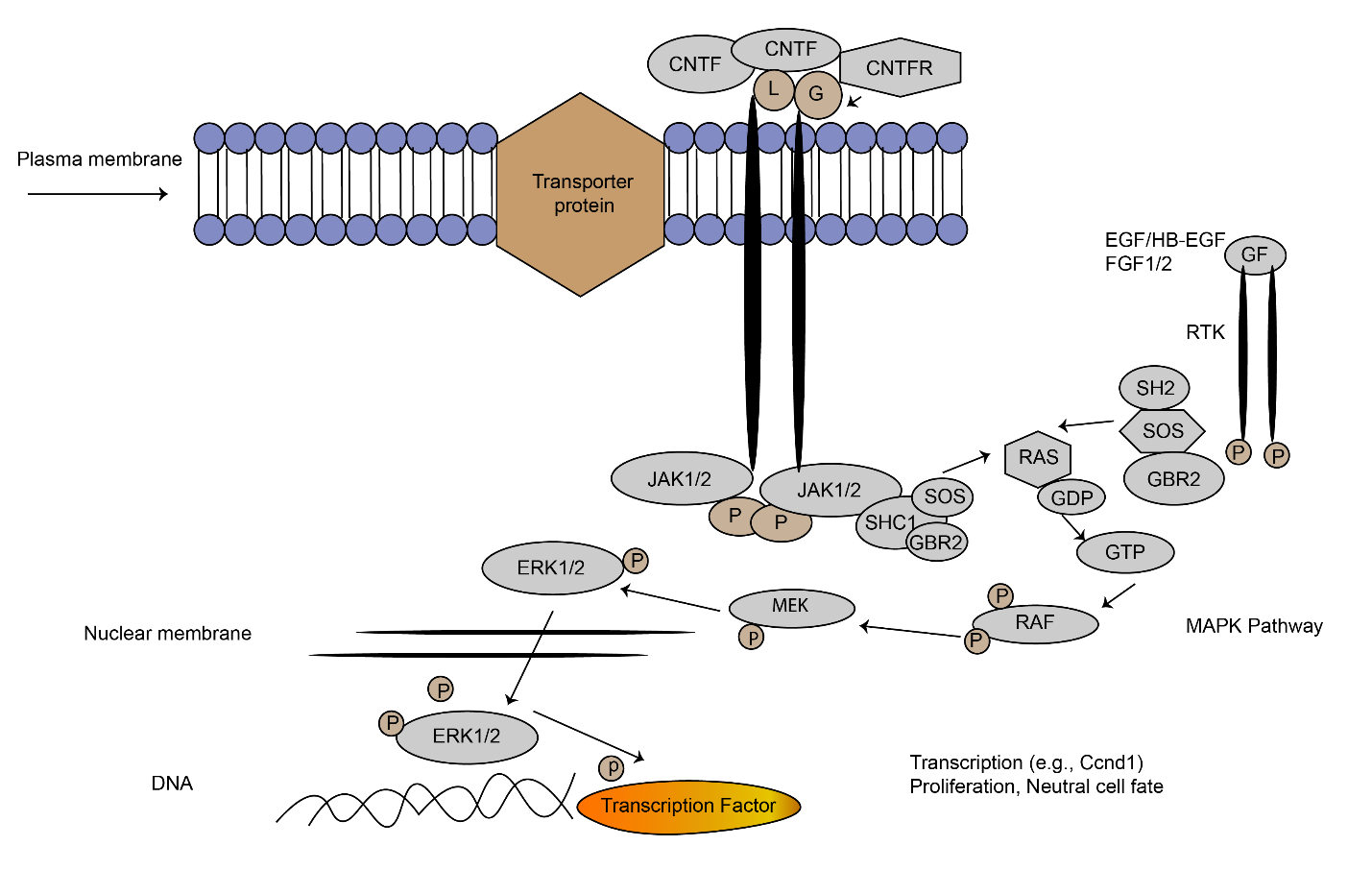
Mitogen activated protein kinase (MAPK) signal transduction.
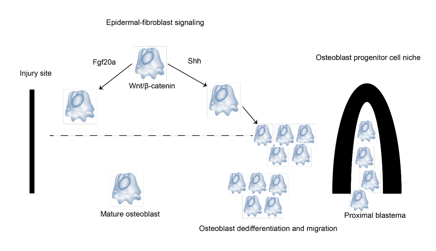
Fin regeneration integrated model.
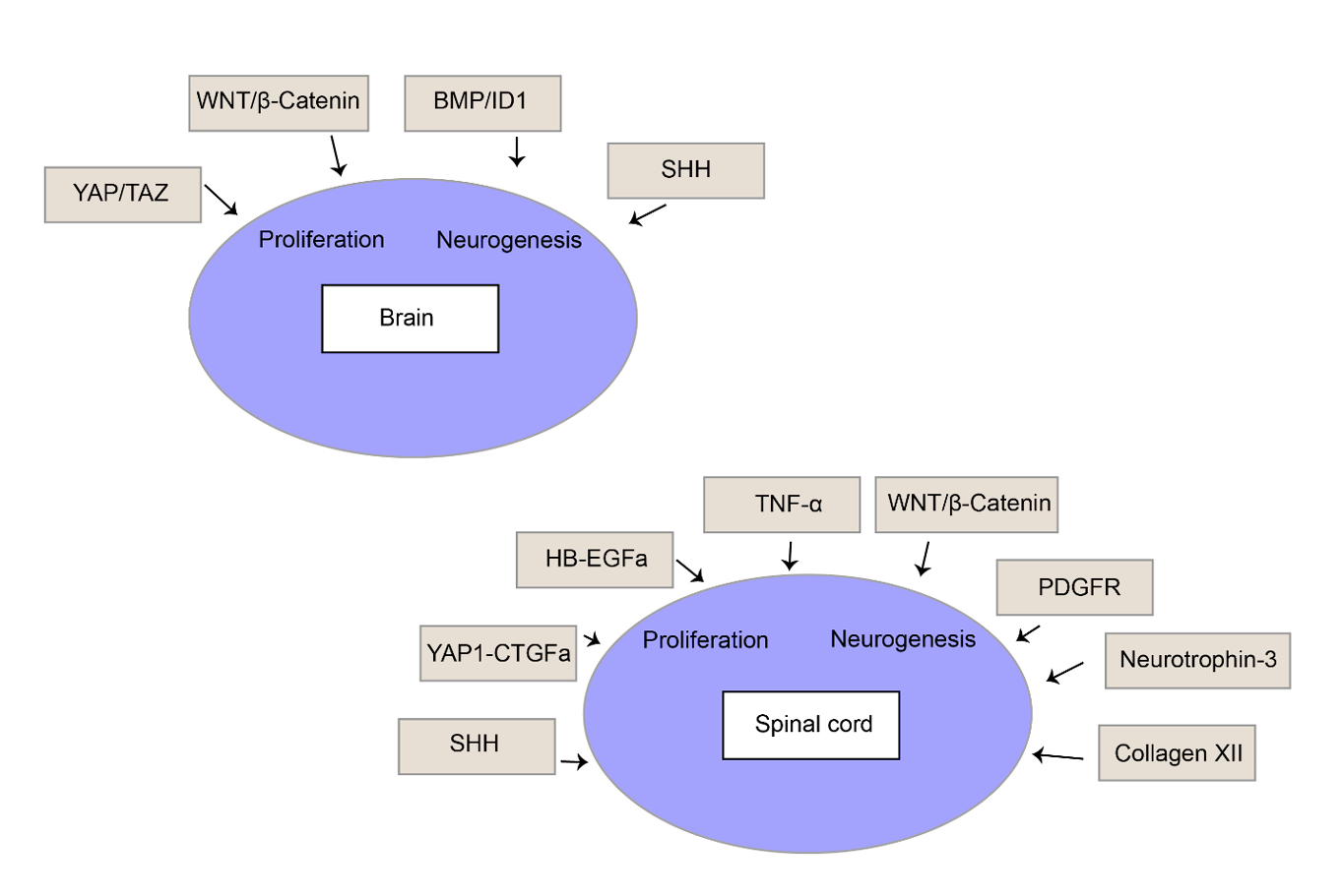
Cell types and pathways controlling CNS regeneration in zebrafish.
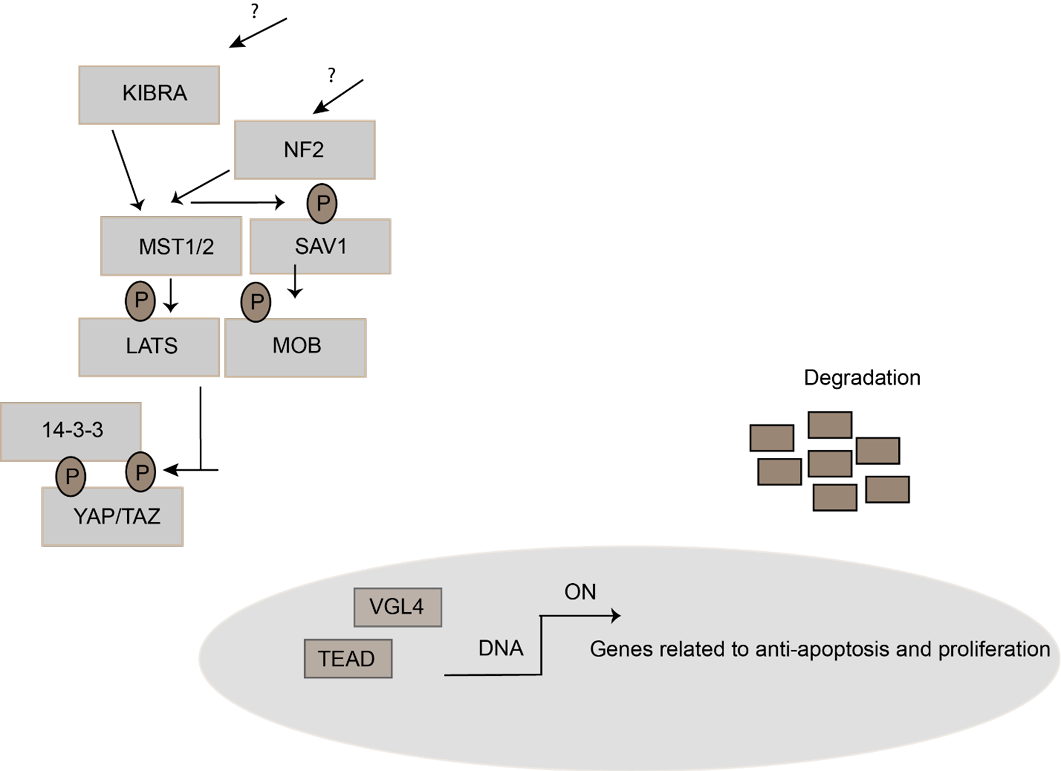
Hippo signaling on high cell density
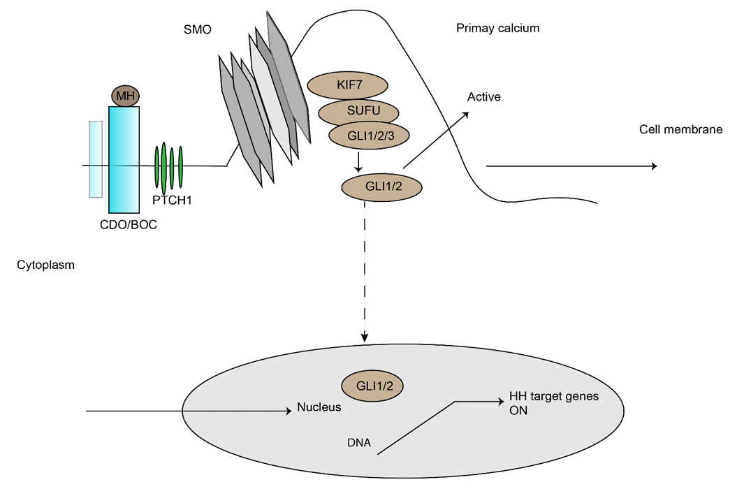
Hedgehog signaling pathway
References
- Adolf, B., P. Chapouton, C.S. Lam, S. Topp, B. Tannhäuser, U. Strähle, M. Götz, and L. Bally-Cuif. 2006. Conserved and acquired features of adult neurogenesis in the zebrafish telencephalon. Dev. Biol., 295(1): 278–293. https://doi.org/10.1016/j.ydbio.2006.03.023[CrossRef]
- Barbosa, J.S. and J. Ninkovic. 2016. Adult neural stem cell behavior underlying constitutive and restorative neurogenesis in zebrafish. Neurogen., 3(1): e1148101. https://doi.org/10.1080/23262133.2016.1148101[CrossRef]
- Barreiro-Iglesias, A., K.S. Mysiak, A.L. Scott, M.M. Reimer, Y. Yang, C.G. Becker, and T. Becker. 2015. Serotonin promotes development and regeneration of spinal motor neurons in zebrafish. Cell Rep., 13(5): 924–932. https://doi.org/10.1016/j.celrep.2015.09.050[CrossRef]
- Bergmann, C. and J.H. Distler. 2016. Canonical Wnt signaling in systemic sclerosis. Lab. Invest., 96(2): 151–155. https://doi.org/10.1038/labinvest.2015.154[CrossRef]
- Bringmann, A., I. Iandiev, T. Pannicke, A. Wurm, M. Hollborn, P. Wiedemann, N.N. Osborne, and A. Reichenbach. 2009. Cellular signaling and factors involved in Müller cell gliosis: Neuroprotective and detrimental effects. Prog. Retin. Eye Res., 28(6): 423–451. https://doi.org/10.1016/j.preteyeres.2009.07.001[CrossRef]
- Bruton, F.A., A. Kaveh, K.M. Ross-Stewart, G. Matrone, M.E. Oremek, E.G. Solomonidis, C.S. Tucker, J.J. Mullins, C.D. Lucas, and M. Brittan. 2022. Macrophages trigger cardiomyocyte proliferation by increasing epicardial vegfaa expression during larval zebrafish heart regeneration. Dev. Cell, 57(12): 1512–1528.e15. https://doi.org/10.1016/j.devcel.2022.05.014[CrossRef]
- Chablais, F. and A. Jaźwińska. 2010. IGF signaling between blastema and wound epidermis is required for fin regeneration. Develop., 137(6): 871–879. https://doi.org/10.1242/dev.043885[CrossRef]
- Chablais, F., and A. Jaźwińska. 2012. The regenerative capacity of the zebrafish heart is dependent on TGFβ signaling. Develop., 139(11): 1921–1930. https://doi.org/10.1242/dev.078543[CrossRef]
- Chablais, F., J. Veit, G. Rainer, and A. Jaźwińska. 2011. The zebrafish heart regenerates after cryoinjury-induced myocardial infarction. BMC Dev. Biol., 11(1): 21. https://doi.org/10.1186/1471-213X-11-21[CrossRef]
- Chapouton, P., B. Adolf, C. Leucht, B. Tannhäuser, S. Ryu, W. Driever, and L. Bally-Cuif. 2006. her5 expression reveals a pool of neural stem cells in the adult zebrafish midbrain. Develop., 133(21): 4293–4303. https://doi.org/10.1242/dev.02573[CrossRef]
- Chapouton, P., R. Jagasia, and L. Bally-Cuif. 2007. Adult neurogenesis in non-mammalian vertebrates. BioEssays, 29(8): 745–757. https://doi.org/10.1002/bies.20615[CrossRef]
- Chapouton, P., P. Skupien, B. Hesl, M. Coolen, J.C. Moore, R. Madelaine, E. Kremmer, T. Faus-Kessler, P. Blader, and N.D. Lawson. 2010. Notch activity levels control the balance between quiescence and recruitment of adult neural stem cells. J. Neurosci., 30(23): 7961–7974. https://doi.org/10.1523/JNEUROSCI.6170-09.2010[CrossRef]
- Cigliola, V., C.J. Becker, and K.D. Poss. 2020. Building bridges, not walls: Spinal cord regeneration in zebrafish. Dis. Model. Mech., 13(5): dmm044131. https://doi.org/10.1242/dmm.044131[CrossRef]
- Di Giaimo, R., T. Durovic, P. Barquin, A. Kociaj, T. Lepko, S. Aschenbroich, C.T. Breunig, M. Irmler, F.M. Cernilogar, G. Schotta, J.S. Barbosa, D. Trümbach, E.V. Baumgart, A.M. Neuner, J. Beckers, W. Wurst, S.H. Stricker, and J. Ninkovic. 2018. The aryl hydrocarbon receptor pathway defines the time frame for restorative neurogenesis. Cell Rep., 25(12): 3241–3251.e5. https://doi.org/10.1016/j.celrep.2018.11.055[CrossRef]
- Didangelos, A., M. Puglia, M. Iberl, C. Sanchez-Bellot, B. Roschitzki, and E.J. Bradbury. 2016. High-throughput proteomics reveal alarmins as amplifiers of tissue pathology and inflammation after spinal cord injury. Sci. Rep., 6(1): 21607. https://doi.org/10.1038/srep21607[CrossRef]
- Diotel, N., R. Rodriguez Viales, O. Armant, M. März, M. Ferg, S. Rastegar, and U. Strähle. 2015. Comprehensive expression map of transcription regulators in the adult zebrafish telencephalon reveals distinct neurogenic niches. J. Comp. Neurol., 523(8): 1202–1221. https://doi.org/10.1002/cne.23733[CrossRef]
- Diotel, N., C. Vaillant, C. Gabbero, S. Mironov, A. Fostier, M.-M. Gueguen, I. Anglade, O. Kah, and E. Pellegrini. 2013. Effects of estradiol in adult neurogenesis and brain repair in zebrafish. Horm. Behav., 63(2): 193–207. https://doi.org/10.1016/j.yhbeh.2012.04.003[CrossRef]
- Edelmann, K., L. Glashauser, S. Sprungala, B. Hesl, M. Fritschle, J. Ninkovic, L. Godinho, and P. Chapouton. 2013. Increased radial glia quiescence, decreased reactivation upon injury and unaltered neuroblast behavior underlie decreased neurogenesis in the aging zebrafish telencephalon. J. Comp. Neurol., 521(13): 3099–3115. https://doi.org/10.1002/cne.23347[CrossRef]
- Fausett, B.V., and D. Goldman. 2006. A role for α1 tubulin-expressing Müller glia in regeneration of the injured zebrafish retina. J. Neurosci., 26(23): 6303–6313. https://doi.org/10.1523/JNEUROSCI.0332-06.2006[CrossRef]
- Ganz, J., J. Kaslin, S. Hochmann, D. Freudenreich, and M. Brand. 2010. Heterogeneity and Fgf dependence of adult neural progenitors in the zebrafish telencephalon. Glia, 58(11): 1345–1363. https://doi.org/10.1002/glia.21012[CrossRef]
- Gemberling, M., T.J. Bailey, D.R. Hyde, and K.D. Poss. 2013. The zebrafish as a model for complex tissue regeneration. Trends Genet., 29(11): 611–620. https://doi:10.1016/j.tig.2013.07.003[CrossRef]
- Gemberling, M., R. Karra, A.L. Dickson, and K.D. Poss. 2015. Nrg1 is an injury-induced cardiomyocyte mitogen for the endogenous heart regeneration program in zebrafish. eLife, 4: e05871. https://doi.org/10.7554/eLife.05871[CrossRef]
- Govindan, J., and M.K. Iovine. 2015. Dynamic remodeling of the extra cellular matrix during zebrafish fin regeneration. Gene Expr. Patterns, 19(1–2): 21–29. https://doi.org/10.1016/j.gep.2015.06.001[CrossRef]
- Grotek, B., D. Wehner, and G. Weidinger. 2013. Notch signaling coordinates cellular proliferation with differentiation during zebrafish fin regeneration. Develop., 140(7): 1412–1423. https://doi.org/10.1242/dev.087452[CrossRef]
- Guillon, E., S. Bretaud, and F. Ruggiero. 2016. Slow muscle precursors lay down a collagen XV matrix fingerprint to guide motor axon navigation. J. Neurosci., 36(9): 2663–2676. https://doi:10.1523/JNEUROSCI.2847-15.2016[CrossRef]
- Haferlach, T., A. Stengel, S. Eckstein, K. Perglerová, T. Alpermann, W. Kern, C. Haferlach, and M. Meggendorfer. 2016. The new provisional WHO entity ‘RUNX1 mutated AML’ shows specific genetics but no prognostic influence of dysplasia. Leukemia, 30(10): 2109–2112. https://doi:10.1038/leu.2016.150[CrossRef]
- Han, P., X.-H. Zhou, N. Chang, C.-L. Xiao, S. Yan, H. Ren, X.-Z. Yang, M.-L. Zhang, Q. Wu, and B. Tang. 2014. Hydrogen peroxide primes heart regeneration with a derepression mechanism. Cell Res., 24(9): 1091–1107. https://doi.org/10.1038/cr.2014.108[CrossRef]
- Hippert, C., A.B. Graca, A.C. Barber, E.L. West, A.J. Smith, R.R. Ali, and R.A. Pearson. 2015. Müller glia activation in response to inherited retinal degeneration is highly varied and disease-specific. PLoS ONE., 10(3): e0120415. https://doi.org/10.1371/journal.pone.0120415.[CrossRef]
- Hirose, K., T. Shiomi, S. Hozumi, and Y. Kikuchi. 2014. Mechanistic target of rapamycin complex 1 signaling regulates cell proliferation, cell survival, and differentiation in regenerating zebrafish fins. BMC Dev. Biol., 14(1): 42. https://doi.org/10.1186/s12861-014-0042-9[CrossRef]
- Hoang, T., J. Wang, P. Boyd, F. Wang, C. Santiago, L. Jiang, S. Yoo, M. Lahne, L.J. Todd, and M. Jia. 2020. Gene regulatory networks controlling vertebrate retinal regeneration. Sci., 370(6519): eabb8598. https://doi.org/10.1126/science.abb8598[CrossRef]
- Huang, Y., M.R. Harrison, A. Osorio, J. Kim, A. Baugh, C. Duan, H.M. Sucov, and C.-L. Lien. 2013. Igf signaling is required for cardiomyocyte proliferation during zebrafish heart development and regeneration. PLoS ONE., 8(6): e67266. https://doi.org/10.1371/journal.pone.0067266[CrossRef]
- Isaacman-Beck, J., V. Schneider, C. Franzini-Armstrong, and M. Granato. 2015. The lh3 glycosyltransferase directs target-selective peripheral nerve regeneration. Neuron, 88(4): 691–703. http://dx.doi.org/10.1016/j.neuron.2015.10.004[CrossRef]
- Itou, J., H. Kawakami, T. Burgoyne, and Y. Kawakami. 2012. Life-long preservation of the regenerative capacity in the fin and heart in zebrafish. Biol. Open, 1(8): 739–746. https://doi.org/10.1242/bio.20121057[CrossRef]
- Jopling, C., E. Sleep, M. Raya, M. Martí, A. Raya, and J.C.I. Belmonte. 2010. Zebrafish heart regeneration occurs by cardiomyocyte dedifferentiation and proliferation. Nature, 464(7288): 606–609. https://doi.org/10.1038/nature08899[CrossRef]
- Kang, J., J. Hu, R. Karra, A.L. Dickson, V.A. Tornini, G. Nachtrab, M. Gemberling, J.A. Goldman, B.L. Black, and K.D. Poss. 2016. Modulation of tissue repair by regeneration enhancer elements. Nature, 532(7598): 201–206. https://doi.org/10.1038/nature17644[CrossRef]
- Kaslin, J., J. Ganz, and M. Brand. 2008. Proliferation, neurogenesis and regeneration in the non-mammalian vertebrate brain. Philos. Trans. R. Soc. B Biol. Sci., 363(1489): 101–122. https://doi.org/10.1098/rstb.2006.2015[CrossRef]
- Kassen, S.C., V. Ramanan, J.E. Montgomery, C.T. Burket, C.G. Liu, T.S. Vihtelic, and D.R. Hyde. 2007. Time course analysis of gene expression during light-induced photoreceptor cell death and regeneration in albino zebrafish. Dev. Neurobiol., 67(8): 1009–1031. https://doi.org/10.1002/dneu.20362[CrossRef]
- Kaur, S., S. Gupta, M. Chaudhary, M.A. Khursheed, S. Mitra, A.J. Kurup, and R. Ramachandran. 2018. let-7 MicroRNA-mediated regulation of Shh signaling and the gene regulatory network is essential for retina regeneration. Cell Rep., 23(5): 1409–1423. https://doi.org/10.1016/j.celrep.2018.04.002[CrossRef]
- Kikuchi, K., J.E. Holdway, A.A. Werdich, R.M. Anderson, Y. Fang, G.F. Egnaczyk, T. Evans, C.A. MacRae, D.Y. Stainier, and K.D. Poss. 2010. Primary contribution to zebrafish heart regeneration by gata4+ cardiomyocytes. Nature, 464(7288): 601–605. https://doi.org/10.1038/nature08804[CrossRef]
- Kishimoto, N., K. Shimizu, and K. Sawamoto. 2012. Neuronal regeneration in a zebrafish model of adult brain injury. Dis. Model. Mech., 5(2): 200–209. https://doi.org/10.1242/dmm.007336[CrossRef]
- Kizil, C., J. Kaslin, V. Kroehne, and M. Brand. 2012. Adult neurogenesis and brain regeneration in zebrafish. Dev. Neurobiol., 72(3): 429–461. https://doi.org/10.1002/dneu.20918[CrossRef]
- Knowlton, M.N., B.M. Chan, and G.M. Kelly. 2003. The zebrafish band 4.1 member Mir is involved in cell movements associated with gastrulation. Dev. Biol., 264(2): 407–429. https://doi:10.1016/j.ydbio.2003.09.001[CrossRef]
- Kroehne, V., D. Freudenreich, S. Hans, J. Kaslin, and M. Brand. 2011. Regeneration of the adult zebrafish brain from neurogenic radial glia-type progenitors. Development, 138(22): 4831–4841. https://doi.org/10.1242/dev.072587[CrossRef]
- Kyritsis, N., C. Kizil, S. Zocher, V. Kroehne, J. Kaslin, D. Freudenreich, A. Iltzsche, and M. Brand. 2012. Acute inflammation initiates the regenerative response in the adult zebrafish brain. Science, 338(6112): 1353–1356. https://doi.org/10.1126/science.1228773[CrossRef]
- Lahne, M., J. Li, R.M. Marton, and D.R. Hyde. 2015. Actin-cytoskeleton- and ROCK-mediated INM are required for photoreceptor regeneration in the adult zebrafish retina. J. Neurosci., 35(47): 15612–15634. https://doi:10.1523/JNEUROSCI.5005-14.2015[CrossRef]
- Lee, H.J., Y. Hou, Y. Chen, Z.Z. Dailey, A. Riddihough, H.S. Jang, T. Wang, and S.L. Johnson. 2020. Regenerating zebrafish fin epigenome is characterized by stable lineage-specific DNA methylation and dynamic chromatin accessibility. Genome Biol., 21(1): 52. https://doi.org/10.1186/s13059-020-1948-0[CrossRef]
- Lepilina, A., A.N. Coon, K. Kikuchi, J.E. Holdway, R.W. Roberts, C.G. Burns, and K.D. Poss. 2006. A dynamic epicardial injury response supports progenitor cell activity during zebrafish heart regeneration. Cell, 127(3): 607–619. https://doi.org/10.1016/j.cell.2006.08.052[CrossRef]
- Lien, C.-L., M. Schebesta, S. Makino, G.J. Weber, and M.T. Keating. 2006. Gene expression analysis of zebrafish heart regeneration. PLoS Biol., 4(8): e260. https://doi.org/10.1371/journal.pbio.0040260[CrossRef]
- Lim, C.H., Q. Sun, K. Ratti, S.-H. Lee, Y. Zheng, M. Takeo, W. Lee, P. Rabbani, M.V. Plikus, J.E. Cain, D.H. Wang, D.N. Watkins, S. Millar, M.M. Taketo, P. Myung, G. Cotsarelis, and M. Ito. 2018. Hedgehog stimulates hair follicle neogenesis by creating inductive dermis during murine skin wound healing. Nat. Commun., 9(1): 4903. https://doi.org/10.1038/s41467-018-07142-9[CrossRef]
- Lim, F.T., S. Ogawa, and I.S. Parhar. 2016. Spred-2 expression is associated with neural repair of injured adult zebrafish brain. J. Chem. Neuroanat., 77: 176–186. http://dx.doi.org/10.1016/j.jchemneu.2016.07.005[CrossRef]
- Lindsey, B.W., A. Darabie, and V. Tropepe. 2012. The cellular composition of neurogenic periventricular zones in the adult zebrafish forebrain. J. Comp. Neurol., 520(10): 2275–2316. https://doi.org/10.1002/cne.23065[CrossRef]
- Luo, J., R.A. Uribe, S. Hayton, A.-A. Calinescu, J.M. Gross, and P.F. Hitchcock. 2012. Midkine-A functions upstream of Id2a to regulate cell cycle kinetics in the developing vertebrate retina. Neural Dev., 7(1): 33. https://doi.org/10.1186/1749-8104-7-33[CrossRef]
- Maynard, A., M. Soretić, and B. Treutlein. 2024. Single-cell genomic profiling to study regeneration. Curr. Opin. Genet. Dev., 87: 102231. https://doi.org/10.1016/j.gde.2024.102231[CrossRef]
- Mercer, S.E., S.J. Odelberg, and H.-G. Simon. 2013. A dynamic spatiotemporal extracellular matrix facilitates epicardial-mediated vertebrate heart regeneration. Dev. Biol., 382(2): 457–469. http://dx.doi.org/10.1016/j.ydbio.2013.08.002[CrossRef]
- Mitra, S., P. Sharma, S. Kaur, M.A. Khursheed, S. Gupta, R. Ahuja, A.J. Kurup, M. Chaudhary, and R. Ramachandran. 2018. Histone deacetylase-mediated Müller glia reprogramming through Her4.1-Lin28a axis is essential for retina regeneration in zebrafish. iScience, 7: 68–84. https://doi.org/10.1016/j.isci.2018.08.008[CrossRef]
- Munderloh, C., G.P. Solis, V. Bodrikov, F.A. Jaeger, M. Wiechers, E. Málaga-Trillo, and C.A. Stuermer. 2009. Reggies/flotillins regulate retinal axon regeneration in the zebrafish optic nerve and differentiation of hippocampal and N2a neurons. J. Neurosci., 29(20): 6607–6615. https://doi.org/10.1523/JNEUROSCI.0870-09.2009[CrossRef]
- Nagashima, M., L.K. Barthel, and P.A. Raymond. 2013. A self-renewing division of zebrafish Müller glial cells generates neuronal progenitors that require N-cadherin to regenerate retinal neurons. Development, 140(22): 4510–4521. https://doi.org/10.1242/dev.090738[CrossRef]
- Nguyen-Chi, M., B. Laplace-Builhe, J. Travnickova, P. Luz-Crawford, G. Tejedor, Q.T. Phan, I. Duroux-Richard, J.-P. Levraud, K. Kissa, G. Lutfalla, C. Jorgensen, and F. Djouad. 2015. Identification of polarized macrophage subsets in zebrafish. eLife, 4: e07288. https://doi.org/10.7554/eLife.07288[CrossRef]
- Noorimotlagh, Z., M. Babaie, M. Safdarian, T. Ghadiri, and V. Rahimi-Movaghar. 2017. Mechanisms of spinal cord injury regeneration in zebrafish: A systematic review. Iran. J. Basic Med. Sci., 20(12): 1287–1296. https://doi.org/10.22038/IJBMS.2017.9620[CrossRef]
- Okada, S., M. Hara, K. Kobayakawa, Y. Matsumoto, and Y. Nakashima. 2018. Astrocyte reactivity and astrogliosis after spinal cord injury. Neurosci. Res., 126: 39–43. https://doi.org/10.1016/j.neures.2017.10.004[CrossRef]
- Petrie, T.A., N.S. Strand, C. Tsung-Yang, J.S. Rabinowitz, and R.T. Moon. 2014. Macrophages modulate adult zebrafish tail fin regeneration. Development, 141(13): 2581–2591. https://doi.org/10.1242/dev.098459[CrossRef]
- Pfefferli, C. and A. Jaźwińska. 2015. The art of fin regeneration in zebrafish. Regenerat., 2(2): 72–83. https://doi.org/10.1002/reg2.33[CrossRef]
- Porrello, E.R., A.I. Mahmoud, E. Simpson, J.A. Hill, J.A. Richardson, E.N. Olson, and H.A. Sadek. 2011. Transient regenerative potential of the neonatal mouse heart. Sci., 331(6020): 1078–1080. https://doi.org/10.1126/science.1200708[CrossRef]
- Poss, K.D. 2010. Advances in understanding tissue regenerative capacity and mechanisms in animals. Nat. Rev. Genet., 11(10): 710–722. https://doi.org/10.1038/nrg2879[CrossRef]
- Poss, K.D., J. Shen, A. Nechiporuk, G. McMahon, B. Thisse, C. Thisse, and M.T. Keating. 2000. Roles for Fgf signaling during zebrafish fin regeneration. Dev. Biol., 222(2): 347–358. https://doi.org/10.1006/dbio.2000.9722[CrossRef]
- Powell, C., F. Elsaeidi, and D. Goldman. 2012. Injury-dependent Müller glia and ganglion cell reprogramming during tissue regeneration requires Apobec2a and Apobec2b. J. Neurosci., 32(3): 1096–1109. https://doi.org/10.1523/JNEUROSCI.5603-11.2012[CrossRef]
- Powell, C., A.R. Grant, E. Cornblath, and D. Goldman. 2013. Analysis of DNA methylation reveals a partial reprogramming of the Müller glia genome during retina regeneration. Proc. Natl. Acad. Sci. U.S.A., 110(49): 19814–19819. https://doi.org/10.1073/pnas.1312009110[CrossRef]
- Purcell, N.H., B.J. Wilkins, A. York, M.K. Saba-El-Leil, S. Meloche, J. Robbins, and J.D. Molkentin. 2007. Genetic inhibition of cardiac ERK1/2 promotes stress-induced apoptosis and heart failure but has no effect on hypertrophy in vivo. Proc. Natl. Acad. Sci. U.S.A., 104(35): 14074–14079. https://doi.org/10.1073/pnas.0610906104[CrossRef]
- Qin, Z., L.K. Barthel, and P.A. Raymond. 2009. Genetic evidence for shared mechanisms of epimorphic regeneration in zebrafish. Proc. Natl. Acad. Sci. U.S.A., 106(23): 9310–9315. https://doi.org/10.1073/pnas.0811186106[CrossRef]
- Ramachandran, R., B.V. Fausett, and D. Goldman. 2010. Ascl1a regulates Müller glia dedifferentiation and retinal regeneration through a Lin-28-dependent, let-7 microRNA signalling pathway. Nat. Cell Biol., 12(11): 1101–1107. https://doi.org/10.1038/ncb2115[CrossRef]
- Ramachandran, R., X.-F. Zhao, and D. Goldman. 2011. Ascl1a/Dkk/β-catenin signaling pathway is necessary and glycogen synthase kinase-3β inhibition is sufficient for zebrafish retina regeneration. Proc. Natl. Acad. Sci. U.S.A., 108(38): 15858–15863. https://doi.org/10.1073/pnas.1107220108[CrossRef]
- Ramachandran, R., X.-F. Zhao, and D. Goldman. 2012. Insm1a-mediated gene repression is essential for the formation and differentiation of Müller glia-derived progenitors in the injured retina. Nat. Cell Biol., 14(10): 1013–1023. https://doi.org/10.1038/ncb2586[CrossRef]
- Reichenbach, A., and A. Bringmann. 2013. New functions of Müller cells. Glia, 61(5): 651–678. https://doi.org/10.1002/glia.22477[CrossRef]
- Russell, C. 2003. The roles of Hedgehogs and Fibroblast Growth Factors in eye development and retinal cell rescue. Vision Res., 43(8): 899–912. https://doi:10.1016/S0042-6989(02)00416-9[CrossRef]
- Sallin, P., A.-S. de Preux Charles, V. Duruz, C. Pfefferli, and A. Jaźwińska. 2015. A dual epimorphic and compensatory mode of heart regeneration in zebrafish. Dev. Biol., 399(1): 27–40. https://doi.org/10.1016/j.ydbio.2014.12.002[CrossRef]
- Sánchez-Iranzo, H., M. Galardi-Castilla, A. Sanz-Morejón, J.M. González-Rosa, R. Costa, A. Ernst, J. Sainz de Aja, X. Langa, and N. Mercader. 2018. Transient fibrosis resolves via fibroblast inactivation in the regenerating zebrafish heart. Proc. Natl. Acad. Sci. U.S.A., 115(16): 4188–4193. https://doi.org/10.1073/pnas.1716713115[CrossRef]
- Schenk, H. and I.A. Drummond. 2025. Kidney development, injury and regeneration—Zebrafish. Curr. Top. Dev. Biol., 163: 307–321. https://doi.org/10.1016/bs.ctdb.2025.01.008[CrossRef]
- Smith, A., F. Avaron, D. Guay, B.d. Padhi, and M. Akimenko. 2006. Inhibition of BMP signaling during zebrafish fin regeneration disrupts fin growth and scleroblast differentiation and function. Dev. Biol., 299(2): 438–454. https://doi.org/10.1016/j.ydbio.2006.08.016[CrossRef]
- Stewart, S., A.W. Gomez, B.E. Armstrong, A. Henner, and K. Stankunas. 2014. Sequential and opposing activities of Wnt and BMP coordinate zebrafish bone regeneration. Cell Rep., 6(3): 482–498. https://doi.org/10.1016/j.celrep.2014.01.010[CrossRef]
- Stewart, S., Z.Y. Tsun, and J.C.I. Belmonte. 2009. A histone demethylase is necessary for regeneration in zebrafish. Proc. Natl. Acad. Sci. U.S.A., 106(47): 19889–19894. https://doi.org/10.1073/pnas.0904132106[CrossRef]
- Stoick-Cooper, C.L., R.T. Moon, and G. Weidinger. 2007. Advances in signaling in vertebrate regeneration as a prelude to regenerative medicine. Genes Dev., 21(11): 1292–1315. https://doi.org/10.1101/gad.1540507[CrossRef]
- Takeda, A., Y. Atobe, T. Kadota, R.C. Goris, and K. Funakoshi. 2015. Axonal regeneration through the fibrous scar in lesioned goldfish spinal cord. Neuroscience, 284: 134–152. https://doi.org/10.1016/j.neuroscience.2014.09.066[CrossRef]
- Tanaka, E.M., and P.W. Reddien. 2011. The cellular basis for animal regeneration. Dev. Cell, 21(1): 172–185. https://doi.org/10.1016/j.devcel.2011.06.016[CrossRef]
- Thomas, J.L., G.W. Morgan, K.M. Dolinski, and R. Thummel. 2018. Characterization of the pleiotropic roles of Sonic Hedgehog during retinal regeneration in adult zebrafish. Exp. Eye Res., 166: 106–115. https://doi.10.1016/j.exer.2017.10.003
- Thomas, J.L., A.H. Ranski, G.W. Morgan, and R. Thummel. 2016. Reactive gliosis in the adult zebrafish retina. Exp. Eye Res., 143: 98–109. https://doi.org/10.1016/j.exer.2015.09.017[CrossRef]
- Thorimbert, V., D. König, J. Marro, F. Ruggiero, and A. Jaźwińska. 2015. Bone morphogenetic protein signaling promotes morphogenesis of blood vessels, wound epidermis, and actinotrichia during fin regeneration in zebrafish. FASEB J., 29(10): 4299–4312. https://doi.org/10.1096/fj.15-272955[CrossRef]
- Thummel, R., J.M. Enright, S.C. Kassen, J.E. Montgomery, T.J. Bailey, and D.R. Hyde. 2010. Pax6a and Pax6b are required at different points in neuronal progenitor cell proliferation during zebrafish photoreceptor regeneration. Exp. Eye Res., 90(5): 572–582. https://doi.org/10.1016/j.exer.2010.02.001[CrossRef]
- Tsang, M. and I.B. Dawid. 2004. Promotion and attenuation of FGF signaling through the Ras-MAPK pathway. Sci. STKE, 2004(228): pe17. https://doi.org/10.1126/stke.2282004pe17[CrossRef]
- Udeh, A., G. Dvoriantchikova, T. Carmy, D. Ivanov, and A.S. Hackam. 2019. Wnt signaling induces neurite outgrowth in mouse retinal ganglion cells. Exp. Eye Res., 182: 39–43. https://doi.10.1016/j.exer.2019.03.004
- Viales, R.R., N. Diotel, M. Ferg, O. Armant, J. Eich, A. Alunni, M. März, L. Bally-Cuif, S. Rastegar, and U. Strähle. 2015. The helix-loop-helix protein id1 controls stem cell proliferation during regenerative neurogenesis in the adult zebrafish telencephalon. Stem Cells, 33(3): 892–903. https://doi.org/10.1002/stem.1883[CrossRef]
- Wagner, D.E., C. Weinreb, Z.M. Collins, J.A. Briggs, S.G. Megason, and A.M. Klein. 2018. Single-cell mapping of gene expression landscapes and lineage in the zebrafish embryo. Sci., 360(6392): 981–987. https://doi.org/10.1126/science.aar4362[CrossRef]
- Wan, J., R. Ramachandran, and D. Goldman. 2012. HB-EGF is necessary and sufficient for Müller glia dedifferentiation and retina regeneration. Dev. Cell, 22(2): 334–347. https://doi.org/10.1016/j.devcel.2011.11.020[CrossRef]
- Wan, J., X.F. Zhao, A. Vojtek, and D. Goldman. 2014. Retinal injury, growth factors, and cytokines converge on β-catenin and pStat3 signaling to stimulate retina regeneration. Cell Rep., 9(1): 285–297. https://doi.org/10.1016/j.celrep.2014.08.048[CrossRef]
- Wang, J., R. Karra, A.L. Dickson, and K.D. Poss. 2013. Fibronectin is deposited by injury-activated epicardial cells and is necessary for zebrafish heart regeneration. Dev. Biol., 382(2): 427–435. https://doi.org/10.1016/j.ydbio.2013.08.012[CrossRef]
- Wehner, D., W. Cizelsky, M.D. Vasudevaro, G. Özhan, C. Haase, B. Kagermeier-Schenk, A. Röder, R.I. Dorsky, E. Moro, F. Argenton, M. Kühl, and G. Weidinger. 2014. Wnt/β-catenin signaling defines organizing centers that orchestrate growth and differentiation of the regenerating zebrafish caudal fin. Cell Rep., 6(3): 467–481. https://doi.org/10.1016/j.celrep.2013.12.036[CrossRef]
- Wehner, D., T.M. Tsarouchas, A. Michael, C. Haase, G. Weidinger, M.M. Reimer, T. Becker, and C.G. Becker. 2017. Wnt signaling controls pro-regenerative Collagen XII in functional spinal cord regeneration in zebrafish. Nat. Commun., 8(1): 126. https://doi.org/10.1038/s41467-017-00143-0[CrossRef]
- Wei, X., S. Fu, H. Li, Y. Liu, S. Wang, W. Feng, Y. Yang, X. Liu, Y.-Y. Zeng, M. Cheng, Y. Lai, X. Qiu, L. Wu, N. Zhang, Y. Jiang, J. Xu, X. Su, C. Peng, L. Han, W.P.-K. Lou, C. Liu, Y. Yuan, K. Ma, T. Yang, X. Pan, S. Gao, A. Chen, M.A. Esteban, H. Yang, J. Wang, G. Fan, L. Liu, L. Chen, X. Xu, J.-F. Fei, and Y. Gu. 2022. Single-cell Stereo-seq reveals induced progenitor cells involved in axolotl brain regeneration. Sci., 377(6610): eabp9444. https://doi.org/10.1126/science.abp9444[CrossRef]
- Zhao, L., R. Ben-Yair, C.E. Burns, and C.G. Burns. 2019. Endocardial Notch signaling promotes cardiomyocyte proliferation in the regenerating zebrafish heart through Wnt pathway antagonism. Cell Rep., 26(3): 546–554.e5. https://doi.org/10.1016/j.celrep.2018.12.048[CrossRef]
- Zhao, X.F., J. Wan, C. Powell, R. Ramachandran, M.G. Myers, and D. Goldman. 2014. Leptin and IL-6 family cytokines synergize to stimulate Müller glia reprogramming and retina regeneration. Cell Rep., 9(1): 272–284. https://doi.org/10.1016/j.celrep.2014.08.047[CrossRef]
- Zupanc, G.K., K.S. Kompass, I. Horschke, R. Ott, and H. Schwarz. 1998. Apoptosis after injuries in the cerebellum of adult teleost fish. Exp. Neurol., 152(2): 221–230. https://doi.org/10.1006/exnr.1998.6853[CrossRef]
Services
Article usage statistics
Loading…