
Titanium dioxide (TiO2) is widely used nanoparticle all over the world. In this study, we have investigated the histopathological effect on zebrafish ovaries after exposure to TiO2 nanoparticles. Adult zebrafish individuals were exposed to 1, 2 and 4 mg/L TiO2 for 5 days, and then their ovaries were evaluated using light and transmission electron microscopy. Numerous degenerated follicles with cytoplasmic vacuolization, mitotic catastrophe in mitochondria , chromatin condensation, mitochondrial vesiculation and dispersion at ooplasm were observed. In mitochondria, mitotic catastrophe , vesiculation, swelling and loss of organization of cristae were detected.Here we showed that TiO2 exposure trigger paraptotic type cell death in zebrafish ovary.
Research Article · Volume 49, Issue 3, 24 May 2017 · Open Access
Exposure of Zebrafish (Danio rerio) to Titanium Dioxide Nanoparticle Causes Paraptosis: Evaluation of Ovarian Follicle Ultrastructure
· · ·
Affiliations & Notes ⌄
- 1Department of Biology, Science and Letters Faculty, Sakarya University, Serdivan, Sakarya, Turkey 2Department of Histology and Embryology, İstanbulFaculty of Medicine, İstanbul University, İstanbul, Turkey , * Corresponding author: cansua@sakarya.edu.tr
Article Info ⌄
- Journal:
- Pakistan Journal of Zoology
- Published:
- 24 May 2017

From the article
- They used female individuals and provided that chronic exposure of zebrafish to 0.1 mg/L TiO2, can significantly impair zebrafish female reproduction.
- They proved that neither nanoTiO2 nor TiO2 bulk showed any toxicity to zebrafish embryos and larvae.
- In this study we found TiO2 nanoparticle exposure inhibit oogenesis and cause cellular cell death in zebrafish.
Selected from the article's own text.
Abstract
INTRODUCTION
Titanium dioxide (TiO2) is one of the most commonly used nanoparticles. They are used in a range of products including sun screen, cosmetics, coatings, paint, plastics, papers, inks, medicine, medicines, pharmaceuticals, food products, toothpaste and building materials ; Kaida et al., 2004; Aitken et al., 2006; . United States Environmental Protection Agency (USEPA) estimated the annual production of TiO2 nanoparticles (nano-TiO2) to be 2000 metric tons in around 2005, with 65% of this production used in products such as cosmetics and sunscreen lotions . TiO2 is generally not considered to be obvious toxic . It is photocatalytic and has the potential to produce cumulative cellular damage . It has been explored for use in water treatment to destroy chemicals such as polychlorinated biphenyls (PCBs), pesticides, and other complex organic contaminants. The conventional sized TiO2 is considered to be physiologically inert and poses little risk to humans, and can be used as food additives (colorants). However, when TiO2 is made at the nanoscale (particle size <100 nm), its biological and environmental effects deserve our emerging attention . It is inevitable for TiO2 NPs to aggregate in water due to its strong interparticle self absorption properties. Dispersed NPs which resulted in toxic effects on the growth of zebrafish . Aquatic organisms can also exposed nanoparticles via food chain. Zhu et al. (2010) provide that nanoscale TiO2 particles (nTiO2) can transfer from Daphnia magna to Danio rerio by dietary exposure.
A new type programmed cell death, paraptosis has been recently identified. Paraptosis, type III programmed cell death is morphlogically different from apoptosis and necrosis. Paraptosis, has been characterized by chromatin condensation, cytoplasmic vacuolization, numerous small vacuoles created in the cytoplasm, widened perinuclear space, and mitochondrial vesiculation and mitochondrial swelling (Sperandio et al., 2000; Danaila et al., 2013). Differently from apoptosis, features such as membrane blebbing and chromatin condensation aren’t seen in paraptosis. Also, this non-apoptotic programmed cell death doesn’t involve apoptotic markers such as caspase activation, apoptotic body formation (Sperandio et al., 2000). In paraptosis like programmed cell death mitotic catastrophe is seen (Bröker et al., 2005; .
In the current study, the toxic effects of TiO2 nanoparticles (<150 nm particle size) were evaluated with light and transmission electron microscopy at zebrafish ovary tissue. Zebrafish is a well known vertebrate model for reproduction and development studies. The zebrafish’s hardiness makes them excellent stress test subjects, as they can survive fairly severe environmental changes without succumbing, surviving long enough to show developmental defects.
MATERIALS AND METHODS
Experimental design
Adult, one year old, zebrafish individuals were obtained from Sakarya University Aquaculture Lab., Esentepe, Turkey were raised in a computer-controlled incubation chamber at 14 h light/10 h dark photoperiod, 28.5 ± 1°C temperature, 7.0 ± 0.5 pH and 61% humidity. They were fed with Artemia sp. TetraMin© Hauptfutter (Tetra Werke, Germany) twice a day. After one week of adaptation period zebrafish were divided into four groups (n=15), one control and 3 experimental groups for 1, 2 and 4 mg/L TiO2 treatment. For investigating the effects of TiO2, fishes were anesthesized with ice water and ovary tissues were dissected after 5 day of the exposure.
Titanium dioxide nanoparticle (<150 nm particle size) was obtained from Sigma Aldrich CAS number 13463-67-7.
Histological sgtudies
The ovaries were fixed in neutral formaldehyde for 24 h. After fixation, tissues were dehydrated in ascending concentrations of ethanol, equilibrated in xylene. The tissues were then embedded in paraffin wax and cut into 5-7 μm sections on a Leica microtome. The sections were mounted on glass slides and stained with hematoxylin and eosin before examination under a Olympus light microscope.
Electron microscopy
For transmission electron microscopy, ovarian tissues were fixed by immersion in a solution containing 4% paraformaldehyde and 0.1% glutaraldehyde in 0.1 M phosphate buffer at pH 7.4 for 4 h. The ovaries were fixed further overnight at 4°C using 2% glutaraldeahyde in 0.1 M phosphate buffer at pH 7.4. After an additional fixation with 1% OsO4 and pre-embedding staining with 1% uranyl acetate, ovaries were dehydrated and embedded in Embed 812 resin. The sectioning was performed using a Leica Ultracut ultramicrotome. Thick sections were stained with tolouidine blue and visualized in a Olympus light microscope to select the area of interest. There after, thin sections were collected and counterstained with 1% uranyl acetate and lead citrate and examined with Jeol transmission electron microscope.
RESULTS
Zebrafish ovary is an extremely dynamic organ in which the follicles undergo asynchronous development. The development of zebrafish oocytes is divided into four stages, based on morphological features .
Control group
In control group, normal ovarian histology was observed. All development stages were monitored. In primary growth phase multiple nucleoli were observed at the germinal vesicle of oocytes (Fig. 1d). In cortical alveoli stage, oocytes were identified due to growing cortical alveoli surrounding the nucleus (Figs. 1b, 1d, 1e). In this stage, increase in size of the oocytes were seen. Zona radiata structure began to emerge. Zona radiata and follicular epithelium structures were clearly monitored (Figs. 1a, 1c, 1d). During vitellogenic stage, the oocytes began to increase in size, due to accumulation of yolk. Zona radiata structure was thicker (Fig. 1f). In mature oocytes, the nucleus was dissolved and the ooplasm which consists of yolk bodies were also monitored (Fig. 1e).
1mg/L TiO2 exposed group
Degeneration and structural changes at mitochondria in both follicular epithelium and ooplasm were monitored (Fig. 2a, 2c, 2d). Deterioration of the integrity of mitochondria was observed (Fig. 2a, 2c, 2d). In mitochondria, swelling and loss of organization of cristae were detected (Fig. 2d). Deterioration at microvilli structure of zona radiata were also established (Fig. 2a). Numerous cytoplasmic vacuoles in ooplasm of the follicles have been identified (Fig. 2b, 2e). In follicles, cytoplasmic vacuolization which is one of the sign of paraptotic cell death were observed (Fig. 2b, 2g). In cortical alveolus stage, unification at cortical alveoli were monitored (Fig. 2b, 2g) and eosinophilic granues were detected (Fig. 2h). Nucleolus organization were lost at primary oocytes (Fig. 2f).
2 mg/L TiO2 exposed group
Besides cytoplasmic vacuolization, opening between zona radiata and follicular epithelium were monitored (Fig. 3a). Chromatin condensation at follicular epithelium (Fig. 3a) and degenerated microvilli were also detected (Fig. 3b). Mitochondrial swelling (Fig. 3c) and loss of organization of cristae were observed (Fig. 3c, 3d). Oocytes were begining to shrink (Fig. 3e, 3h). In cortical alveolus stage, vacuolization were severe (Fig. 3f) and many vacuoles were monitored under zona radiata layer (Fig. 3e). In vitellogenic oocyte, unification at cortical alveoli were detected (Fig. 3g).
4 mg/L TiO2 exposed group
Degeneration and vacuolization at follicular epithelium were observed (Fig. 4a, 4b). Opening between zona radiata and follicular epithelium was also seen at this group too (Fig. 4a, 4b). Microvilli structure were degenerated (Fig. 4b). Multilamellar vesicle formation were detected at ooplasm and follicular epithelium cytoplasm (Fig. 4b, 4e). Cytoplasmic vacuolization were monitored at both follicular epithelium and ooplasm (Fig. 4a, 4b, 4e, 4g, 4h). Disrupted mitochondria were established. Mitotic catastrophe, the signature characteristic of paraptosis were monitored at mitochondria (Fig. 4c). Altered morphology and loss of organization of cristae were detected at mitochondrium (Fig. 4d). In cortical alveolus stage, dispersion at ooplasm were monitored (Fig. 4f). Unification at the cortical alveoli structure were detected at follicles (Fig. 4g, 4h).
DISCUSSION
Nanoparticles are potential aquatic pollutants and entering waterways. They also have risks human health via water and aquatic systems . Evaluation the toxicological effects of these materials has a great importance in aquatic organisms.
Nanoparticle exposure cause histopathological changes in tissues. Chen et al. (2011) evaluated the effects of TiO2 nanoparticles on growth and some histological parameters of zebrafish after a long term exposure (2-6 months). They detected distinct nanoparticle accumulation and morphological alternation at gill. They monitored a hyperplasia-like thickening of the primary lamellae in gill filaments.
Nanoparticle exposure aslo inhibit reproduction. In particular TiO2 nanoparticles have been used in crop production, dietary supplements, food additives, food packaging components, medicine, toothpastes, sunscreens, cosmetics, and waste water treatment. This widespread use of TiO2 nanoparticles has inevitably led to harmful biological responses in humans and animals . Yön and Akbulut (2014) conducted a study about histopathological effects of bisphenol A on zebrafish ovary and they showed bisphenol A slowed down oogenesis in zebrafish. Similarly, Wang et al. (2011) investigated chronic exposure effects of TiO2 nanoparticles on zebrafish reproduction. They used female individuals and provided that chronic exposure of zebrafish to 0.1 mg/L TiO2, can significantly impair zebrafish female reproduction. They found 29.5% reduction in the cumulative number of zebrafish eggs after 13 weeks of nTiO2 exposure. The distribution of follicular developmental stages was skewed by the TiO2 treatment toward the immature stage, as evident by an increase in stage I follicles and some reduction in other stages, especially, stage IV follicles. Distinctively we investigated histological and ultrastructural effects of TiO2 nanoparticle. We proved that TiO2 nanoparticle exposure inhibit oogenesis via causing cell death, which is also corroborated by Wang et al. (2011).
Zhu et al. (2008) examined developmental and toxic effects of TiO2 exposure for 96 h at 1-500 mg/L concentration on zebrafish embryo and larvae. They proved that neither nanoTiO2 nor TiO2 bulk showed any toxicity to zebrafish embryos and larvae. Toxic response may be different in embryo. This response may be due to three-layered acellular envelope, called chorion which protects embryo before hatching.
Many studies have shown that TiO2 treatment cause cell death in many cells. Cell death in human bronchial epithelial cells induced by titanium dioxide exposure was proved by Chen et al. (2008). Nanoparticle aggregates also cause oxidative stress in zebrafish embryos. Faria et al. (2014) exhibited TiO2 aggregates impaired embryo growth and generated oxidative stress in the absence of solar simulated radiation. Cell death can be trigger by oxidative stress formation. Disruption of antioxidant defense mechanisms and generation of reactive oxygen species by cytoplasmic degeneration and nuclear destruction were reported on hepatocytes treated by TiO2 nanoparticles (Alarifi et al., 2013).
Several studies indicated that TiO2 has direct effects on ovarian functions. In a study that investigation the effects of TiO2 on rat ovarium showed mitochondrial swelling and loss of cristae, condensation of nuclear material and apoptosis . These results are consistent with our study.
CONCLUSION
In this study we found TiO2 nanoparticle exposure inhibit oogenesis and cause cellular cell death in zebrafish. Paraptosis is a programmed cell death type which has been recently defined. There are few information about it and many the details haven’t been illuminated. As far as known about paraptosis we can say that our data are exactly match feature of paraptosis and TiO2 exposure cause paraptosis type cell death in zebrafish ovaries.
Statement of conflict of interest
Authors have declared no conflict of interest.
Figures
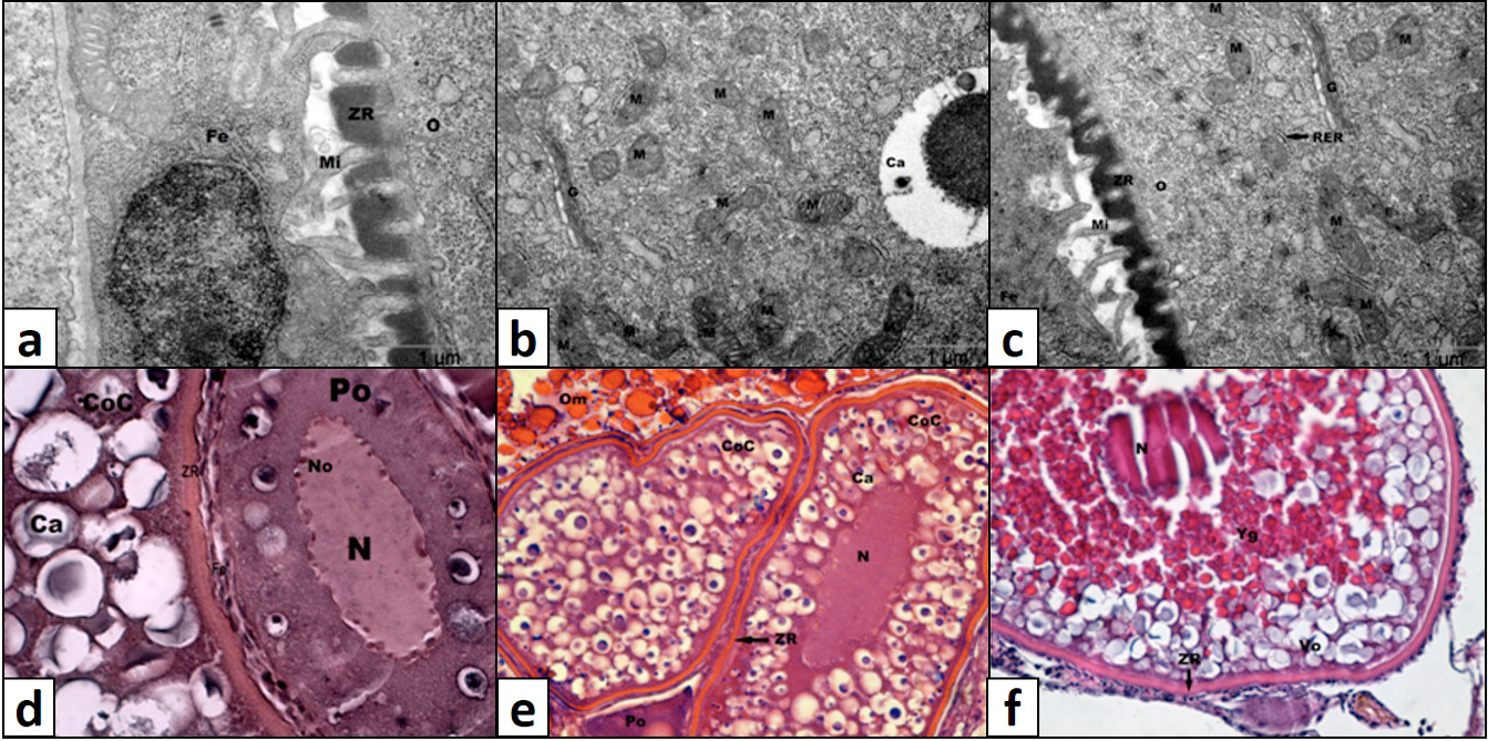
Hıstological and electron micrographic structure of ovary of control group; a,b,c, Electron micrograph of follicles; d,e,f, light micrograph of follicules. Fe, follicular epithelium. Mi, microvilli; ZR, zona radiata; O, ooplasm; M, mitochondrium; G, golgi; RER, rough endoplasmic reticulum; Po, primary oocyte; Ca, cortical alveoli; N, nucleus; No, nucleolus; CoC, cortical alveolus stage; Vo, vitellogenic oocyte; Om, mature oocyte; Yg, yolk granule. Magnification: d, x100; e, x20; f, x40.
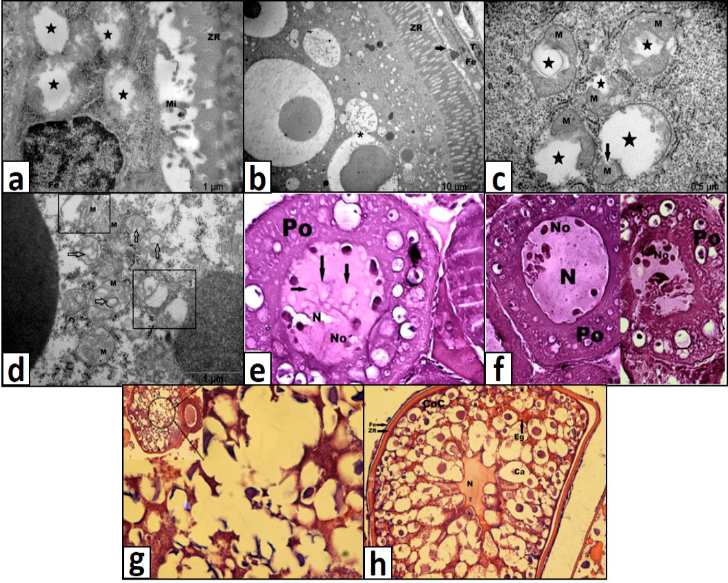
Hıstological and electron micrographic structure of ovary of 1 mg/L TiO2 exposed group; a, degeneration at microvili structure and mitochondrial deterioration (star) at follicular epithelium cells; b, cytoplasmic vacuolization and unification (asterix) of cortical alveoli; c, mitochondrial deterioration (star); d, mitochondrial swelling (square) and loss of organization of cristae (arrow) at mitochondrium; e, cytoplasmic vacuolization (black arrow) at primary oocytes; f, loss of nucleolus organization at primary oocyte; g, severe cytoplasmic vacuolization and degeneration at cortical alveoli structure; h, Eosinophilic granules accumulation at cortical alveolus stage ZR, zona radiata; Mi, microvilli; Fe, follicular epitheium; T, teca cells; M, mitochondrium,;N, nucleus; No, nucleolus; Ca, cortical alveoli; Eg, eosinophilic granule. Magnification: e, f,g, x100; h x40.
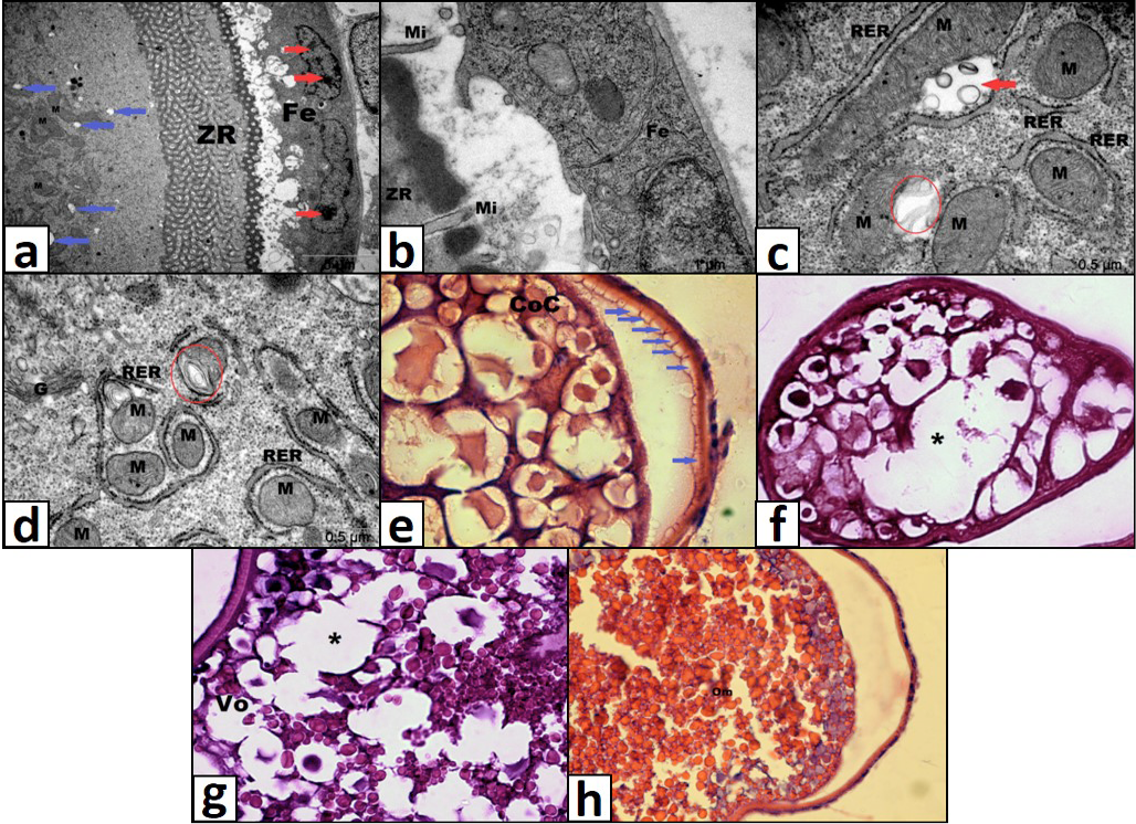
Lıght microscopic and electron micrographic structure of ovary of 2 mg/L TiO2 exposed group. A, chromatin condensation (red arrow) and cytoplasmic vacuolization (blue arrow); b, opening between zona radiata and follicular epithelium and degeneration at microvili structure; c, mitochondrial swelling (red arrow) and loss of organization of cristae (circle); d, loss of organization of cristae (circle); e, vacuolization under zona radiata (blue arrows); f, cytoplasmic vacuolization; g, unification of cortical alveoli (asterix); h, shrinking of mature oocyte. ZR, zona radiata; Fe, follicular epithelium; Mi, microvilli; M, mitochondria; G, golgi; RER, rough endoplasmic reticulum; CoC, cortical alveolus stage; Vo, vitellogenic oocyte; Om, mature oocyte. Magnification: e, f, g) x40, h) 20 x.
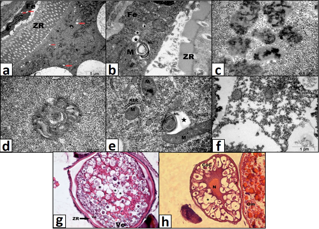
Lıght microscopical histological and electron micrographic strucure of ovary of 4 mg/L TiO2 exposed group. A, cytoplasmic vacuolization (red arrow) at follicular epithelium and ooplasm; b, multilamellar vesicle and cytoplasmic vacuolization (star); c, mitotic catastrophe; d, altered mitochondrium morphology and loss of cristae organization; e, cytoplasmic vacuolization (star) and multilamellar vesicle; f, dispersion at ooplasm; g, degenerated vitellogenic oocyte; h, unification of cortical alveoli (asterix). ZR, zona radiata; Fe, follicular epithelium; M, mitochondrium; ML, multilamellar vesicle; RER, rough endoplasmic reticulum; Ve, vitelline envelope; Vo, vitellogenic oocyte; N, nucleus; CoC, cortical alveolus stage; Om, mature oocyte. Magnification: g, h, x20.
References
- Aıtken, R.J., Chaudhry, M.Q., Boxall, A.B.A. and Hull, M., 2006. Manufacture and use of nanomaterials: current status in the UK and global trends. Occup. Med., 56: 300-306. https://doi.org/10.1093/occmed/kql051[CrossRef]
- Alarıfı, S., Alı, D., Al-Doaıss, A.A., Alı, B.A., Ahmed, M. and Al-Khedhaıry, A.A., 2013. Histologic and apoptotic changes induced by titanium dioxide nanoparticles in the livers of rats. Int. J. Nanomed., 8: 3937–3943.
- Amatruda, J.F., Shepard, J.L., Stern, H.M. and Zon, L.I., 2002. Zebrafish as a cancer model system. Cancer Cell, 1: 229–231. https://doi.org/10.1016/S1535-6108(02)00052-1[CrossRef]
- Bar-Ilan, O., Chuang, C.C., Schwahn, D.J., Yang, S., Joshı, S., Pedersen, J.A., Hamers, R.J., Peterson, R.E. and Heıdeman, W., 2013. TiO2 nanoparticle exposure and illumination during zebrafish development: mortality at parts per billion concentrations. Environ. Sci. Technol., 47: 4726-4733. https://doi.org/10.1021/es304514r[CrossRef]
- Broker, L.E., Kryut, F.A.E. and Gıaccone, G., 2005. Cell death independent of caspases: A review. Clin. Cancer Res., 11: 3155-3162. https://doi.org/10.1158/1078-0432.CCR-04-2223[CrossRef]
- Caruso, R., Fedele, F., Lucıuanò, R., Branca, G., Parısı, C., Paparo, D. and Parısı, A., 2011. Mitotic catastrophe in malignant epithelial tumors: The Pathologist’s viewpoint. Ultrastruct. Pathol., 35: 66–71. https://doi.org/10.3109/01913123.2010.543753[CrossRef]
- Chen, E., Ruvalcaba, M., Araujo, L., Chapman, R. and Chun Chın, W., 2008. Ultrafine titanium dioxide nanoparticles induce cell death in human bronchial epithelial cells. J. exp. Nanosci., 3: 171-183. https://doi.org/10.1080/17458080802412430[CrossRef]
- Chen, J., Dong, X., Xın, Y. and Zhao, M., 2011. Effects of titanium dioxide nano-particles on growth and some histological parameters of zebrafish (Danio rerio) after a long term exposure. Aquat. Toxicol., 101: 493–499. https://doi.org/10.1016/j.aquatox.2010.12.004[CrossRef]
- Danaıla, L., Popescu, I., Paıs, V., Rıga, D., Rıga, S. and Paıs, E., 2013. Apoptosis, paraptosis, necrosis, and cell regeneration in posttraumatic cerebral arteries. Chirurgia, 108: 319-324.
- Daughton, C.G., 2004. Non-regulated water contaminants: emerging research. Environ. Impact Assess. Rev., 24: 711–732. https://doi.org/10.1016/j.eiar.2004.06.003[CrossRef]
- Farıa, M., Navas, J.M., Soares, A.M.V.M. and Barata, C., 2014. Oxidative stress effects of titanium dioxide nanoparticle aggregates in zebrafish embryos. Sci. Tot. Environ., 470: 379-389. https://doi.org/10.1016/j.scitotenv.2013.09.055[CrossRef]
- Ispas, C., Andreescu, D., Patel, A., Goıa, D.V., Andreescu, S. and Wallace, K.N., 2009. Toxicity and developmental defects of different sizes and shape nickel nanoparticles in zebrafish. Environ. Sci. Tech., 43: 6349–6356. https://doi.org/10.1021/es9010543[CrossRef]
- Kaıda, T., Kobayashı, K., Adachı, M. and Suzukı, F., 2004. Optical characteristics of titanium oxide interference film and the film laminated with oxides and their applications for cosmetics. J. Cosmet. Sci., 55: 219–220.
- Kıng, K.L. and Cıdlowskı, J.A., 1995. Cell cycle and apoptosis: common pathways to life and death. J. cell. Biochem., 58: 175-180. https://doi.org/10.1002/jcb.240580206[CrossRef]
- Koç, N.D., Aytekın, A. and Yuce, R., 2008. Ovary maturation stages and histological investigation of ovary of the zebrafish (Danio rerio). Braz. Arch. Biol. Tech., 51: 513-522. https://doi.org/10.1590/S1516-89132008000300010[CrossRef]
- Moore, M.N., 2002. Biocomplexity: the post-genome challenge in ecotoxicology. Aquat. Toxicol., 59: 1–15. https://doi.org/10.1016/S0166-445X(01)00225-9[CrossRef]
- Moore, M.N., Depledge, M.H., Readman, J.W. and Leonard, P., 2004. An integrated biomarker-basedstrategy for ecotoxicological evaluation of risk in environmental management. Mutat. Res., 552: 247–268. https://doi.org/10.1016/j.mrfmmm.2004.06.028[CrossRef]
- Moore, M.N., 2006. Do nanoparticles present ecotoxicological risks for the health of the aquatic environment? Environ. Int., 32: 967–976. https://doi.org/10.1016/j.envint.2006.06.014[CrossRef]
- Park, J.H., Kım, S. and Bard, A.J., 2006. Novel carbon-doped TiO2 nanotube arrays with high aspect ratios for efficient solar water splitting. Nano Lett., 6: 24–28. https://doi.org/10.1021/nl051807y[CrossRef]
- Sperandıo, S., deBelle, I. and Bredesen, D.E., 2000. An alternative, nonapoptotic form of programmed cell death. Proc.natl. Acad. Sci. U.S.A., 97: 14376- 14381. https://doi.org/10.1073/pnas.97.26.14376[CrossRef]
- USEPA United States Environmental Protection Agency, 2009. Nanomaterial case studies: Nanoscale titaniumdioxide in water treatment and in topical sunscreen. EPA/600/R-09/057. Washington, DC.
- Wang, J.J., Sanderson, B.J. and Wang, H., 2007. Cyto- and genotoxicity of ultrafine TiO2 particles in cultured human lymphoblastoid cells. Mutat. Res., 628: 99–106. https://doi.org/10.1016/j.mrgentox.2006.12.003[CrossRef]
- Wang, J., Zhu, X., Zhang, X., Zhao, Z., Lıu, H., George, R., Wılson-Rawls, J., Chang, Y. and Chen, Y., 2011. Disruption of zebrafish (Danio rerio) reproduction upon chronic exposure to TiO2 nanoparticles. Chemosphere, 83: 461–467. https://doi.org/10.1016/j.chemosphere.2010.12.069[CrossRef]
- Wolf, R., Matz, H., Orıon, E. and Lıpozencıc, J., 2003. Sunscreens: The ultimate cosmetic. Acta Dermatovenerol. Croat., 11: 158–162.
- Yön, N.D. and Akbulut, C., 2014. Histological Changes in Zebrafish (Danio rerio) Ovaries Following Administration of Bisphenol A. Pakistan J. Zool., 46: 1153-1159.
- Zhu, X., Zhu, L., Duan, Z., Qı, R., Lı, Y. and Lang, Y., 2008. Comparative toxicity of several metal oxide nanoparticle aqueous suspensions to zebrafish (Danio rerio) early developmental stage. J. Environ. Sci. Hlth. A Tox. Hazard Subst. Environ. Eng., 43: 278-284. https://doi.org/10.1080/10934520701792779[CrossRef]
- Zhu, X., Wang, J., Zhang, X., Chang, Y. and Chen, Y., 2010. Trophic transfer of TiO2 nanoparticles from daphnia to zebrafish in a simplified freshwater food chain. Chemosphere, 79: 928–933. https://doi.org/10.1016/j.chemosphere.2010.03.022[CrossRef]
- Zhao, X., Sheng, L., Wang, L., Hong, J., Yu, X., Sang, X., Sun, Q., Ze, Y. and Hong, F., 2014. Mechanisms of nanosized titanium dioxide-induced testicular oxidative stress and apoptosis in male mice. Part. Fibre Toxicol., 11: 1-14. https://doi.org/10.1186/s12989-014-0047-3[CrossRef]
Services
Article usage statistics
Loading…