Research Article · Volume 12, Issue 5, 14 March 2024 · Open Access
Age and Gender-Dependent in Nitric Oxide Postnatal Change Activity in the Rat Brain
· · · · · · ·
Affiliations & Notes ⌄
1Laboratory of Biology and Health, Neurosciences, Neuroimmunology and Behaviour Unit, Faculty of Science Ibn Tofail University, Kenitra, Morocco
2Department of Psychiatry and Behavioral Neurobiology, University of Alabama at Birmingham Heersink School of Medicine, Birmingham, AL 35233, USA. *Correspondence | Mouloud Lamtai and Laila Ibouzine-Dine, Laboratory of Biology and Health, Neurosciences, Neuroimmunology and Behaviour Unit, Faculty of Science Ibn Tofail University, Kenitra, Morocco
In summary, this biochemical investigation reveals more precise information on NO kinetics in the prefrontal cortex, the striatum, the hippocampus and the hypothalamus of female and male rats.
It also revealed the timing of critical changes in NO levels during postnatal development.
Selected from the article's own text.
Abstract
The postnatal period represents the critical window referring to specific intervals of time during which the brain’s development exhibits increased sensitivity to specific types of learning and external influences. This period is usually associated with the development of age-related neurodevelopmental processes. Nitric oxide (NO) is an essential signaling molecules that play an important role in physiological processes including neuronal survival and differentiation, neurogenesis, synaptogenesis, pruning, and brain plasticity. In the present study, we aimed to determine the activity of NO during the early and late critical periods of structural and functional maturation of the central nervous system (CNS). In this study, we focused on the prefrontal cortex, hippocampus, striatum, and hypothalamus of male and female “Wistar” rat pups, as these areas have differential developing states. NO level was determined on postnatal days (PND) 1, 2, 3, 4, 7, 8, 9, 10, 11, 12, 14, 15, 16, and 17. The supernatant nitrite/nitrate (NOx) concentration was assessed after the reduction of nitrates to nitrites using the Griess reaction. Results revealed three distinct periods during postnatal development. In the first period, between PND1 and PND4, NO expression in both sexes increased, reaching a peak at PND8. The second period (PND8 to PND10) showed a slight decrease in NO expression. In the last period (PND11 to PND17), NO expression showed a high level. In summary, NO exhibited variations across the four examined areas, suggesting a complicated regulation of NO production during neonatal and infantile neurodevelopment.
Keywords | Nitric oxide, Postnatal development, Neuronal physiology, Signaling pathways, Neurotransmitter and free radicals, Sex-dimorphic, Rat, Age, Brain
INTRODUCTION
Nitric oxide (NO) a free radical, is produced by NO synthase (NOS) through a reaction that coverts L-arginine and oxygen into citrulline and NO . NOS are complex proteins found in a family of three isoforms which differ in their structure and distribution. Neuronal NOS (NOS1/nNOS), expressed in a subpopulation of neurons and endothelial NOS (NOS3/eNOS) expressed in endothelial cells, both being Ca2+ calmodulin-dependent and constituve enzymes, while the third isoform inducible NOS (NOS2/iNOS) is primarily present in immune system cells and its activation is Ca2+ calmodulin independent ; .
In the brain, endogenous NO acts as a diffusible neurotransmitter mainly produced by nNOS, activated upon glutamate binding to postsynaptic N-methyl-D-aspartate (NMDA) receptors ; . Since Nnos has been identified at different time points in various cell types of the central nervous system (CNS) , in certain areas of the brain, it is only temporarily expressed ; and there is a gradual increase in others during embryonic and postnatal development. Additionally, during the first month of postnatal life, histochemical and immunocytochemical research have characterized the distribution of NO-producing neurons in the rat corpus callosum structure (cc) .
Previous studies have indicated that the stage of postnatal development plays a crucial role in determining the biological activity of NO within developmental processes such as neurogenesis, neuronal survival and differentiation, synaptic transmission especially in the cortex and hippocampus. This has been demonstrated in several studies ; ; ; ; ; ; .
In rodents like rats and mice, numerous developmental processes occur during the period from birth to weaning and sexual maturity. Within these species, neurogenesis remains notably active during the initial two weeks after birth . Although, However, it has been shown that the NO generated by neuronal NOS inhibits neurogenesis in the adult brain . In addition, research by Luo et al. suggest that NO generation in neurons may be the source of the inhibitory effect that nNOS enhances and not from neural stem cells (NSCs) .
The aim of this study is to enhance our understanding of NO’s role in diverse developmental mechanisms by studying its kinetics. Additionally, we want to study differences in NO levels between male and female rats, examining the effects of age and gender on NO levels.
MATERIALS AND METHODS
Experimental animals
In our investigation, we utilized male and female Wistar rat pups. Initially, a total of 14 pregnant female Wistar rats were obtained from the Laboratory of Biology and Health at Ibn Tofail University. These rats were individually housed in standard plexiglass cages (430*290*210mm) under constant condition, maintaining a temperature of 24°C and a relative humidity of 50-60%. A 12-hour light-dark cycle, and the rats had unrestricted access to both food and water. The pregnant females gave birth to a total of 112 pups, distributed between 56 males and 56 females. The newborn pups were monitored daily at 9:30 a.m., and the day of birth was designated as postnatal day 0 (PND0) for each rat pup.
All experiments adhered to the ethical guidelines outlined by the National Institutes of Health for the appropriate use of laboratory animals in research. The protocols employed in these experiments were approved by the Animal Ethics Committee of Ibn Tofail University (ITU). The subsequent biochemical analyses were carried out at the Biology and Health Laboratory of ITU.
Biochemical analyses
Tissue processing
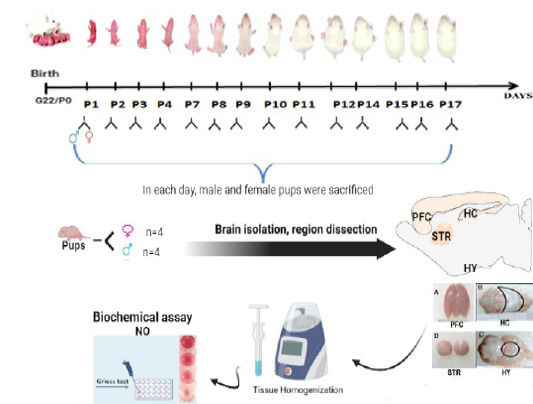
A total of 112 Wistar rats, both male and female, were used, with 8 pups for each day (4 males and 4 females). Rat pups were sacrificed by decapitation at each of PND 1, 2, 3, 4, 7, 8, 9, 10, 11, 12, 14, 15, 16, 17, and the prefrontal cortex, hippocampal, striatum, and hypothalamus from each hemisphere were individually dissected and homogenized using a Dounce homogenizer in a lysis buffer kept at an ice-cold temperature (RIPA lysate solution +1mM PMSF). The homogenates were centrifuged for 15 minutes at 14,000g and were subsequently stored at -80°C.
Nitrite/Nitrate assay
The conversion of NO to nitrite and nitrate is assumed to increase nitrite formation in the aqueous solutions of biological systems. Nitrite is the only stable product that remains after NO undergoes auto-oxidation. Consequently, assessing its concentrations in serum and tissue homogenates is widely acknowledged as a reliable indicator of NO activity ; . In the present study, we quantified nitrite concentration using the diazotization method, which is based on the Griess reaction, in rat brain tissue homogenates (prefrontal cortex, hippo campus, striatum, and hypothalamus). This indirect assay serves as a means to assess NO production .
Tissue samples (500μl) were dispensed into tubes, and an equivalent volume of Griess reagent (1% sulphanilamide (1 ml) and 0, 1% N-1-naphthylethylenediamine dihydrochloride (1 ml) in 2, 5% orthophosphoric acid) was added to each tube. Following a 30 minute incubation at room temperature, absorbance was measured at 540 nm. The nitrite concentrations in the tissue homogenates were determined through linear regression analysis, utilizing standard calibration curves generated with sodium nitrite. Tissue nitrite levels were expressed in μmol/g tissue. Figure 1 provides a Schematic representation of the experiment design.
Statistical analysis
All data were analyzed by two-way ANOVA, followed by Bonferroni’s post hoc test for comparison between groups. The data are represented as mean ± standard error of the mean (SEM) and illustrated by figures produced by the Graph Pad Prism 8 software (Graph Pad Software Inc., La Jolla, California, United States). Significant differences were considered for p < 0.05.
RESULTS and DISCUSSION
Kinetics of NO release in rat brain as a function of the type of regions and gender
Pre-frontal cortex
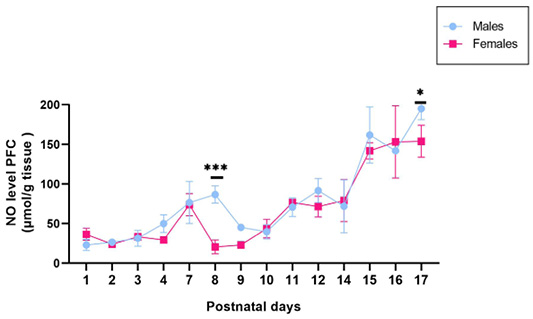
We analyzed here the time-dependent formation of NO in the Pre-frontal cortex (Figure 2). No activity increased from P1 to P7 in males and females and then decreased from P7 to P9 in female than in male rats, the activity at these points was significantly different in both sexes. From PND 9 to PND 17, the level of NO increases progressively in both sexes. The level of NO at P17 was higher than P1 in both groups.
The two-way ANOVA identified a main effect of gender on NOx activities (F(1, 84) =11,39 ; p = 0,0011) additionally to significant differences in interaction between age and sex (F(13, 84) = 3,488; p = 0,0002).
Striatum
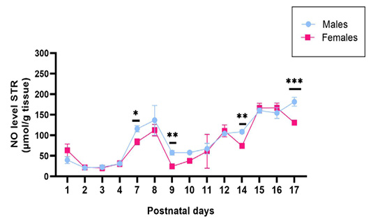
In general, NO activity showed a slight increase from PND 4 to PND 8 in female and male rats with significant difference was observed between the genders on PND7, and then decreased at P9. From PND9 to PND17, the level of NO increased progressively with significant difference was observed between the genders on PND9, PND14 and PND17. Males had a significantly higher concentration of NO compared to females during these time points (Figure 3).
ANOVA showed that sex differences had a significant impact on NOx activities in striatum (F(1, 84) =19,52 ; p < 0,0001). There was also significant interaction between Age and sex (F(13, 84) = 5,459; p < 0,0001 ).
Hippocampus
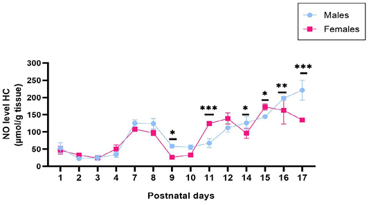
NO levels increased both in male and female rats, without sex differences, between postnatal days 1 and 8 and then decreased at PND9 with significant difference between male and female.
Starting from PND10, the NO peak returned to very high levels with a significant difference observed between the genders among all ages except for PND12 (Figure 4). The two-way ANOVA identified a main effect of gender on NOx activities (F(1, 84) =11,55 ;p = 0,001) additionally to significant differences in interaction between age and sex (F(13, 84) = 14,32 ; p < 0,0001).
Hypothalamus
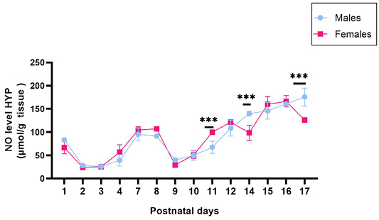
At PND1, the NO showed a slight increase in both sexes. Thereafter, the NO level stabilized in both sexes until PND4. From P7 to P8, the NO activity increased in males and females and then decreased at P9. Between the genders, there was no significant difference during this time.
After PND9, the NO level augmented progressively until 12 days of age. From then on, values varied in the females with a significant difference was observed between the genders at PND14, while in males NO level continued to increase, to become higher at 17 days with significant difference between males and female pups (Figure 5). ANOVA indicated no significant effect of sex differences on NOx activities in hypothalamus (F(1, 84) = 0,3254 ; p = 0,5699). But age and sex interacted significantly (F(13, 84) =9,714; p < 0,0001).
Based on the results of previous studies, we have examined the changes in NO production during early and late postnatal development in the prefrontal cortex, hippocampus, striatum, and hypothalamus of rats using histological techniques to visualize NO metabolite such as nitrite and nitrate. This study was performed during the postnatal period (P1–P17), with a group of male and female Wistar rats born normally and sacrificed in each day.
The present results indicate that NO exhibited different variations across the four areas examined. The amount of NO produced depends on the area of the brain, it is most obvious in the cortex, hippocampus, hypothalamus, substantia nigra, and amygdala ; . Additionally, our study found that NO levels change during postnatal development in an age-dependent manner. During this postnatal development period, three distinct periods can be distinguished. In the first period, between PND1 and PND4, NO expression in both sexes increased, reaching a peak at PND8. The second period (PND8 to PND10) showed a slight decrease in NO expression. In the last period (PND11 to PND17), NO expression showed a high level. These changes in NO expression correlate with two stages of postnatal development in rats (Neonatal 0-6 d, infantile 7-17 d) ; . Therefore, these finding indicate that the NO production change may be associated with neuronal maturation during postnatal development.
We first observed for this first period, P1–P8 progressive increase of NO in both sexes across the two areas hippocampus and hypothalamus except the STR and PFC area. In addition, the result shows gender differences at P7 in STR and at P8 in PFC. It may be possible that this progressive increase could further be attributed to the level of nNOS expression; early reports have linked increased levels of NO with the origin of nNOS production. According to a study by Luo and colleagues, Neural Stem cell derived nNOS is localized in the nuclus and expressed at a much lower amount than in neurons, which suggests that NO diffuses slowly outside NSCs and may act via a nuclear signaling molecule . Similarly, a study by Fernandez et al. (2003) found that the synthesis of NO is associated with an increase in immune reactivity, expression, and constitutive NOS activities during the few postnatal days, with a peak occurring on PND5 . Furthermore, sex differences at P7 and P8 might be caused by an increase in factors that stimulate NO production in male than female.
In the rat neocortex, nicotinamide adenine dinucleotide phosphate-diaphorase (NADPH-d) cells are a unique population of neurons that develop quite quickly and mature early in the developmental process. It is generally acknowledged that only around 98% of NADPH-d positive neurons in the cortex have nNOS . A study used histochemical techniques of NADPH-diaphorase to evaluate the postnatal development of nitrergic neurons in the male rat pre-frontal cortex found that neurons begin to express NADPH-d at birth and differentiate during a relatively short period . While another study by Zhang and al. found that at the age of PND2, the striatum of control pups had a dense distribution of nNOS immunoreactive cells with no obvious sex difference . A study found a correlation between elevated nNOS expression in hypothalamic neurons and elevated catecholamine expression in the brain .
Additionally, a study found that Brain-derived neurotrophic factor (BDNF) stimulates the production of NO in the soma and dendrites of hippocampal neurons . Several investigations have demonstrated the postsynaptic localization of both nNOS and TrKB receptors in the postsynaptic density of glutamatergic synapses in the hippocampus . On the other hand, our results during the second period from postnatal days P8 to P10 are in concurrence with earlier studies that found a correlation between a decrease in NO and a decrease in synaptic density .
A literature survey revealed that the regulation of brain network development occurs through plasticity and activity-dependent mechanisms that control the continuous formation and elimination of spine synapses. NO is involved in these aspects of structural plasticity . In the rat brain, synaptogenesis begins during the initial postnatal week and it is subsequently followed by a period of pruning . This synaptogenesis is accompanied by robust astrogenesis and is possibly enhanced by the early liberation from astrocytes of synapse-forming factors that include thrombospondins 1 and 2 .
Activation of the synapse, achieved by the involvement of NMDA receptors and calcium influx, could trigger to the activation on nNOS closely linked with postsynaptic density protein 95 (PSD-95) within the postsynaptic density. NO would be released as a result of this process and the initiation of a cGMP- PKG cascade in nearby dendrites . NO is released in a calcium-dependent manner by some NADPH-d-positive cells in response to glutamate activation of NMDA receptors, suggesting that this neuroactive molecule may contribute to the generation of axonal projections, the elimination of redundant connections, and/or the formation of axonal synapsis in the late stages of development.
During synapse formation in the early postnatal days (PND 8–10), there is an increase in microglia’s interaction with developing dendritic spines causing a change in the synapse’s consequent elimination . Aldo, reduced NO production in the second phase may result from microglia’s role in the final neuronal network’s creation, which includes supporting in the pruning of excess neurons and synapses and promoting cell differentiation (P8 to P10).
In addition to synapse development, Ca2+ transients, actin buildup, and the production of dendritic filopodia are all induced by the contact between microglia and dendrites of layer 2/3 pyramidal neurons in the developing somatosensory cortex. It is interesting that this filopodia production only occurred during the period of robust synaptogenesis (P8-P10) and not occur at later postnatal ages (P12-P14 and P26-P30) .
In vitro and in vivo findings indicates that microglia have the capacity to decrease the number of neural precursor cells within proliferative zones in the neocortex of primates and rodents. This reduction in neural precursor cells aligns with the aggregation of microglia and coincides with the onset of developmental cell death in diverse brain regions. The first few weeks after birth are the only times when synapses are eliminated by microglia .
A study by Rörig and colleagues has shown in rodents that between PND6 and PND10, NO may affect electrical coupling, coordination of transcriptional activity, synchronization of metabolic states, and electrical connection between adjacent neurons ; . As known, NO functions as a paracrine messengerin newly generated neurons to regulate the growth and differentiation of mouse brain neural progenitor cells (NPC) .
Our data in the third period (P11 to P17) suggested that there are regional differences in the timing of NO production in males and females. In the hippocampus and hypothalamus, our results show gender difference at P11 which NO expression was higher in females than in males in contrast at P14 and PND17 female showed low expression of NO. Consistent with these findings, a study indicates that a majority of neuronal populations expressing nNOS also exhibit the presence of estrogen receptor alpha (ERα) in the hypothalamus at PND11 . Similarly, recent research has shown that E2 increases nNOS in the brain. Furthermore, it has been suggested that observed sex differences in NO release are related to differential modulation of nNOS expression as well as activity . In addition, the early-life expression of nNOS in the hypothalamus shows that NO plays a part in the maturation of gonadotropin-releasing hormone (GnRH) by regulating GnRH mRNA expression . A microRNA switch regulates the rise in hypothalamic GnRH production puberty a phenomenon that might be crucial for sexual maturation .
According to Prevot, the first groups of ovarian follicles that may ovulate during puberty begin to form at P12 in conjunction with a decline in circulating levels of estrogen-binding alpha-fetoprotein between PND12 and PND16 . The impact of sex hormones on microglial activities in these rat’s hippocampal and hypothalamus regions could be another explanation for the sex differences we observed in our study and it may be possible that changes in NO level coincide with robust synaptogenesis and astrogenesis. Previous studies have shown that at PND11–16, the hippocampus has shown the largest rise in the number of GFAP-positive cells . Another study have demonstrated that in mice, microglial cell numbers dramatically rise in many brain regions across the first two postnatal weeks, peaking in density at PND14 .
CONCLUSION
In summary, this biochemical investigation reveals more precise information on NO kinetics in the prefrontal cortex, the striatum, the hippocampus and the hypothalamus of female and male rats. It also revealed the timing of critical changes in NO levels during postnatal development. These findings reinforce the role of neurodevelopmental processes in the control of NO production in newborn and infant animals.
ACKNOWLEDGMENTS
I would like to express my thanks to the Neurosciences, Neuroimmunology, and Behavioral unit for their support in writing this article.
NOVELTY STATEMENT
We investigate the NO kinetics and its function in many developmental processes. While NO’s role in neurogenesis and other developmental processes has been studied in the past, our research focuses on the temporal dynamics of NO generation in particular brain regions during early and late postnatal development. In addition, while gender and age differences in NO levels have not been thoroughly investigated in the context of developmental neurobiology, our study investigates these possibilities. We provide new insights into the impact of age and gender on the regulation of NO in the brain by comparing the NO levels in male and female rats at various phases of postnatal development.
Our research examines gender and age impacts and provides a novel viewpoint on the function of NO in developmental pathways and provides a foundation for further research on neurogenesis in pathological conditions.
AUTHOR’S CONTRIBUTION
All authors contributed equally to the manuscript.
Abbreviations
BDNF, Brain-derived neurotrophic factor; CC, corpus callosum; CNS, central nervous system; GnRH, gonadotropin releasing hormone; NADPH-d, nicotinamide adenine dinucleotide phosphate diaphorase; NMDA, N-methyl-d-aspartate; NO, nitric oxide; NOS, NO synthase; NPC, neural progenitor cells; NSCs, neural stem cells; PND, postnatal day.
Conflict of interest
The authors have declared no conflict of interest.
Figures
Figure 1:.
Experimental design. Male and female Wistar rat pups were sacrificed by decapitation at each of Postnatal Days (PND) 1, 2, 3, 4, 7, 8, 9, 10, 11, 12, 13, 14, 15, 16, 17. The prefrontal cortex (PFC), hippocampus (HC), striatum (STR), and hypothalamus (HY) were separately dissected and homogenized for the measurement of NO concentrations by using the diazotization method based on the Griess reaction.
Figure 2:.
The graphic shows the Kinetics of the NO release in the Pre-frontal cortex area from male and female animals, for each postnatal day analyzed (PND1 to PND 17). Error bars represent the standard deviation of the means. The significance level is 0.05.*p < 0.05.
Figure 3:.
The graphic shows the Kinetics of the NO release in the Striatum area from male and female animals, for each postnatal day analyzed (PND1 to PND17). Error bars represent the standard deviation of the means. The significance level is 0.05.*p < 0.05.
Figure 4:.
The graphic shows the Kinetics of the NO release in the Hippocampus area from male and female animals, for each postnatal day analyzed (PND1 to PND 17). Error bars represent the standard deviation of the means. The significance level is 0.05.*p < 0.05.
Figure 5:.
The graphic shows the Kinetics of the NO release in the Hypothalamus area from male and female animals, for each postnatal day analyzed (PND1 to PND 17). Error bars represent the standard deviation of the means. The significance level is 0.05.*p < 0.05.
References
Barbaresi P, Mensà E, Sagrati A, Graciotti L (2020). Postnatal development of the distribution of nitric oxide-producing neurons in the rat corpus callosum. Neurosci. Res., 151: 15–30. https://doi.org/10.1016/j.neures.2019.02.005[CrossRef]
Bryan NS, Grisham MB (2007). Methods to detect nitric oxide and its metabolites in biological samples. Free Radic. Biol. Med., 43(5): 645–657. https://doi.org/10.1016/j.freeradbiomed.2007.04.026[CrossRef]
Carrascal L, Gorton E, Pardillo-Díaz R, Perez-García P, Gómez-Oliva R, Castro C, Nunez-Abades P (2020). Age-dependent vulnerability to oxidative stress of postnatal rat pyramidal motor cortex neurons. Antioxidants, 9(12): 1–21. https://doi.org/10.3390/antiox9121307[CrossRef]
Catalani A, Sabbatini M, Consoli C, Cinque C, Tomassoni D, Azmitia E, Angelucci L, Amenta F (2002). Glial fibrillary acidic protein immunoreactive astrocytes in developing rat hippocampus. Mech. Ageing. Dev., 123(5): 481–490. https://doi.org/10.1016/S0047-6374(01)00356-6[CrossRef]
Chachlaki K, Garthwaite J, Prevot V (2017a). The gentle art of saying NO: How nitric oxide gets things done in the hypothalamus. Nat. Rev. Endocrinol., 13(9): 521–535. https://doi.org/10.1038/nrendo.2017.69[CrossRef]
Chachlaki K, Malone SA, Qualls-creekmore E, Münzberg H, Giacobini P, Ango F (2017b). Phenotyping of nNOS Neurons in the Postnatal and Adult Female Mouse Hypothalamus. J. Comp. Neurol., 525(15): 3177–3189. https://doi.org/10.1002/cne.24257[CrossRef]
Chachlaki K, Prevot V (2020). Nitric oxide signalling in the brain and its control of bodily functions. Br. J. Pharmacol., 177(24): 5437–5458. https://doi.org/10.1111/bph.14800[CrossRef]
Chao CC, Hu S, Molitor TW, Shaskan EG, Peterson PK (1992). Activated microglia mediate neuronal cell injury via a nitric oxide mechanism. J. Immunol., 149(8): 2736–2741. https://doi.org/10.4049/jimmunol.149.8.2736[CrossRef]
Chong C, Ai N, Ke M, Tan Y, Huang Z, Li Y, Lu J, Ge W, Su H (2017). Roles of nitric oxide synthase isoforms in neurogenesis. Mol. Neurobiol., 55(3): 2645-2652. https://doi.org/10.1007/s12035-017-0513-7[CrossRef]
Christopherson KS, Ullian EM, Stokes CCA, Mullowney CE, Hell JW, Agah A, Lawler J, Mosher DF, Bornstein P, Barres BA (2005). Thrombospondins are astrocyte-secreted proteins that promote CNS synaptogenesis. Cell, 120(3): 421–433. https://doi.org/10.1016/j.cell.2004.12.020[CrossRef]
Chung YH, Kim YS, Lee WB (2004). Distribution of neuronal nitric oxide synthase-immunoreactive neurons in the cerebral cortex and hippocampus during postnatal development. J. Mol. Histol., 35(8–9): 765–770. https://doi.org/10.1007/s10735-004-0667-2[CrossRef]
Contestabile A, Ciani E (2004). Role of nitric oxide in the regulation of neuronal proliferation, survival and differentiation. Neurochem. Int., 45(6): 903–914. https://doi.org/10.1016/j.neuint.2004.03.021[CrossRef]
Cunningham CL, Martínez-Cerdeño V, Noctor SC (2013). Microglia regulate the number of neural precursor cells in the developing cerebral cortex. J. Neurosci., 33(10): 4216–4233. https://doi.org/10.1523/JNEUROSCI.3441-12.2013[CrossRef]
Cyr, AR, Lauren VH, Sruti SS, Brian SZ (2020). Nitric oxide and endothelial dysfunction. Crit. Care Clin., 36(2): 307–321. https://doi.org/10.1016/j.ccc.2019.12.009[CrossRef]
Fernández AP, Alonso D, Lisazoaín I, Serrano J, Leza JC, Bentura ML, López JC, Encinas JM, Fernández-Vizarra P, Castro-Blanco S, Martínez A, Martinez-Murillo R, Lorenzo P, Pedrosa JA, Peinado MA, Rodrigo J (2003). Postnatal changes in the nitric oxide system of the rat cerebral cortex after hypoxia during delivery. Dev. Brain. Res., 142(2): 177–192. https://doi.org/10.1016/S0165-3806(03)00068-3[CrossRef]
Fernando A, Justo O, Kimie C (2022). The modulation of neuroinflammation by inducible nitric oxide synthase. J Cell. Commun. Signal., 16(2): 155–158. https://doi.org/10.1007/s12079-021-00663-x[CrossRef]
Garay PA, Hsiao EY, Patterson PH, Mcallister AK (2013). Brain, behavior, and immunity maternal immune activation causes age and region-specific changes in brain cytokines in offspring throughout development. Brain Behav. Immun., 31: 54–68. https://doi.org/10.1016/j.bbi.2012.07.008[CrossRef]
Husi H, Ward MA, Choudhary JS, Blackstock WP, Grant SGN (2000). Proteomic analysis of NMDA receptor-adhesion protein signaling complexes. Nat. Neurosci., 3(7): 661-669. https://doi.org/10.1038/76615[CrossRef]
Hvizdosova N, Tomasova L, Bolekova A, Kolesar D, Kluchova D (2014). Nitrergic neurons during early postnatal development of the prefrontal cortex in the rat: Histochemical study. Acta Histochem., 116(5): 736–739. https://doi.org/10.1016/j.acthis.2014.01.001[CrossRef]
Imura T, Kanatani S, Fukuda S, Miyamoto Y, Hisatsune T (2005). Layer-specific production of nitric oxide during cortical circuit formation in postnatal mouse brain. Cereb. Cortex., 15(3): 332–340. https://doi.org/10.1093/cercor/bhh135[CrossRef]
Kharazia VN, Schmidt HHHW, Weinberg RJ (1994). Type I nitric oxide synthase fully accounts for nadph-diaphorase in rat striatum, but not cortex. Neuroscience, 62(4): 983–987. https://doi.org/10.1016/0306-4522(94)90337-9[CrossRef]
Kolarow R, Kuhlmann CRW, Munsch T, Zehendner C (2014). BDNF-induced nitric oxide signals in cultured rat hippocampal neurons: Time course, mechanism of generation, and effect on neurotrophin secretion. Front. Cell. Neurosci., 7(8): 323. https://doi.org/10.3389/fncel.2014.00323[CrossRef]
Kourosh-arami M, Hosseini N, Mohsenzadegan M, Komaki A, Joghataei MT (2020). Neurophysiologic implications of neuronal nitric oxide synthase. Rev. Neurosci., 31(6): 617–636. https://doi.org/10.1515/revneuro-2019-0111[CrossRef]
Kuppusamy P, Ohnishi ST, Numagami Y, Ohnishi T, Zweier JL (1995). Three-dimensional imaging of nitric oxide production in the rat brain subjected to ischemia-hypoxia. J Cereb. Blood Flow. Metab., 15(6): 899–903. https://doi.org/10.1038/jcbfm.1995.114[CrossRef]
Li J, Chen S, Lin RCS, Smith SS (1997). Cerebellar nitric oxide synthase is expressed within granule cell patches innervated by specific mossy fiber terminals: A developmental profile. Dev. Neurosci., 19(3): 274–282. https://doi.org/10.1159/000111216[CrossRef]
Luo CX, Jin X, Cao CC, Zhu MM, Wang B, Chang L, Zhou QG, Wu HY, Zhu DY (2010). Bidirectional regulation of neurogenesis by neuronal nitric oxide synthase derived from neurons and neural stem cells. Stem Cells, 28(11): 2041–2052. https://doi.org/10.1002/stem.522[CrossRef]
Maccallini C, Amoroso R (2023). Neuronal nitric oxide synthase and post-translational modifications in the development of central nervous system diseases: Implications and regulation. Molecules, 28(18): 1–14. https://doi.org/10.3390/molecules28186691[CrossRef]
Messina A, Langlet F, Chachlaki K, Roa J, Rasika S, Jouy N, Gallet S, Gaytan F, Parkash J, Tena-Sempere M, Giacobini P, Prevot V (2016). A microRNA switch regulates the rise in hypothalamic GnRH production before puberty. Nat. Neurosci., 19(6): 835–844. https://doi.org/10.1038/nn.4298[CrossRef]
Miyamoto A, Wake H, Ishikawa AW, Eto K, Shibata K, Murakoshi H, Koizumi S, Moorhouse AJ, Yoshimura Y, Nabekura J (2016). Microglia contact induces synapse formation in developing somatosensory cortex. Nat. Commun., 25(7): 12540. https://doi.org/10.1038/ncomms12540[CrossRef]
Moreno-López B, Romero-Grimaldi C, Noval JA, Murillo-Carretero M, Matarredona ER, Estrada C (2004). Nitric oxide is a physiological inhibitor of neurogenesis in the adult mouse subventricular zone and olfactory bulb. J. Neurosci., 24(1): 85–95. https://doi.org/10.1523/JNEUROSCI.1574-03.2004[CrossRef]
Nikonenko I, Nikonenko A, Mendez P, Michurina TV, Enikolopov G, Muller D (2013). Nitric oxide mediates local activity-dependent excitatory synapse development. Proc. Natl. Acad. Sci. U.S.A., 110(44). https://doi.org/10.1073/pnas.1311927110[CrossRef]
Portillo F, Moreno-López B (2020). Nitric oxide controls excitatory/inhibitory balance in the hypoglossal nucleus during early postnatal development. Brain Struct. Funct., 225(9): 2871–2884. https://doi.org/10.1007/s00429-020-02165-9[CrossRef]
Prevot V (2015). Puberty in mice and rats. Knobil Neills Physiol. Reprod. Two-Vol Set 2: 1395–1439. https://doi.org/10.1016/B978-0-12-397175-3.00030-2[CrossRef]
Roerig B, Feller MB (2000). Neurotransmitters and gap junctions in developing neural circuits. Brain Res. Rev., 32(1): 86–114. https://doi.org/10.1016/S0165-0173(99)00069-7[CrossRef]
Rörig B, Sutor B (1996). Nitric oxide-stimulated increase in intracellular cGMP modulates gap junction coupling in rat neocortex. Neuroreport, 7(2): 569–572. https://doi.org/10.1097/00001756-199601310-00046[CrossRef]
Semple BD, Blomgren K, Gimlin K, Ferriero DM, Noble-haeusslein LJ (2013). Progress in neurobiology brain development in rodents and humans: Identifying benchmarks of maturation and vulnerability to injury across species. Prog. Neurobiol., Jul-Aug: 106-107. https://doi.org/10.1016/j.pneurobio.2013.04.001[CrossRef]
Tagliaferro P, Ramos AJ, Lopez-Costa JJ, López EM, Brusco A (2003). Changes in the postnatal development on nitric oxide system induced by serotonin depletion. Dev. Brain. Res., 146(1–2): 39–49. https://doi.org/10.1016/j.devbrainres.2003.09.006[CrossRef]
Taranukhin AG, Yamova LA, Chernigovskaya EV (2006). Participation of catecholamines and NO in regulation of apoptosis of nonapeptidergic neurons of neonatal rat pups. J. Evol. Biochem. Physiol., 42(2): 217–225. https://doi.org/10.1134/S0022093006020141[CrossRef]
Tremblay MĚ, Lowery RL, Majewska AK (2010). Microglial interactions with synapses are modulated by visual experience. PLoS Biol., 8(11). https://doi.org/10.1371/journal.pbio.1000527[CrossRef]
Vercelli AE, Cracco CM (1994). Effects of eye enucleation on NADPH-diaphorase positive neurons in the superficial layers of the rat superior colliculus. Dev. Brain. Res., 83(1): 85–98. https://doi.org/10.1016/0165-3806(94)90182-1[CrossRef]
Tripathi, Kumar M, Ojha SK, Kartawy M, Hamoudi W, Choudhary A, Stern S, Aran A, Amal H (2023). The no answer for autism spectrum disorder. Adv. Sci., 10(22). https://doi.org/10.1002/advs.202205783[CrossRef]
Xue B, Johnson AK, Hay M (2007). Sex differences in angiotensin II-induced hypertension. Braz. J. Med. Biol. Res., 40(5): 727–734. https://doi.org/10.1590/S0100-879X2007000500018[CrossRef]
Zeiss CJ (2021). Comparative milestones in rodent and human postnatal central nervous system development. Toxicol. Pathol., 49(8): 1368–1373. https://doi.org/10.1177/01926233211046933[CrossRef]
Zghari O, Lamtai M, Azirar S, El-Brouzi MY, Benmhammed H, El-Hessni A, Ouichou A, Mesfioui A (2023). Neuroprotective effects of melatonin against neurotoxicity induced by intrahippocampal injection of aluminum in male Wistar rats: Possible involvement of oxidative stress pathway. Adv. Anim. Vet. Sci., 11(5). https://doi.org/10.17582/journal.aavs/2023/11.5.711.719[Full text][CrossRef]
Zhang J, Jing Y, Zhang H, Bilkey DK, Liu P (2018). Maternal immune activation leads to increased nNOS immunoreactivity in the brain of postnatal day 2 rat offspring. Synapse, 72(1): 1–14. https://doi.org/10.1002/syn.22011[CrossRef]
Ziaja M, Pyka J, Ciombor J, Płonka PM (2005). Kinetics of nitric oxide release in neonatal and mature rat brain during endotoxemia, as studied by diethyldithiocarbamate spin trapping. Curr. Top. Biophys., 29(12): 73–82.