Research Article · Volume 14, Issue 4, 15 April 2026 · Open Access
A Novel Turpentine-Induced Model of Femoral Head Aseptic Necrosis in Rabbits
· ·
Affiliations & Notes ⌄
1¹Department of Veterinary Surgery, Moscow State Academy of Veterinary Medicine and Biotechnology, Moscow
2Russia
3²Department of Animal Anatomy and Histology, Moscow State Academy of Veterinary Medicine and Biotechnology, Moscow, Russia. *Correspondence | T.E. Deneche, Department of Veterinary Surgery, Moscow State Academy of Veterinary Medicine and Biotechnology, Moscow
Although effective in inducing necrosis, many of these models are associated with significant systemic toxicity, high mortality rates, and limited reproducibility.
The present study demonstrates that intra-articular administration of turpentine provides a reliable and reproducible experimental model of femoral head aseptic necrosis in rabbits.
The proposed model consistently reproduces clinical signs, radiological changes, gross pathological findings, and histopathological features characteristic of osteonecrosis, while exhibiting reduced systemic toxicity and zero mortality.
This experimental approach may serve as a valuable platform for future investigations into the pathogenesis of femoral head aseptic necrosis and for the evaluation of surgical and pharmacological treatment strategies.
Selected from the article's own text.
Abstract
Aseptic necrosis of the femoral head (Legg–Calvé–Perthes disease) is a severe orthopedic disorder associated with disruption of local blood supply, resulting in ischemia, progressive destruction of bone tissue, and degeneration of articular cartilage. Experimental animal models are essential for studying disease pathogenesis and evaluating therapeutic approaches; however, many existing models are limited by high systemic toxicity and poor reproducibility.The aim of this study was to develop and validate a novel, low-toxicity experimental model of femoral head aseptic necrosis in rabbits using intra-articular administration of turpentine. Twelve adult Californian rabbits were randomly divided into two equal groups. Group 1 received an intra-articular injection of a calcium chloride–ethanol mixture, while Group 2 received intra-articular turpentine (0.3 mL per hip joint). Clinical observation, hematological and biochemical analyses, radiography, computed tomography, surgical examination, and histological analysis were performed. Histological sections were prepared at a thickness of 5–7 µm and stained with hematoxylin and eosin, Alcian blue, and Van Gieson stains (Table 1). Rabbits treated with turpentine developed consistent lameness and characteristic radiological and histopathological features of femoral head osteonecrosis. Imaging revealed bone rarefaction, trabecular deformation, and acetabular involvement. Histological examination demonstrated destruction of articular cartilage, hypertrophic and necrotic chondrocytes, depletion of glycosaminoglycans confirmed by Alcian blue staining, and exposure of subchondral bone. No mortality was recorded, and systemic toxicity was limited. Intra-articular administration of turpentine provides a reliable, reproducible, and relatively safe experimental model of femoral head aseptic necrosis in rabbits. This model may be useful for future studies investigating disease mechanisms and evaluating surgical and pharmacological treatment strategies.
Keywords: Aseptic necrosis, femoral head, rabbits, turpentine, histology, experimental model
INTRODUCTION
Aseptic necrosis of the femoral head, also known as Legg–Calvé–Perthes disease, is a severe orthopedic disorder belonging to the group of Osteochondropathies ; ; . It is characterized by disruption of the local blood supply to the femoral head, leading to ischemia, impaired nutrition of bone and articular cartilage, and subsequent noninfectious necrosis of epiphyseal bone tissue ; ; . Progressive structural damage of the femoral head results in joint incongruity, pain, lameness, and significant functional impairment of the affected limb ; .
In veterinary practice, femoral head aseptic necrosis is most commonly diagnosed in young, small-breed dogs, typically between four and eight months of age . Diagnosis is based on clinical signs, including progressive lameness and pain on hip joint manipulation, as well as characteristic radiographic changes ; ; ; . Pathogenetically, the disease progresses through several stages: Vascular compromise and ischemia, followed by necrosis of bone tissue, degeneration of articular cartilage, collapse of the femoral head, and secondary degenerative joint changes ; ; .
Ischemia and disturbance of microcirculation play a central role in the initiation and progression of femoral head aseptic necrosis ; . Reduced blood perfusion leads to hypoxia, chondrocyte death, degradation of the extracellular matrix, and loss of mechanical stability of the subchondral bone ; . Several vascular and hematological hypotheses have been proposed to explain the development of osteonecrosis of the femoral head including intravascular coagulation, endothelial dysfunction, and thrombophilia ; ; .
Surgical intervention remains the primary treatment option for advanced cases of femoral head aseptic necrosis. Femoral head and neck excision arthroplasty is widely used to relieve pain and restore limb function by forming a functional fibrous pseudoarthrosis ; . Histological examination of the excised femoral head provides definitive confirmation of the diagnosis and allows detailed evaluation of cartilage degeneration, subchondral bone remodeling, and cellular changes associated with ischemic injury.
Due to the limited availability of clinical material and the ethical constraints of experimental studies in companion animals, experimental models play a crucial role in advancing the understanding of disease pathogenesis and in evaluating novel therapeutic strategies. However, many existing experimental models of femoral head osteonecrosis rely on aggressive chemical agents, mechanical vascular occlusion, or systemic corticosteroid administration. Although effective in inducing necrosis, these approaches are frequently associated with high systemic toxicity, increased mortality, and limited reproducibility ; ; .
Rabbit models have gained increasing attention in orthopaedic research due to their suitable size, well-developed hip joint anatomy, and favorable response to experimental manipulation. Turpentine is known to induce a localized inflammatory response when administered in controlled doses. We hypothesized that intra-articular administration of turpentine could indirectly disrupt the local microcirculation of the femoral head through inflammation-mediated ischemia, leading to reproducible aseptic necrosis while minimizing systemic toxic effects.
Therefore, the aim of the present study was to develop and validate a novel turpentine-induced experimental model of femoral head aseptic necrosis in rabbits and to confirm the resulting pathological changes using clinical observation, imaging techniques, and histological analysis. The proposed model is intended to provide a reliable and biologically safe platform for future experimental investigations of Perthes disease and for the evaluation of surgical and pharmacological treatment strategies.
MATERIALS AND METHODS
Animals and ethical approval
The experimental study was conducted using twelve clinically healthy adult Californian rabbits aged 15–16 months and weighing 4.8–5.2 kg. All experimental procedures involving animals were carried out in accordance with the European Convention for the Protection of Vertebrate Animals Used for Experimental and Other Scientific Purposes (ETS No. 123). Animals were housed under standard laboratory conditions with free access to food and water throughout the experimental period.
Experimental design
The rabbits were randomly divided into two equal groups (n= 6 per group).
Group 1 (conventional model): Animals received an intra-articular injection of a mixture consisting of 0.5 mL of 10% calcium chloride solution and 0.5 mL of 96% ethanol into each hip joint.
Group 2 (experimental model): Animals received intra-articular injections of turpentine at a dose of 0.3 mL per hip joint.
The dose of turpentine was selected based on preliminary observations to ensure reliable induction of femoral head necrosis while minimizing systemic toxicity.
Induction of femoral head aseptic necrosis
All intra-articular injections were performed under general anesthesia. Premedication consisted of Zoletil (2 mg/kg, intramuscularly) combined with Dexdomitor (5 µg/kg, intramuscularly). The hip joint area was aseptically prepared prior to injection. After administration, animals were monitored daily for changes in behavior, gait, limb loading, and general clinical condition.
Clinical hematological and biochemical evaluation
Clinical examination focused on the assessment of lameness, joint pain, abnormal posture, and changes in physical activity. Blood samples were collected before intra-articular injection and three weeks post-administration. Hematological parameters included leukocyte count and erythrocyte sedimentation rate. Biochemical analysis included alanine aminotransferase, aspartate aminotransferase, alkaline phosphatase, bilirubin, creatinine, urea, and total protein.
Radiographic and computed tomography examination
Radiographic examination of the hip joints was performed before the experimental intervention and repeated three weeks after injection. Computed tomography was conducted at three weeks post-injection to evaluate femoral head architecture, trabecular integrity, and acetabular involvement. Imaging findings were assessed for bone rarefaction, deformation, and osteolytic changes.
Surgical procedure
Following confirmation of femoral head pathology, femoral head resection arthroplasty was performed for diagnostic confirmation. Premedication included Zoletil (2 mg/kg, intramuscularly) and Dexdomitor (5 µg/kg, intramuscularly). Additionally, meloxicam (0.1 mg/kg), cefazolin (25 mg/kg), and tranexamic acid (15 mg/kg) were administered prior to induction. Anesthesia was induced with propofol (5 mg/kg intravenously) and maintained with isoflurane (1–2%). Epidural anesthesia was achieved using bupivacaine (1 mg/kg). Postoperative management included antibiotic therapy, analgesia, and daily wound care.
Histological analysis
Excised femoral heads were fixed in buffered formalin, decalcified, dehydrated through graded ethanol concentrations, embedded in paraffin, and sectioned using a rotary microtome. Histological sections were prepared at a thickness of 5–7 µm. Sections were stained with hematoxylin and eosin for general morphology, Alcian blue for detection of glycosaminoglycans, and Van Gieson stain for visualization of collagen fibers in articular cartilage.
Statistical analysis
Considering the exploratory and model-validation design of this study and the relatively small sample size, the analysis focused on descriptive interpretation of the findings. Outcomes were assessed based on clinical observations, imaging results, and histopathological evaluation (Table 1).
RESULTS
Clinical observations and mortality
Following intra-articular administration of turpentine, rabbits in the experimental group developed clear clinical signs within the first week. All animals exhibited varying degrees of pelvic limb lameness, characterized by reduced weight-bearing, altered gait, reluctance to move, and decreased physical activity. In several cases, abnormal limb posture and signs of pain during manipulation of the hip joint were observed. No neurological deficits were detected. Importantly, no mortality was recorded in either experimental group throughout the study period (0% mortality).
Hematological and biochemical findings
Three weeks after intra-articular injection, rabbits in the turpentine-treated group demonstrated hematological changes consistent with an inflammatory response. These included leukocytosis, neutrophilia, and an increased erythrocyte sedimentation rate. Biochemical analysis revealed mild and transient systemic alterations, including moderate elevation of liver enzymes and bilirubin levels, as well as slight increases in creatinine and urea in some animals. These findings indicated limited systemic toxicity compared to the conventional chemical induction model.
Table 1: Overview of the experimental design for turpentine-induced femoral head necrosis in rabbits.
Stage
Procedure
Details
1. Injection
Intra-articular injection
Turpentine injected into both hip joints to induce aseptic necrosis
2. Baseline Assessments
X-ray imaging
Performed before injection
Blood analysis
Clinical and biochemical blood tests before injection
3. 3-Week Assessments
CT scan
Conducted at 3 weeks post-injection
X-ray imaging
Repeat X-ray at 3 weeks
Blood analysis
Repeat clinical and biochemical blood tests
4. Surgery
Resection arthroplasty
Surgical removal of the femoral head to confirm necrosis
5. Final Evaluation
Histological analysis
H andE, Alcian Blue, and Van Gieson staining
Radiographic and computed tomography findings
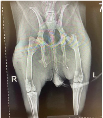
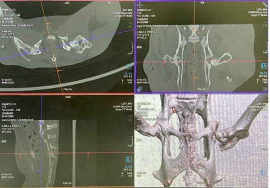
Radiographic examination of the hip joints performed three weeks after turpentine administration revealed areas of bone rarefaction, irregularity of the femoral head contour, and widening of the joint space. Computed tomography provided more detailed visualization of the pathological changes, demonstrating heterogeneous bone density, trabecular deformation, focal osteolysis, and signs of acetabular involvement. These imaging findings were consistent with advanced osteonecrotic changes of the femoral head (Figures 5 and 6).
Gross pathological findings
Gross examination of the hip joint during surgical exposure revealed visible deformation of the femoral head, irregularity and flattening of the articular surface, and areas of cartilage thinning or focal loss. The joint capsule appeared thickened, and periarticular soft tissues exhibited signs of inflammatory reaction. These macroscopic findings confirmed the presence of pronounced pathological changes associated with aseptic necrosis.
Histopathological findings
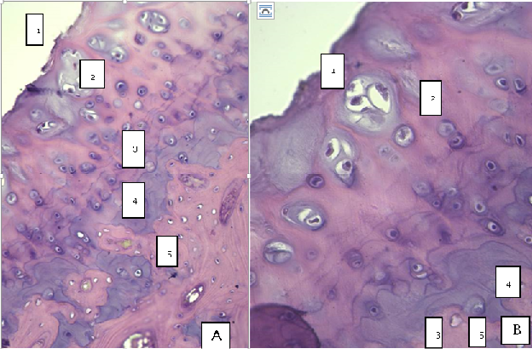
Histological examination of femoral head specimens from the turpentine-treated rabbits demonstrated characteristic features of aseptic necrosis. The articular cartilage showed extensive structural damage, with complete loss of the superficial zone and marked disruption of the middle zone. Residual cartilage fragments appeared deformed and irregular.
Distinct populations of hypertrophic chondrocytes and necrotic chondrocytes were identified within the cartilage matrix, representing different stages of cellular degeneration rather than interchangeable processes. Fissures, delamination of the cartilage matrix, and fragmentation were frequently observed, resulting in an uneven articular surface.
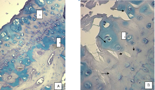
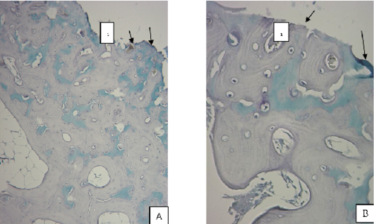
Depletion of glycosaminoglycans was confirmed using Alcian blue staining, which demonstrated markedly reduced staining intensity within the extracellular matrix of the cartilage. Preservation of Alcian blue positivity was observed only in localized areas surrounding isogenic cell groups and within the deep cartilage zone, indicating focal retention of glycosaminoglycans. In several specimens, complete loss of cartilage coverage resulted in direct exposure of the underlying subchondral bone (Figures 1 and 4)
Discussion
The present study demonstrates that intra-articular administration of turpentine provides a reliable and reproducible experimental model of femoral head aseptic necrosis in rabbits ; ; . The combination of clinical signs, imaging findings, gross pathological changes, and histological features observed in this study confirms successful induction of osteonecrotic lesions consistent with those described in Legg–Calvé–Perthes disease ; ; .
Previous experimental models of femoral head aseptic necrosis have predominantly relied on aggressive chemical agents, mechanical interruption of blood supply, or systemic corticosteroid administration. Although effective in inducing necrosis, many of these models are associated with significant systemic toxicity, high mortality rates, and limited reproducibility. In contrast, the turpentine-induced model used in the present study resulted in consistent pathological changes without recorded mortality and with only mild, transient systemic biochemical alterations, indicating a comparatively safer experimental approach.
The histopathological changes observed in this study are in close agreement with those reported in previous experimental and clinical investigations of femoral head osteonecrosis. Loss of the superficial and middle zones of articular cartilage, presence of hypertrophic and necrotic chondrocytes, depletion of glycosaminoglycans, and exposure of subchondral bone have been described as hallmark features of aseptic necrosis in both spontaneous and experimentally induced models.
Radiographic and computed tomography findings further support the relevance of the proposed model ; ; ; . Bone rarefaction, trabecular deformation, osteolytic changes, and acetabular involvement observed in the current study correspond closely to imaging features reported in naturally occurring Perthes disease and in other experimental models ; ; ; . The presence of acetabular changes suggests advanced joint involvement, which is particularly important for evaluating surgical interventions such as femoral head resection arthroplasty ; .
The underlying mechanism of necrosis in the proposed model appears to be indirect vascular compromise mediated by a localized inflammatory response. Intra-articular administration of turpentine induces inflammation of periarticular tissues, which likely leads to disruption of microcirculation and ischemia of the femoral head. Prolonged ischemia subsequently results in chondrocyte death, extracellular matrix degradation, and collapse of subchondral bone. This inflammation-mediated ischemic mechanism closely resembles the multifactorial pathogenesis observed in clinical cases of femoral head aseptic necrosis ; ; .
An important advantage of the present model is its simplicity and reproducibility. The use of a single intra-articular injection at a controlled dose allows for consistent induction of osteonecrotic changes while minimizing animal loss and severe systemic complications. This makes the model suitable for future experimental studies aimed at investigating disease progression, evaluating surgical techniques, and testing novel pharmacological therapies.
Nevertheless, the study has certain limitations. The sample size was relatively small, and quantitative statistical analysis was not performed due to the descriptive nature of the experimental design. In addition, long-term follow-up beyond the initial experimental period was not included. Future studies should incorporate larger experimental groups, extended observation periods, and molecular or biomechanical analyses to further validate and refine the proposed model.
CONCLUSIONS
The present study demonstrates that intra-articular administration of turpentine provides a reliable and reproducible experimental model of femoral head aseptic necrosis in rabbits. The proposed model consistently reproduces clinical signs, radiological changes, gross pathological findings, and histopathological features characteristic of osteonecrosis, while exhibiting reduced systemic toxicity and zero mortality. This experimental approach may serve as a valuable platform for future investigations into the pathogenesis of femoral head aseptic necrosis and for the evaluation of surgical and pharmacological treatment strategies.
Limitations and Recommendations
The present study is limited by a relatively small sample size and the absence of quantitative statistical analysis. Future studies should include larger experimental groups, extended follow-up periods, and quantitative biomechanical or molecular analyses to further validate and refine the proposed experimental model.
ACKNOWLEDGMENT
The authors gratefully acknowledge the technical assistance provided by the staff of the Department of Veterinary Surgery and the Department of Animal Anatomy and Histology at the Moscow State Academy of Veterinary Medicine and Biotechnology. We thank the laboratory personnel for their support in conducting the histological examinations and imaging procedures. We also acknowledge the institutional facilities and equipment that made this experimental study possible.
NOVELTY STATEMENT
This study presents a novel experimental approach using intra-articular turpentine administration to induce femoral head aseptic necrosis in rabbits. Unlike conventional models that rely on aggressive chemical agents or systemic corticosteroids associated with high toxicity, this model produces consistent osteonecrotic changes with minimal systemic effects, zero mortality, and good reproducibility. This approach provides a safe and reliable platform for future studies on disease pathogenesis and therapeutic interventions.
AUTHOR’S CONTRIBUTION
T.E. Deneche conceived and designed the study, performed the experimental procedures, and drafted the manuscript. E.N. Borkhunova performed histological analysis and interpretation of pathological findings. I.D. Lyaskovskiy contributed to data analysis, manuscript revision, and final approval of the submitted version. All authors read and approved the final manuscript.
Ethical approval
All experimental procedures involving animals were conducted in accordance with the European Convention for the Protection of Vertebrate Animals Used for Experimental and Other Scientific Purposes (Strasbourg, 1986; ETS No. 123) and were approved by the local institutional ethics committee of the Moscow State Academy of Veterinary Medicine and Biotechnology.
Data availability statement
The data generated and analyzed during the current study are available from the corresponding author upon reasonable request.
Generative AI and AI assisted technology statement
The authors declare that no generative AI and AI assisted technology was used in the creation of this manuscript.
Conflict of interest
The authors have declared no conflict of interest regarding the publication of this article.
Figures
Figure 1:.
Experimental model of femoral head aseptic necrosis (rabbit 3, group 2). A: The cartilage surface is rough (1), the deformed middle cartilage zone is visible (2) with hypertrophied and pycnotic chondrocytes; the mineralization front is represented by several lines (3); mineralized cartilage (4); subchondral bone (5). B: Higher magnification of the same preparation. Hematoxylin and eosin staining. A (×100), scale bar = 200 µm; B (×400), scale bar = 50 µm.
Figure 2:.
Experimental model of femoral head aseptic necrosis (rabbit 3, group 2). Distribution of glycosaminoglycans in cartilage. A: Significant glycosaminoglycan content is observed in the deformed middle zone (territorial and interterritorial matrix of superficial layers, 1) and in mineralized cartilage (2); subchondral bone (3). Areas with high glycosaminoglycan content are stained green. B: Marked deformation of articular cartilage with loss of superficial and most of the middle layers; fissures and delamination of the extracellular matrix are evident (arrows). Alcian blue staining. A (×100), scale bar = 200 µm; B (×400), scale bar = 50 µm.
Figure 3:.
Experimental model of femoral head aseptic necrosis. Area of complete destruction of articular cartilage with exposure of subchondral bone (1) showing fissures (arrows). Residual cartilaginous matrix is focally observed (green staining). Alcian blue staining. A (×100), scale bar = 200 µm; B (×400), scale bar = 50 µm.
Figure 4:.
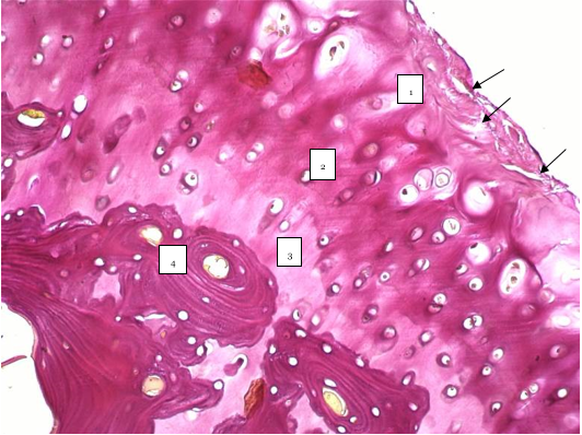
Experimental model of femoral head aseptic necrosis. Destructive changes in the articular cartilage matrix with cracks (arrows) and fraying. Middle zone (1), deep zone (2), mineralized cartilage zone (3), and subchondral bone (4). Van Gieson staining (×200), scale bar = 100 µm.
Figure 5:.
Radiographic examination performed three weeks after turpentine administration revealed femoral head deformation, bone rarefaction, flattening of the acetabular socket, and widening of the joint space.
Figure 6:.
Computed tomography performed three weeks after turpentine administration demonstrated trabecular deformation and acetabular involvement.
References
Aithal HP, Singh A (2024). Textbook of Vet. Ortho. surg. 1st edn. CBS Publishers and Distributors, New Delhi.
Cardoso CB, Tudury EA, Silva JRM, Pereira WL, Araújo FP (2018). Avascular necrosis of the femoral head in dogs: clinical and radiographic findings. Vet and Comparative Ortho. Traumatol, 31(3): 215-222.
Duganets IV (2018). Osteochondropathies in small animals: clinical and pathological aspects. J. Vet. Saint Petersburg, 1: 1-5.
Glueck CJ, Freiberg RA and Wang P (2014). Role of thrombophilia in osteonecrosis of the femoral head. Clini. Orthop. Related Research, 472(3): 903-912.
Herring JA (1994). Legg–Calvé–Perthes disease. J. Bone Jt. Surg. Am., 76(3): 448-458. https://doi.org/10.2106/00004623-199403000-00017[CrossRef]
Johnston SA and Tobias KM (2018). Vet sur: small animal. 2nd edn. Elsevier Saunders, St. Louis.
Kerachian MA Harvey EJ Cournoyer D Chow TY and Séguin C (2006). Avascular necrosis of the femoral head: vascular hypotheses. Endothelium, 13(4): 237-244. https://doi.org/10.1080/10623320600904211[CrossRef]
Kim HKW and Herring JA (2011). Pathophysiol, classifications, and natural history of Perthes disease. Orthop. Clin. North Am., 42(3): 285-295. https://doi.org/10.1016/j.ocl.2011.04.007[CrossRef]
Little DG Kim HKW Rogan T and Sussman MD (2011). Pathophysiology and treatment of Legg–Calvé–Perthes disease. J. Bone Jt. Surg. Am., 93(20): 1959-1970.
Mont MA Jones LC and Hungerford DS (2006). Nontraumatic osteonecrosis of the femoral head: ten years later. J. Bone Jt. Surg. Am., 88(5): 1117-1132. https://doi.org/10.2106/JBJS.E.01041[CrossRef]
Smith JO Kothari A Hing CB and Back DL (2021). Experimental models of avascular necrosis of the femoral head: a systematic review. Bone Jt. Res., 10(2): 79-88.
Wang CJ Wang JW Ko JY and Huang CC (2003). Experimental ischemic osteonecrosis of the femoral head in rabbits. Clinl. Orthop. Related Res., (414): 282-291.
Zhao DW Hu YC Yu T Wang W Yang L and Wang BJ (2020). Pathogenesis of osteonecrosis of the femoral head: cellular and molecular mechanisms. Orthop. Surg., 12(2): 404-413.
Zhou YX Zhang H Zhao XH and Yu XC (2020). Animal models of femoral head osteonecrosis: a review. J.Orthop. Transl., 21: 49-57.