
The Journal of Advances in Parasitology


Research Article
Blood Parasites of Camels from Central Regions of Iran: Comparative Evaluation of Various Detection Techniques and Serum Protein Components
Alisina Karimi1, Sadegh Rahbari2, Ali Yousefi3
1Department of Pathobiology, College of Veterinary, Tehran Science and Research Branch; 2Department of Pathobiology, Science and Research Branch; 3Department of Pathobiology, College of Veterinary, Tehran Science and Research Branch, Islamic Azad University, Tehran, Iran.
Abstract | The 227 blood samples of camels (male: 130, female: 97) were examined from different regions of central areas of Iran between March 2011 and April 2012. Of the examined samples, 4.85% were found to be infected by two species of blood parasites (Trypanosoma evansi, Dipetalonema evansi). Comparison of various techniques, viz., formal gel, mercuric chloride, wet smears and thin smears for detection of T. evansi showed the sensitivity and specificity 44- 100%, 100- 93%, 88- 100%, 88- 100% respectively, where the hematocrit centrifuge test was used as a gold standard. The results of all techniques used for detection of microfilaria were similar. Statistical analysis did not show any significant relation between blood parasite infection and variables such as age, gender and season (p >0.05) but comparison of serum protein components in infected camels with T. evansi showed significant difference except α2-globulin and β-globulin (p <0.05).
Keywords | Camel, Blood parasite, Trypanosoma evansi, Dipetalonema evansi, Iran
Editor | Muhammad Imran Rashid, Department of Parasitology, University of Veterinary and Animal Sciences, Lahore, Pakistan.
Received | January 28, 2015; Revised | March 22, 2015; Accepted | March 25, 2015; Published | March 30, 2015
*Correspondence | Ali Yousefi, Islamic Azad University, Tehran, Iran; Email: a.usefi@srbiau.ac.ir
Citation | Karimi A, Rahbari S, Yousefi A (2015). Blood parasites of camels from central regions of Iran: comparative evaluation of various detection techniques and serum protein components. J. Adv. Parasitol. 2(1): 1-4.
DOI | http://dx.doi.org/10.14737/journal.jap/2014/2.1.1.4
ISSN | 2311-4096
Copyright © 2015 Karimi et al. This is an open access article distributed under the Creative Commons Attribution License, which permits unrestricted use, distribution, and reproduction in any medium, provided the original work is properly cited.
INTRODUCTION
Camel is an important multipurpose animal and since the old times, it has been used for transportation and produce milk, wool and meat in arid and semi-arid areas of the world. Nowadays, according to industry development and technology in all fields, this animal has lost its former importance and now the main target of camels breeding is for production of meat.
They are exposed with various endo and ecto parasites which cause substantial economic losses and some of them have zoonotic importance such as Sarcoptes scabiei (Beesley, 1998) and Linguatula serrata (Lloyd, 1998).
Several studies have been documented on the occurrence of blood parasites in camel from different regions of Iran, viz., Sistan and Balochestan, Fars, Hormozgan (Rahbari and Bazargani, 1995), Semnan (Hamedani et al., 2012), Zabol (Ranjbar-Bahadori and Afshari-Moghadam, 2009) and Kerman (Radfar et al., 2006).
According to Iran Ministry of Agriculture statistics there are about 149600 camels in the country and due to the entry of imported camels and their role in the transmission of pathogens, importance of research in the field of factors affecting the production such as blood parasites in camel breeding areas will be felt.
The aims of our study were to determine the prevalence of blood parasites by using of different diagnostic techniques and evaluation of serum protein components changes in naturally infected camels with Trypanosoma evansi in the camels from different regions of central areas of Iran.
MATERIAL AND METHODS
Sample Collection
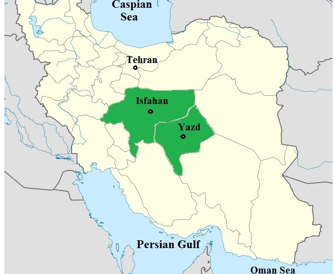
From March 2011 to April 2012, a total of 227 blood samples of camels (male: 130, female: 97) from different regions of central areas (Isfahan province and Yazd province) of Iran were collected (Figure 1).
Two blood samples were collected from each camel through jugular vein. First 5 ml sample was collected into a sterile test tube containing 5 ml of alsever’s solution and was stores at 4°C for later use. Subsequently, 10 ml of the blood sample was collected in another tube to separate serum.
Wet and Thin Smears Preparation
The direct wet smears were prepared by placing the blood samples on a glass slide and cover with a cover-slip to spread blood as a monolayer of cells and the examined with the high dry objective (40 X). The blood thin smears were prepared, air-dry briefly, fixed in methyl alcohol and stained with Geimsa and examined for parasites using an oil immersion objective (100 X).
Hematocrit Centrifuge Technique
Sample of blood is introduced into a capillary tube (coated with heparin) the end sealed with a putty, and the tube centrifuged at 10,000 RPM for five minutes to sediment the cells then looking for directly at the buffy coat layer in under the 4X lens objective.
Formal Gel Test
It is carried out by adding two drop of formalin solution 40% to 1 ml of serum, the test is positive if the serum coagulates immediately and turns white. In negative reactions, the serum remains unchanged or coagulation may take up to 30 minutes to appear.
Mercuric Chloride Test
One drop of serum added to 1ml diluted solution of mercuric chloride (1: 25000). If the serum turns to opalescent, the test is positive. If the result is negative, it will not change until 15 minutes.
Modified Knott Method
One ml blood was added into tube containing 9 ml of 2% formalin and mixed up. The tube was allowed to stand for 15 min to get complete hemolysis. The mixture was then centrifuged at 1,500 RPM for 5 min. The supernatant was discarded and sediment was stained with of methylene blue (1:1000) then the mixture was put on glass slide and examined.
Total Serum Protein and Electrophoresis
Serum total protein level was estimated using the standard protein estimation kit (Zist Shimi Co., Iran) by the method of Bradford (1976). Optical density of the test solutions was measured spectrophotometrically at 545 nm. For serum protein components estimation, cellulose acetate electrophoresis method for the separation of serum proteins was used and levels were determined by densitometry.
Statistical Analysis
Data management was performed using SPSS V19.0 (SPSS Inc., Chicago, Illinois, USA) statistical software. The Chi-square test was used for determining the significance of association between blood parasitic infections and variables such as; age, sex, season (A p-value level of significance was 0.05). Duncan’s multiple range test (DMRT) was used for comparison of total serum protein mean and serum globulin mean in the infected and uninfected camels.
RESULTS
Blood samples collected from 227 camels (Camelus dromedaries) from different regions of central areas of Iran revealed two genera of parasites including Trypanosoma evansi and Dipetalonema evansi (Table 1).
Considering hematocrit centrifuge test as a gold standard for detection of T. evansi and all other techniques showed the sensitivity and specificity respectively were as follows; formal gel (44- 100%), mercuric chloride (100- 93%), wet smears (88- 100%) and thin smears (88- 100%).
Table 1: Prevalence of blood parasites of 227 camels from central areas of Iran by different diagnostic methods
|
Methods |
Trypanosoma evansi |
Dipetalonema evansi |
||
|
Number of infected |
% Infection |
Number of infected |
% Infection |
|
|
Hematocrit centrifuge |
9 |
3.96 |
- |
- |
|
Formal gel |
4 |
1.76 |
- |
- |
|
Mercuric chloride |
24 |
10.57 |
- |
- |
|
Wet smears |
8 |
3.52 |
2 |
0.88 |
|
Thin smears |
8 |
3.52 |
2 |
0.88 |
|
Modified Knott's |
- |
- |
2 |
0.88 |
In the comparison of sensitivity and specificity of different techniques used for the detection of microfilaria of Dipetalonema evansi, modified Knott’s test was used as the gold standard. The results of all other techniques were similar. Data on the prevalence of blood parasites based on the age, sex and season are presented in Table 2. In general 4.85% of blood samples were infected with hemoparasite. The highest prevalence was observed at T. evansi (3.96%). According to the data in the Table 2 and the statistical analysis (the Chi-square test) did not show any significant relation between blood parasite infection and variables such as age, gender and season (p >0.05).
Table 2: Prevalence of blood parasites in camels from central areas of Iran based on age, sex and season
|
Group |
Number |
Number of infected |
% Infection |
Chi-Square |
P- value |
|
Age |
0.369 |
0.832 |
|||
|
1-5 years |
32 |
2 |
6.25 |
- |
- |
|
6-10 years |
67 |
3 |
4.48 |
- |
- |
|
11-18 years |
128 |
6 |
4.69 |
- |
- |
|
Gender |
1.006 |
0.316 |
|||
|
Male |
130 |
8 |
6.15 |
- |
- |
|
Female |
97 |
3 |
3.09 |
- |
- |
|
Season |
7.035 |
0.071 |
|||
|
Spring |
63 |
0 |
0 |
- |
- |
|
Summer |
67 |
5 |
7.46 |
- |
- |
|
Autumn |
43 |
3 |
6.98 |
- |
- |
|
Winter |
54 |
3 |
5.56 |
- |
- |
|
Total |
227 |
11 |
4.85 |
- |
- |
Table 3: The data of the mean ± standard error of the mean for serum proteins components (μg/dl) in infected and non-infected camels with Trypanosoma evansi
|
Parameter |
Infected |
Uninfected |
|
Total protein* |
6.62±0.05 |
6.36±0.06 |
|
Albumin* |
2.38±0.12 |
3.08±0.14 |
|
Globulin* |
4.23±0.16 |
3.27±0.18 |
|
α1-globulin* |
0.10±0.04 |
0.47±0.05 |
|
α2-globulin |
0.72±0.11 |
0.84±0.12 |
|
β-globulin |
0.84±0.07 |
0.69±0.08 |
|
γ-globulin* |
2.55±0.13 |
1.16±0.15 |
|
A/G* |
0.58±0.06 |
0.96±0.07 |
* - This result is significant at the p=0.05 level
The results of total protein value and estimation of serum protein components in serum samples from the two groups of animals (infected, uninfected) are shown in Table 3. There was significant difference in all serum protein components parameters of infected camels with T. evansi except α2-globulin and β-globulin (p <0.05).
DISCUSSION
Several studies have been documented on blood parasites of camels in the world and among them Babesia sp, Theileria sp, T. evansi and microfilaria were reported. In our investigation, two species of hemoparasites, T. evansi and D. evansi were found in blood samples.
Trypanosoma evansi causes a trypanosomiasis known as “surra”. It affects a large number of wild and domesticated animals in different parts of the world (Singh and Singla 2013; Bal et al., 2012; Mullen, 2002). There is extensive variation in the pathogenicity of various strains and the susceptibility of different host species (Bal et al., 2012). The disease may reveal as an acute or chronic form, and in the latter case may persist for several months, possibly years (Mullen, 2002). Hence surra causes a major constraint to camel’s productivity in decreasing of milk and meat yield, also premature births and abortions in pregnant females and treatment cost (Elamin et al., 1999; Boid et al., 1985; Reid, 2002).
Outbreaks of T. evansi infections have been reported in various areas of world in different species of animals (Kumar et al. 2012). As per the available information on the prevalence is concerned in Iran 19.47 and 1.6 per cent prevalence has been reported from Zabol (Ranjbar-Bahadori and Afshari-Moghadam, 2009) and Kerman provinces (Radfar et al., 2006), respectively. Different rates of occurrence has been seen in different countries from time to time viz., Nigeria (27%) (Pacholek et al., 2001); Chad (30%) (Losos, 1980); Mauritania (24%) (Dia et al., 1997); Ethiopia (21%) (Zeleke and Bekele, 2001); Kenya (28%) (Njiru et al., 2001); Sudan (33%) (Elamin et al., 1999); India (22%) (Pathak et al., 1993); Jordan (33%) (Al-Rawashdeh et al., 2000) which are higher prevalences than our study. Our results are close to the findings of Rahbari and Bazargani (1995), Hamedani et al (2012) in Iran and Abd-Elmaleck et al (2014) in Egypt who recorded 7.7%, 2.63% and 3.06% prevalence respectively.
Dipetalonema evansi is a filarial nematode which affects camels and occurs in the spermatic cord, pulmonary arteriole, right auricle, lymph nodes and mesentery. Clinical symptoms depend upon the location of adult worm but usually hypertrophic sclerosis and aneurysm are the common lesions in this infection (Parsani et al., 2008; Dakkak and Ouhelli, 1987; Nagaty, 1947). Few studies have been carried out on filariasis of camels in world such as in Iran (Rahbari and Bazargani, 1995; Sazmand et al., 2013), Sudan (Elamin et al., 1999) while results of our study were similar to Ranjbar-Bahadori and Afshari-Moghadam (2009) in Zabol.
Boid et al (1980) in their study showed that significant increased levels of γ-globulin on infected camels with T. evansi which were similar to our study. On the other hand, formal gel and mercuric chloride tests were used by Luckins et al (1979) for detection of T. evansi in 226 camels and showed the sensitivity and specificity; 77-100%, 63-100% respectively which results differ from our study.
ACKNOWLEDGEMENTS
We would like to thank Dr. Hadi Miranzadeh for instrumental assistance on this project.
CONFLICT OF INTEREST
The authors declare that there is no conflict of interests.
REFERENCES


