
The Journal of Advances in Parasitology


Case Report
The Journal of Advances in Parasitology 1 (3): 39 – 40Cryptosporiodiosis in a Nellore Doe Associated with Hydrocephalus Foetus
YV Pridhvidhar Reddy1, Sirigireddy Sivajothi2*, Bhavanam Sudhakara Reddy3
- Department of Obstetrics and Gynaecology
- Department of Veterinary Parasitology
- T.V.C.C. (Veterinary Medicine), College of Veterinary Science, Sri Venkateswara Veterinary University, Proddatur, Andhra Pradesh- 516360, India
*Corresponding author: sivajothi579@gmail.com
ARTICLE CITATION:
Reddy YVP, Sivajothi S, Reddy BS (2014). Cryptosporiodiosis in a Nellore Doe Associated with Hydrocephalus Foetus. J. adv. Parasitol. 1 (3): 39 – 40.
Received: 2014–05–06, Revised: 2014–07–03, Accepted: 2014–07–04
The electronic version of this article is the complete one and can be found online at
(
http://dx.doi.org/10.14737/journal.jap/2014/1.3.39.40
)
which permits unrestricted use, distribution, and reproduction in any medium, provided the original work is properly cited
ABSTRACT
Congenital foetal anomalies can be divided into heritable, toxic, nutritional and infectious categories. Although uncommon inherited congenital anomalies are probably seen in all breeds of small ruminants but reports in doe are meager. Caesarean operation was performed to the goat with incomplete cervical dilatation. Up on caesarean two foetuses were recovered of which, a dead hydrocephalus foetus with enlarged head and one normal foetus. Hydrocephalus foetus had increased cerebrospinal fluid, enlarged skull and atrophied brain. Faecal sample examination of one day age old live foetus and doe showed cryptosporidium oocysts. This paper reports the Cryptosporidiosis associated with hydrocephalus foetus in a doe and presence of Cryptosporidiosis to a day old foetus.
Hydrocephalus is an excessive cerebrospinal fluid accumulation in the cranial cavity. Several pathologies are encountered in the goat’s embryo and foetus that they occur due to infectious and non-infectious reasons at pre-implantation, embryonic and fetal periods (Semecan et al., 2012). Cryptosporidiosis is one of the zoonotic diseases caused by a protozoan parasite of the genus Cryptosporidium. Although the disease affects mainly the gastro-intestinal tract of a wide range of vertebrate hosts, significant effects of infections have been noted mainly in humans and calves. Cryptosporidiosis was recorded in calves as young as 4 days and in the gazella, the condition was observed among day-old neonates previously (Kanyari et al., 2002). The present communication reports the Cryptosporidiosis associated with hydrocephalus foetus in a doe which was delivered by Caesarean operation.
A doe of 5 years age old was presented to the Veterinary Dispensary, Yerraguntla (which is native of corresponding author) with history of severe abdominal straining, reduced intake of water and feed from previous night. Doe had the mating history of five months. Up on clinical examination it had restless, colic signs, raised rectal temperature (105.6°F), pulse (115/min) and respiratory rate (68/min). Peripheral blood smears and faecal samples were collected from the doe for routine laboratory examination. Vaginal examination revealed incomplete cervical dilation. Complete cervical dilatation was not observed even 30 minutes after administration of Valethamate Bromide (20 mg/doe). Based on history and clinical presentation and response to the above therapy emergency caesarean was done.
Caesarean was performed by ventral paramedian approach under xylazine sedation and local analgesia (2% Lignocaine). Up on caesarean one normal foetus and another with large cranium were recovered (Figure-1).
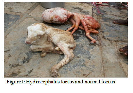
In this normal foetus was live and abnormal foetus with enlarged head was dead at the time of Caesarean. Following caesarean the doe was treated with Ceftriaxone inj (500 mg I.M. for 5 days), 25 % Dextrose (250 ml, I.M. for 2 days), and Meloxicam inj (3 ml, I.M. for 3 days) and an herbal uterotonic. On examination, abnormal foetus had soft skull which is more prone for pressure. Cranial bones were thin with defects, filled with fibrous membrane. There is an abnormal increase in the amount of cerebrospinal fluid within the cranial cavity that is accompanied by expansion of cerebral ventricles, enlargement of the skull and especially forehead and atrophy of brain. Normal foetus had normal activities no other abnormalities were noticed after Caesarean.
Faecal samples from both doe and day old normal foetus were collected into plastic containers directly from the rectum. The smears of faeces were made on glass slides and dried in room temperature and stained by modified Ziehl-Neelsen staining method, as described by Henriksen and Pohlenz, (1981). The stained smears were finally examined microscopically under oil immersion objective of compound microscope. Same procedure was used for diagnosis of Cryptosporiodiosis in neonatal dairy calves in Andhra Pradesh (Sivajothi et al., 2014). Samples collected from the normal foetus and doe revealed presence of cryptosporidiosis.

The present case was diagnosed as hydrocephalous foetus associated with Cryptosporidiosis.
The incidence of dystocia in goats has been reported about (7%) from reproductive diseases (Abdul Rehman et al., 2000). The causes of dystocia have been reported either due to maternal or fetal in origin. Recent studies reveal that the fetal causes of dystocia were more common than the maternal causes (Hussain and Zaid, 2010). The cause of hydrocephalus is diverse and includes genital factors, developmental anomalies intrauterine or prenatal infection or bleeding in the brain (Woo et al., 2010). Hydrocephalus occurs when there is resistance in CSF passage that causes a higher pressure gradient between CSF proximal and distal to the obstruction and moreover alternate pathways of CSF absorption are unable to reduce the increased CSF volume within the ventricles and return it to normal range (Thomas, 2010; Zhao et al., 2010). It has been reported that the disruption of CSF absorption is connected with the duration of hydrocephalus (Zhao et al., 2010). Congenital hydrocephalus also occur secondary to wide range of nervous system anomalies including meningomyelocele, chiari malformation, Dandy-walker syndrome and cerebral hypoplasia (Thomas, 2010). Congenital hydrocephalous in domestic animals according to some authors is inherited through an autosomal recessive gene, although the role in its origin may be played by viral infections of foetus and dietary factors (Bester et al., 1976).
Cryptosporidium is considered as one of the major enteric protozoan parasite in goat kids and morbidity could be high in outbreaks of cryptosporidiosis in kids (Paul et al., 2014). Apart from mortality (up to 40%), cryptosporidiosis causes decline in productivity, retarded growth, decreased feed efficiency, delayed maturity, loss of fertility and overall financial loss in the form of treatment of ailing animals (Paraud and Chartier, 2012). In this study day age old foetus had cryptosporidiosis along with the infected doe. The infective stages in this case would have to be the merozoites which systemically invade the maternal tissues including the circulatory system resulting in the fetal infection. The exact route of fetal infection may be oral by ingestion of merozoites possibly contained in amniotic fluids or via the blood (Kanyari et al., 2002).
REFERENCES
Abdul-Rahman LY, Al-Janabi AS, Asofi MK (2000). Study of some reproduction aspects of the mature local Iraqi goats. The Veterinarian, 10 (1): 47 - 60.
Bester RC, Cimprich RE, Evans LH (1976). Hydrocephalus in an 18-month old colt. Am. J. Vet. Med. Assoc. 168: 1041 - 1042.
Henriksen SA, Pohlenz JFL. (1981). Staining of cryptosporidia by a modified Ziehl-Neelsen technique. Acta Veterinary Scandvica 22: 594 - 596.
PMid:6178277
Hussain SO, Zaid NW (2010). Dystocia in goats, causes and treatment. Al-Qadisiya J. Vet. Med. Sci. 9(1) 11 - 15.
Kanyari PWN, Oyejide AO, Alak JIB, Anderson DL, Wilson ST, Srivastava K (2002). Cryptosporidium Parvum: Experimental Transplacental Transmission In Murine Hosts. Israel J. Vet. Med. 57 (2). http://www.isrvma.org/article/57_2_3.htm).
Paraud C, Chartier C (2012). Cryptosporidiosis in small ruminants. Small Ruminant Res. 103: 93 – 97.
http://dx.doi.org/10.1016/j.smallrumres.2011.10.023
Paul S, Sharma DK, Boral R, Mishra AK, Nayakwadi S, Banerjee PS, Pawaiya RVS (2014). Cryptosporidiosis in Goats: a Review. Adv. Anim. Vet. Sci.2 (3S): 49 – 54.
http://dx.doi.org/10.14737/journal.aavs/2014/2.3s.49.54
Roberts SJ (1971). Veterinary Obstetrics and genital diseases, 2nd ed. CBS Publishers, New Delhi, India, Pp, 69.
Sivajothi S, Reddy BS, Rayulu VC (2014). Cryptosporiodiosis in neonatal dairy calves. Adv. Applied Sci. Res. 5(1):74 - 76.
Semecan A, Kaymaz M, Fındık M, Rişvanlı A, Köker A. (2012) Çiftlik Hayvanlarında Doğum ve Jinekoloji. 589 - 590, Medipres Matbaacilik Ltd. Sti., Malatya.
Thomas WB (2010) : Hydrocephalus in dogs & cats. Veterinary Clinics of North America, Small Animal Practice, 40, 143-159.
http://dx.doi.org/10.1016/j.cvsm.2009.09.008
PMid:19942061
Woo Dc, Choi CB, Nam JW, Ryu KN, Jahng GH, Lee DW, Kim HY, Ahn KJ, Choc BY (2010). Quantitative analysis of hydrocephalic ventricular alterations in Yorkshire terrices using magnetic resonance imaging. Veterinarian medicina 55: 125 - 132.
Zhao K, Sun H, Shan Y, Mao BY, Zhang H (2010). Cerebrospinal fluid absorption disorders of arachnoid villi in canine model of hydrocephalus. Neurology India 58: 371 - 376.
http://dx.doi.org/10.4103/0028-3886.65601
PMid:20644263


