
The Journal of Advances in Parasitology


Research Article
The Journal of Advances in Parasitology 1(1): 4 – 5Study on Paramphistomiosis in Cattle at Sonatala Upazila, Bogra, Bangladesh
Kazal Krishna Ghosh1*, Tamanna Jahan Mony2, Muhammad Shah Jalal1, Muhammad Sirajul Islam2,
*Corresponding author: kazal_krishna@yahoo.com
ARTICLE CITATION:
Ghosh KK, Mony TJ, Jalal MS and Islam MS (2014). Study on paramphistomiosis in cattle at sonatala upazila, bogra, Bangladesh. (abulmoschus esculentus l.). J. Adv. Parasitol. 1(1): 4 – 5
Received: 2013–10–22, Revised: 2013–12–03, Accepted: 2013–12–11
The electronic version of this article is the complete one and can be found online at
(http://dx.doi.org/10.14737/journal.jap/2014/1.1.4.5)
which permits unrestricted use, distribution, and reproduction in any medium, provided the original work is properly cited
ABSTRACT
The study aimed at investigating the occurrence of Paramphistomiosis in association with age, sex and breed of sick cattle brought for treatment at Upazila Veterinary Hospital, Sonatala, under Bogra district, Bangladesh. Coproscopy was conducted to monitor the occurrence of Paramphistomiosis. Out of total 107 examined cattle, 32 (29.90%) were found positive for Paramphistomiosis. It was also observed that, rate of infections in young cattle (younger than two years) were higher (34%) than adult (over two years) (26.31%). Higher infection was observed in male cattle (33.33%) than females (27.11%) and the frequency of Paramphistomiosis was lower in local–bred (24.28%) than cross–bred cattle (40.54%). So effective control measures should be taken to minimize this problem.
INTRODUCTION
Parasitic diseases are one of the major causes of hindering the livestock development around the globe including Bangladesh. It has been estimated that about 10% animals die annually due to parasitic diseases (Mia and Kibria, 1993). Gastrointestinal parasites are a major constraint to health and productivity in grazing livestock production systems (Fox, 1997). Various species of Paramphistomum cause a disease called Paramphistomosis which affects production, since these parasites provoke lower nutrition utilization, a loss of weight and a decrease in milk production, and ultimately causing great economic losses (Rangel–Ruiz et al., 2003). These parasites wander in the duodenal mucosa resulting in severe erosions. In heavy infection, they cause enteritis characterized by oedema, haemorrhages and ulceration whereas adult flukes in the fore–stomach are well tolerated. The immature fluke causes high degree of morbidity as well as mortality (Panda, 1985). Death due to immature Paramphistomes is very high and may be as high as 80–90% in domesticated ruminants ( Juyal et al., 2003; Ilha et al., 2005). Given the fact that the parasitic infection causes high economic losses of livestock in Bangladesh, the present study was undertaken to observe the frequency of Paramphistomiosis in cattle at Sonatala, under Bogra district
MATERIALS AND METHODS
This study was carried out in 107 cattle brought for treatment at Upazila Veterinary Hospital, Sonatala, Bogra from 17 November, 2012 to 16 January, 2013. A prototype questionnaire regarding age, sex, breed, clinical history, presenting sings of study individual was designed to collect the objectives oriented data from each cattle. Faecal samples of cattles were collected in a clean plastic container and direct smear and sedimentation techniques were employed for faecal analysis. The egg of Paramphistomum was identified on the basis of its characteristic morphological feature (Soulsby, 1982).
RESULTS AND DISCUSSION
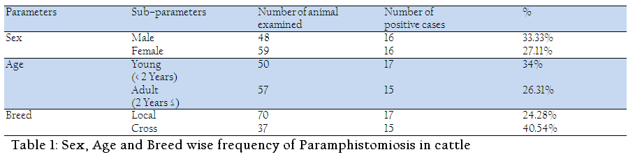
Out of 107 cattle examined, 32(29.90%) were found positive with Paramphistomiasis. Uddin et al. (1994) reported higher infection rate (56.66%) than the results present here. It might be due to the variation in study place, feed, de–worming, and managements of animals. The occurrence of Paramphistomiasis was slightly higher in male (33.33%) than females (27.11%) (Table–1). In contrast, Saifuzzaman (1996) reported that the infection rate in male was 45.54%, which was lower than in female 55.56%. The lower infection in female might be due to the social practice of keeping female under better management and feeding conditions in comparison to males, which are generally let loose to graze freely in pastures.As presented in Table–1, Paramphistomiosis rate was higher in younger cattle (34%) than adult (26.31%). Similarly, Juyal et al. (2003) showed higher frequency in young (61.36%) than adult (49.36%), however, Sarder et al. (2006) reported that frequency of Paramphistomum increases with the age. This variation might be due to the high susceptibility and low resistance at young age. The frequency of this parasitic infection was observed higher (40.54%) in cross–bred cattle than local–bred (24.28%). Sarder et al. (2006) reported that the frequency of Paramphistomiosis is 45.27% in native bred and 51.11% in cross bred cattle which is in line with the current findings
CONCLUSION
The result of this study will give an overall idea about the prevalence of Paramphistomiosis in cattle at the study area. However, this study will provide foundations for further extensive studies related to these infections which are necessary to design preventive and control measures against Paramphistomiosis in Bangladesh.
ACKNOWLEDGEMENT
Authors are thankful to the Upazila Livestock Officer, Sonatala Upazila Veterinary Hospital, Bogra and Department of Parasitology, Chittagong Veterinary and Animal Sciences University, for providing necessary facilities and help for the experiment.
REFERENCES
Fox M (1997). Pathophysiology of infection with gastro–intestinal nematodes in domestic ruminants: Recent developments. Vet. Parasitol. 72: 285–297.
http://dx.doi.org/10.1016/S0304-4017(97)00102-7
Ilha MR, Loretti AP and Reis AC (2005). Wasting and mortality in beef cattle parasitized by Eurytrema coelamaticum in the state of Parana, southern Brazil. Vet. Parasitol. 133: 49–60.
http://dx.doi.org/10.1016/j.vetpar.2005.02.013
PMid:16046069
Juyal PD, Kasur K, Hassan SS and Paramjit K (2003). Epidemiological status of Paramphistomiasis in domestic ruminants in Punjab. J. of Para. 231–235.
Mia NH and Kibria (1993). A study of livestock for second plan. Planning Commission, Dhaka.
Panda PG (1985). Outbreak of immature Amphistomiasis in cattle in India. Ind. J. of Vet. Sci. 5: 364–375.
Rangel–Ruiz LJ, Albores–Brahms and Gamboa–Anguilar J (2003). Seasonal trends of Paramphistomum cervi in Tabasco, Mexico. Vet. Parasitol. 116: 217–232.
http://dx.doi.org/10.1016/j.vetpar.2003.07.002
PMid:14559164
Saifuzzaman ABM (1996). Incidence and seasonal variation of helminth parasites in cattle of Chandia thana in Comilla district. Thesis, Department of Parasitology,Bangladesh Agricultural University, Mymensingh.
Sarder SA, Ehsan MA, Anower AKMM, Rahman MM and Islam MA (2006). Incidence of liver flukes gastro–intestinal parasites in cattle, Bang. J. of Vet. Med. 4(1): 39–42.
Soulsby EJL (1982). Helminthes arthropod and protozoa of domesticated animal (7th Ed), Iowa State University, Iowa University Press, 64–71.
Uddin KH, Miah MF and Taimure JJFA (1994). Gastro–intestinal parasitosis in calves under traditional management in Bangladesh. Bang. Vet. J. 35(1–2): 9–18.


