

Seuratascaris schmackeri sp. nov. (Nematoda: Ascarididae) from the Chinese Frog Odorrana schmackeri Boettger, 1892 (Amphibia: Anura) Based on Morphological and Molecular Evidence


Seuratascaris schmackeri sp. nov. (Nematoda: Ascarididae) from the Chinese Frog Odorrana schmackeri Boettger, 1892 (Amphibia: Anura) Based on Morphological and Molecular Evidence
Ying Liu1,2, Ji-Yong Fang2, Na Zheng3 and Hai-Long Wu1*
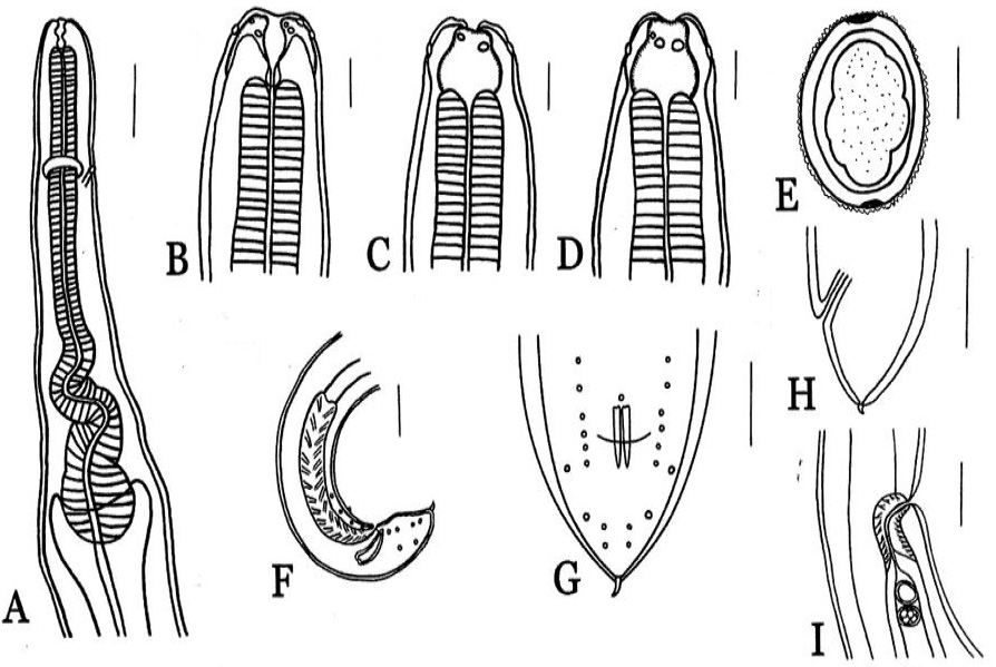
Seuratascaris schmackeri sp. nov. from Odorrana schmackeri in China. (A) anterior part of male, laterial view, showing oesphagus, nerve-ring, excretory pore, intestinal caecum. (B) anterior part of male, laterial view, showing interlabial and postlabial groove. (C) anterior part of male, dorsal view, showing dorsal lip with two pipillars. (D) anterior part of female, lateral-ventral view, showing lateral-ventral lip with one papillae and amphid. (E) egg. (F) posterior end of male, lateral view. (G) posterior end of male, ventral view. (H) posterior end of female, lateral view. (I) region of Vulva, lateral view. Scale bars: A= 200μm; B, C, D= 500μm; E= 50μm; F, G, H = 200μm; I = 300μm.
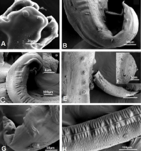
Scanning eletron micrographs of Seuratascaris schmackeri sp. nov. from Odorrana schmackeri in China, male. (A) cephalic extremity, dorsal view, dorsal lip with two papillae, subventral lip with denticles, showing interlabial and postlabial groove. (B) tail extremity, laterial view, spicule. (C) posterior end, lateral view, precloacle adcloacle papillae and herringbone cuticle apperance. (D) image of precloacle papillae. (E) cloacle region, lateral view, adcloacle papillae, a big median papillae in front of cloacle. (F) image of median papillae in front of cloacle, lateral view. (G) posterior end, ventral view, postcloacle papillae. (H) middle of body, lateral view, lateral alae.

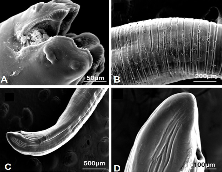
Scanning eletron micrographs of Seuratascaris schmackeri sp. nov. from Odorrana schmackeri in China, female. (A) cephalic extremity, lateral view, lateral-ventral lip with one oval papillae and one small round papillae and amphid, lip with denticles, showing interlabial and postlabial groove. (B) middle of body, lateral view. (C) posterior end, lateral view, anus. (D) posterior end, lateral view.


