The Journal of Advances in Parasitology
Case Report
Asphyxial Death by Oestrus ovis in a Pneumonic Goat
Md. Ahaduzzaman1*, Md. Shafiqul Islam2, Sharmin Akter1, Md. Jashim Uddin1, Shad Md. Omar Sharif1, Abdul Mannan1
1Department of Medicine and Surgery; 2Department of Pathology and Parasitology, Chittagong Veterinary and Animal Sciences University, Chittagong-4202, Bangladesh.
Abstract | Nasal myiasis is an unusual disease in goats caused by the infestation of nasal cavities by the larvae of Oestrus ovis. A buck was presented with clinical history of nasal discharge, sneezing and onset of episodes of neurological symptoms such as ataxia, staggering gait, convulsions and paddling of the legs. The animal was treated with ivermectin @ 0.2 mg/kg and amoxicillin @10 mg/kg body weight with supportive therapy, but the response to the treatment was grave owing to asphyxia. On post-mortem, 13 larvae of Oestrus ovis were recovered from the nasal sinus and their morphological study was performed. The carcass showed severe congestion of the lungs accompanied by frothy exudation in the trachea. This report presents the first case of goat nasal myiasis in Bangladesh.
Keywords | Oestrus ovis, Goat, Ivermectin, Pneumonia, Nasal myiasis
Editor | Muhammad Imran Rashid, Department of Parasitology, University of Veterinary and Animal Sciences, Lahore, Pakistan.
Received | July 14, 2015; Revised | August 03, 2015; Accepted | August 04, 2015; Published | August 10, 2015
*Correspondence | Md. Ahaduzzaman, Chittagong Veterinary & Animal Sciences University (CVASU), Bangladesh; Email: [email protected]
Citation | Ahaduzzaman M, Islam MS, Akter S, Uddin MJ, Sharif SMO, Mannan A (2015). Asphyxial death by Oestrus ovis in a pneumonic goat. J. Adv. Parasitol. 2(3): 48-51.
DOI | http://dx.doi.org/10.14737/journal.jap/2015/2.3.48.51
ISSN | 2311-4096
Copyright © 2015 Ahaduzzaman et al. This is an open access article distributed under the Creative Commons Attribution License, which permits unrestricted use, distribution, and reproduction in any medium, provided the original work is properly cited.
Larvae of flies belonging to the Oestridae family include several genera infesting the nasopharyngeal cavities and respiratory organs during their migration, cause obligatory myiasis in sheep and goat. The adult female fly is larviparous, lay down a number of first-instar larvae around or juxtaposition the nostrils of sheep and goats. To complete their normal life cycle in goat, the larvae migrate to the nasal and frontal sinus mucosa followed by undergoes two moult. Within 2 to 12 months, the fully grown third instar larvae are expelled and pupate on the ground. (Blood et al., 1986; Papadopoulos et al., 2006). This parasite is found worldwide and infestation of O. ovis is usually not life threatening, but occasionally there is a bacterial infection that causes encephalitis and death of the host. On the other hand, secondary infections often give rise to mucopurulent discharge, respiratory distress and sneezing fits in the affected sheep and goats (Urquhart et al., 1996). Although nervous form of O. ovis infestation in sheep and goats is rare because of larval penetration into ethmoid bones, and reach cranial cavity resulting in abnormal neurological signs such as a staggering gait, mania and sudden death (Shivasharanappa et al., 2011). These neurological sign and symptoms are usually confused with the infection of Coenurus cerebralis because of which this disease is also familiar as ‘False Gid’. This infection can result in economic losses in sheep and goat reared for meat and dairy produce. Despite the fact that both sheep and goat can be host of this parasite, infection prevalence and larval burdens are generally inflated in sheep than in goats after either natural or experimental infestation (Duranton et al., 1995). Furthermore little is known about the development of immunity for whether auto recovery or not, but it is possible that some animals are in immune deficient during infection. In the same coin respiratory co-infections are common in sheep and O. ovis has been suggested to have an immunosuppressive effect with consequent association with respiratory pathogens (Dorchies et al., 1993). The present documentation, reports rare cases of goat nasal sinus infestation with migrating O. ovis larvae resulting in sudden onset of episodes of neurological signs and later on asphyxia death.
A 65 kg body weight Jamunapari buck was presented with clinical history of nasal discharge, sneezing and onset of episodes of neurological symptoms such as ataxia, staggering gait, convulsions, circling and paddling of the legs and expelled of single larvae with a sneeze. Clinical examination of the animals revealed elevated body temperature
Table 1: Hemato biochemical parameters of infected buck
|
Name of the test |
Result |
Normal range |
Name of the test |
Result |
Normal range |
|
Serum calcium |
9.15 |
9.7-12.4 mg/dl |
Haemoglobin |
6.0 |
8-12 gm% |
|
S. Magnesium |
3.95 |
1.8-2.3 mg/dl |
ESR (wintrobe tube method) |
3.0 |
0 in 1st hour |
|
S. Phosphorus |
2.55 |
4.2-9.1 mg/dl |
PCV |
31 |
50-70% |
|
S. Total protein |
74.0 |
64-70 mg/dl |
Total count of RBC |
3.59 |
8-18 million/cumm |
|
S. Glucose |
303.75 |
50-75 mg/dl |
Total count of WBC |
10.25 |
8-12(Thousand/cumm) |
|
S. GPT |
76.6 |
6-19 U/L |
Lymphocytes |
20 |
22-35% |
|
S. GOT |
6.9 |
167-513 U/L |
Neutrophils |
75 |
30-48% |
|
S. Creatinine |
3.6 |
1.0-1.8 mg/dl |
Eosinophils |
03 |
1-8% |
|
S. Albumin |
23.1 |
27-39 g/dl |
Monocytes |
02 |
0-4% |
|
Basophils |
0 |
0-1% |
|||
(105°F), heart rate 140/minute, respiration rate 80/minute and good body condition. 5 ml of blood was collected from the jugular vein for routine examination, including serum calcium, magnesium, phosphorous, total protein, glucose, SGPT, SGOT, creatinine and albumin (Table 1).
In animals, treatment was instituted immediately after brief clinical examination as warranted by the acuteness of the condition. Treatment with Ivermectin @ 0.2 mg/kg body weight subcutaneously was used to combat against oestrosis; amoxicillin @ 10mg/kg body weight and ketoprofen @ 3.3 mg/kg body weight to treat pneumonic condition. Despite therapy, episodes of neurological signs increased with time, the animal became laterally recumbent and finally succumbed to the disease within one day of the initiation of treatment.
The goat was necropsies thoroughly immediately after death and macroscopic observations, including in the nasal, paranasal sinuses and cranial cavities were recorded. A sagittal section was made in the skull using the electric saw (Figure 2). The live larvae were carefully removed from the nasal cavities, paranasal sinuses, and collected for further morphological examinations.
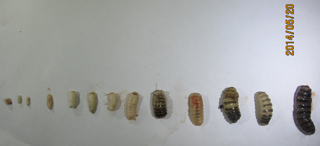
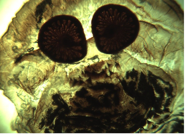
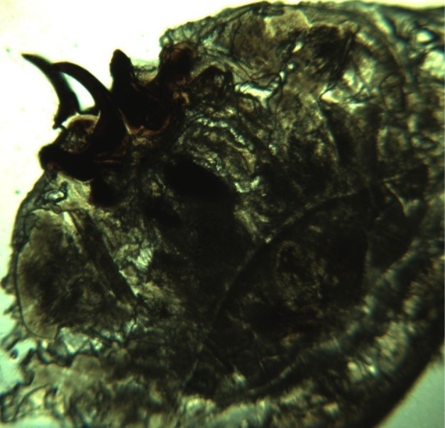
On necropsy, 13 larvae were recovered from the nasal sinuses. The larvae was confirmed as Oestrus ovis using the Morphology for identification such as yellowish white, tapering anteriorly with prominent step posteriorly, each segment has a dark/black band dorsally (Figure 1). Microscopically, characteristic oral hook and stigmal plate with radially arranged respiratory holes of O. ovis were noticed (Figure 3, 4 and 5). The carcass showed severe congestion of the lungs with frothy exudation in the trachea. The lung surface showed pneumonic lesions along with the presence of whitish patches and foamy appearances. Patchy areas of congestion were also found on the serosal surface of the rumen and reticulum. There was no other significant gross lesion in the heart and spleen.
Oestrosis, that develops from the first to the third stage larva which is obligate parasites of the nasal and sinus cavities of sheep and goats. In their normal development in goat, the larvae migrate to the nasal mucosa and nasal and frontal sinus mucosa where they undergo 2 moults. We found larvae that were migrated to the dorsal turbinates and frontal sinuses and two were sneezed out. Macrocyclic lactones such as ivermectin @ 0.2 mg/Kg body weight and Closantel @7.5 mg/Kg body weight are claimed to be effective in controlling the buildup of heavy infestations and for the removal of overwintering larvae of O. ovis (Dorchies et al., 2000). However, treatment was not effective, although
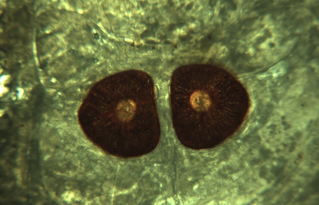
Figure 4:D–shaped closed, dark black coloured, stigmal plates with radially arranged respiratory holes (under 4x microscopy)
the larvae yet not migrate and reach the aberrant locations like the cranial cavity. The episodes of abnormal hyper excitability signs in the affected buck were caused possibly due to the injury to the nasal mucosa by the migration of O. ovis larvae, as mechanical activity of oral hooks and spines as well as the proteolytic enzymes present in the secretory and excretory products of these larvae were responsible for these changes (Tabouret et al., 2003). Secondary infections often lead to muco-purulent discharge, respiratory distress and sneezing fits as observed (Urquhart et al., 1996). In the blood serum examination, it’s shown that there was increased level of serum magnesium, total protein, glucose, SGPT, creatinine and decreased level of Serum calcium, phosphorus, SGOT, and albumin. However, elevated level of total proteins (80-90 mg/dl) and reduced glucose level (25-30.8 mg/dl) was found previously in the case of nasal bot (Sharma et al., 2014).
Congestion is an excess of blood contained within blood vessels in a part of the body due to a passive process. Grossly, it appears as dark blue-red tinge colour. Suspected agonal congestion was found on the serosal surface of the rumen and reticulum and which may be due to asphyxia.
Oestrosis not always but hardly turn into lethal to the host. In the present study, one buck was infected with O. ovis. This report documents the first known case of asphyxial death by nasal myiasis in a goat of Bangladesh by O. ovis. Possible human infections living in close proximity have also to be taken into consideration as cases are not unknown in the endemic territory.
ACKNOWLEDGEMENT
Special thanks to Aranzazu Meana, European Veterinary Parasitology College (EVPC) for his suggestive opinion to identify the parasite.
CONFLICT OF INTEREST
The authors declare that they have no conflict of interest.
Author’s Contribution
Md. Ahaduzzaman and Abdul Mannan treated the patient at the hospital. Md. Ahaduzzaman, Abdul Mannan, Md. Shafiqul Islam, Md. Jashim Uddin and Shad Md. Omar Sharif performed the postmortem examination. Sharmin Akter and Abdul Mannan carried out the hematological evaluation. Md. Shafiqul Islam prepared the histopathological slide for microscopic experimentation. Md. Ahaduzzaman and Sharmin Akter designed the research work, drafted and revised the manuscript. All authors read and approved the final version of the manuscript.
REFERENCES


