Advances in Animal and Veterinary Sciences
Research Article
Tegumental Alterations on Raillietina galli, an Intestinal Parasite of Fowl, after Treatment with Millettia taiwaniania Extract
Kholhring Lalchhandama*
Department of Life Sciences, Pachhunga University College, Aizawl 796 001, Mizoram, India.
Abstract | Millettia taiwaniana is a multipurpose medicinal plant used in the treatment of several diseases. The root bark is used by the Mizo people of India and Myanmar for the treatment of intestinal helminthiasis. The study aims to reveal the anthelmintic effects on a parasitic tapeworm of fowl, Raillietina galli, using scanning electron microscopy. The tapeworms were treated in vitro with the methanol extract of the root bark. The treated cestodes were fixed, dehydrated, and dried. They were then observed using scanning electron microscopy. There was massive tegumental damage throughout the body surface. Erosion and shrinkage were seen on the scolex and body segments. There were numerous pits due to disintegration of the tegumental layer. Folds and blebs were formed on the large gravid proglottids. M. taiwaniana extract exhibits powerful anthelmintic effects similar to those established of standard anthelmintic drugs.
Keywords | Anthelmintic, Millettia taiwaniana, Raillietina galli, Microscopy, Tegument
Received | May 25, 2019; Accepted | July 19, 2019; Published | November 01, 2019
*Correspondence | Kholhring Lalchhandama, Department of Life Sciences, Pachhunga University College, Aizawl 796 001, Mizoram, India; Email: [email protected]
Citation | Lalchhandama K (2019). Tegumental alterations on raillietina galli, an intestinal parasite of fowl, after treatment with millettia taiwaniania extract. Adv. Anim. Vet. Sci. 7(11): 1010-1014.
DOI | http://dx.doi.org/10.17582/journal.aavs/2019/7.11.1010.1014
ISSN (Online) | 2307-8316; ISSN (Print) | 2309-3331
Copyright © 2019 Lalchhandama. This is an open access article distributed under the Creative Commons Attribution License, which permits unrestricted use, distribution, and reproduction in any medium, provided the original work is properly cited.
INTRODUCTION
Millettia taiwaniana Hayata is a perennial shrub of family Fabaceae that has gained attention in pharmaceutical investigations. It is widely used in traditional practices in different cultures of Southeast Asia as a medicinal plant for the treatment of clinical conditions including cancer, anaemia and immune diseases (Harrison et al., 2011). The roots and seeds are commonly used as insecticides and as fish poison. As haemotopoietic agent, the seed extract has been used as supplementary medicine for the treatment of blood cancer, and it is also known to be directly anticarcinogenic (Ye et al., 2012). Among the Mizo people of India and Myanmar, the root bark is a potent fish poison and antiparasitic agent (Lalchhandama et al., 2008).
Important bioactive compounds such as chalcones, dihydroflanonol, isoflavonoids, and rotenoids are identified from the seed (Ye et al., 2008; Deyou et al., 2017). 4-Hydroxylonchocarpin and deguelin from the seeds have high antiinflammatory activities by inhibiting synthesis of nitric oxide, activity of inducible NO synthase (iNOS) and expression of iNOS gene (Ye et al., 2014). Millepachine has been among the promising lead compounds showing broad-spectrum activities against different cancer cells (Wu et al., 2016; Yang et al., 2018).
Species of Millettia are known for their abundant contents of rotenoids. Rotenone, cis-12a-hydroxyretenone, cis-12a-hydroxyrot-2′-enonic acid, and rot-2′-enonic acid have insecticidal properties. Isoflavones, β-sitosterol, oleanolic acid, karanjin, and pachycarin A to C are also isolated (Chen et al., 1999; Lu et al., 1999; Shao et al., 2001). Barbigerone, a rotenoid derivative, is an established anticarcinogen (Qiu et al., 2015). Isoflavonoids such as auriculasin, 6,8-diprenylorobol, erysenegalensein E, isoerysenegalensein E, furowanin A and B, and millewanins G (1) and H (2) from the leaves showed strong antiestrogenic activity (Okamoto et al., 2006).
It was previously reported that M. taiwaniana root bark extract had anthelmintic activity by causing dose-dependent efficacy on poultry tapeworm (Roy et al., 2008), and the anthelmintic activity was associated with structural damages and biochemical alterations in the parasites (Lalchhandama et al., 2008). Anthelmintic drugs are best evaluated by studying their effects on the fine structure of the worms. Therefore, this study attempted to assess the effects of the plant extract on the tegument of a parasitic cestode.
MATERIALS AND METHODS
Plant Extract
M. taiwaniana Hayata was collected from Aizawl, India, and identified (PUC-BOT-M-036) as reported earlier (Roy et al., 2008). The root bark was washed, slice into small pieces, and dried. Extract was done with methanol in a Soxhlet apparatus and concentrated in a vacuum rotary evaporator (Buchi Rotavapor® R-215).
In vitro Survival Test
Live adult cestodes, Raillietina galli Yamaguti, 1935, were recovered from the intestines of fowls (Gallus gallus domesticus Linnaeus, 1758). They were collected in neutral phosphate-buffered saline (PBS) maintained at 37±1ºC. One group was treated with albendazole (20 mg/ml), the second with M. taiwaniana root extract (20 mg/ml dissolved in PBS with 1% DMSO), and the third group was maintained as control in PBS with 1% DMSO. Survival time was recorded, and data were analysed using Student’s t-test, and the level of significance taken at p < 0.05.
Scanning Electron Microscopy
Plant extract-treated cestode was washed with PBS and then fixed in 4% neutral-buffered formaldehyde at 4°C for 4 hours. 0.1 M Sodium cacodylate (pH 7.2) was used as buffer. Secondary fixation was done with 1% osmium tetroxide (OsO4) at 4°C for 1 h. The fixative was buffered again with sodium cacodylate. The specimens were dehydrated through increasing concentrations of propanone up to pure propanone. After complete dehydration, they were then treated with tetramethylsilane, Si(CH₃)₄, for 15 minutes and left to dry in an air-drying chamber at 25°C. They were mounted on metal stubs and sputter coated with gold in JFC-1100 (JEOL Ltd., Tokyo, Japan) ion-sputtering chamber. Finally, they were observed under a JSM-6360 scanning electron microscope (JEOL Ltd., Tokyo, Japan) at an electron accelerating voltage of 20 kV.
RESULTS
In vitro anthelmintic efficacy of albendazole and M. taiwaniana extract against R. galli are shown in Table 1. Both the drug and the plant extract showed significant (p < 0.05) dose-dependent anthelmintic activity at all concentrations tested. Normalised against the survival value of control, it took 23.76 ± 1.93 h and 4.39 ± 0.88 h for albendazole to completely kill the parasites at the lowest and highest concentrations respectively. The plant extract took 063.09 ± 2.05 h and 012.91 ± 0.88 h at similar concentrations.
Table 1: Efficacy of albendazole and M. taiwaniana root bark extract on R. galli.
| Treatment | Dose (mg/ml) | Normalised survival time (hour) in mean ± SD | t value | t critical value |
| Control | 0 | 100.00 ± 2.56 | NA | NA |
| Albendazole | 1.25 | 023.76 ± 1.93* | 58.32 |
2.26 |
| 2.5 | 020.24 ± 0.58* | 74.53 | 2.45 | |
| 5 | 016.30 ± 0.66* | 77.66 | 2.45 | |
| 10 | 012.15 ± 0.61* | 81.85 | 2.45 | |
| 20 | 004.39 ± 0.88* | 86.56 |
2.45 |
|
|
M. taiwaniana extract |
1.25 | 063.09 ± 2.05* | 26.17 | 2.23 |
| 2.5 | 046.90 ± 1.36* | 31.57 | 2.26 | |
| 5 | 039.48 ± 1.52* | 49.87 | 2.31 | |
| 10 | 022.48 ± 1.09* | 68.29 | 2.36 | |
| 20 | 012.91 ± 0.88* | 78.85 | 2.45 |
*Significantly different at p < 0.05 against control; NA = not applicable; n = 6.
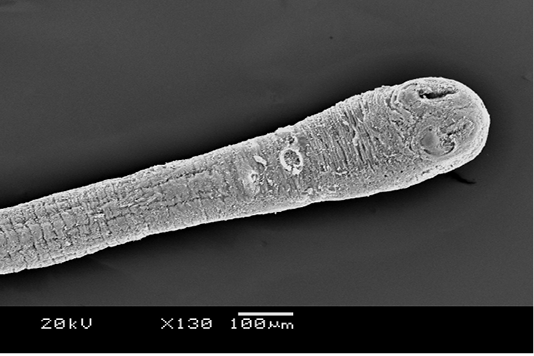
Figure 1: Scanning electron microscopy of the anterior portion of R. galli treated with M. taiwaniana extract. The terminal bulb is the scolex and the stalk is the neck. Two suckers are seen on the scolex. x 130, scale bar is 100 µm.
Structural changes on the body of R. galli after exposure to M. taiwaniana extract was examined by scanning electron microscopy. The plant extract exerted typical anthelmintic effects by causing extensive damage on the tegument throughout the body surface. The effect on the anterior part of the body is shown in Figure 1. There is severe constriction of the tegument with numerous irregular folds. Erosion and scarring are evident throughout. The attachment organs (suckers) are completely damaged and all the spines are sloughed off. There are large pits at the centre of each sucker indicating heavy erosions. Proglottids (body segments) of the neck region indicate with extensive erosion of the tegument (Figure 2). There are no signs of smooth hair-like microtriches, which are otherwise the surface components of proglottids. Minutes pits and holes are formed at many places. There is an unusual distention on a portion of the mature proglottids (Figure 3). The distended proglottids indicate extensive erosion with pit formations, along with severe shrinkage of the tegument. Under magnification of the enlarged proglottid, distorted and degenerated microtriches are visible (Figure 4). The last gravid proglottid is particularly shrunk and crooked as shown in Figure 5. The tegument looks as if it is forcefully wrung. Shrinkage is associated numerous folds along the length of the body. The tegumental folds are actually due to severe constriction which produced many blisters or blebs on the surface (Figure 6). These blebs represent degenerated microtriches.
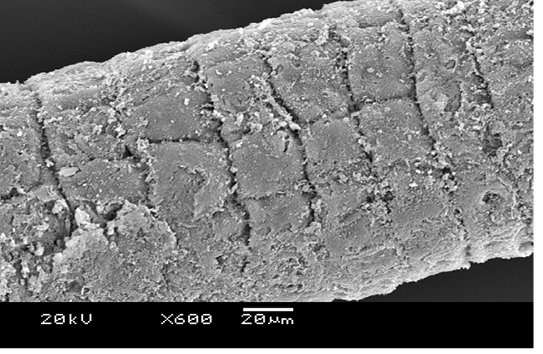
Figure 2: Scanning electron microscopy of the neck of R. galli treated with M. taiwaniana extract. Body segments or proglottids are heavily eroded. x 600, scale bar is 20 µm.
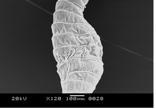
Figure 3: Scanning electron microscopy of mature proglottids of R. galli treated with M. taiwaniana extract. Abnormal enlargement of the body is evident. x 120, scale bar is 100 µm.
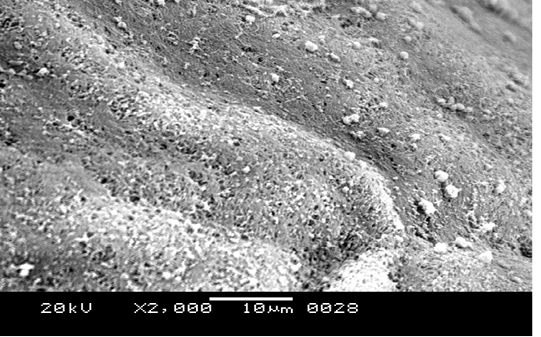
Figure 4: Scanning electron microscopy of a mature proglottids of R. galli treated with M. taiwaniana extract. A fuzzy surface depicts degenerated microtriches. x 2000, scale bar is 10 µm.
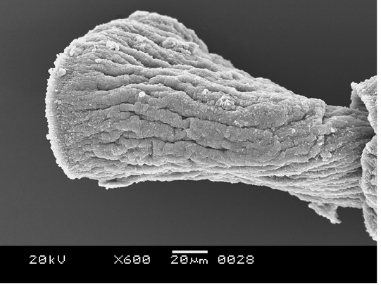
Figure 5: The last gravid proglottid of R. galli treated with M. taiwaniana extract. Shrinkage with a series of longitudinal folds is visible. x 600, scale bar is 20 µm.

Figure 6: Magnification of the tegument of a gravid proglottid of R. galli treated with M. taiwaniana extract. Tegumental folds are composed of blisters or blebs. x 4000, scale bar is 5 µm.
DISCUSSION
The body surface of cestode is called tegument and is fully covered with hairy microtriches. Without internal digestive and complex nervous system, the tegument acts as a primary absorptive and sensory site, and for this the tegument is the principal target site of anthelmintic drugs. Different anthelmintics are shown to induce surface damages of varying degrees on different cestode. These drugs enter cestode bodies by passive diffusion, and as such, the effects of anthelmintic agents are studied and established upon structural changes they cause on teguments (Rana and Misra-Bhattacharya, 2013; Taman and Azab, 2014).
Albendazole and flubendazole causes rostellar distortion, eruption of swellings or blebs on the tegument, defacement of the microtriches, and increased vesiculation on the human tapeworm, Echinococcus granulosus (Elissondo et al., 2006). Albendazole and praziquantel combination caused upon E. granulosus and Mesocestoides corti resulted in extensive malformation of the suckers, erosion of the tegument and disintegration of microtriches (Li et al., 1996; Markoski et al., 2006). Albendazole also caused severe shrinkage and tegumental collapse in Raillietina echinobothrida (Lalchhandama, 2010).
The fine structure of the body surface of R. galli and related species is elucidated (Bâ et al., 1995; Lalchhandama, 2009). M. taiwaniana extract showed anthelmintic effects on R. galli. Suckers were most conspicuously destroyed on the scolex while the rostellum is hardly affected. in the present study also showed considerable structural damages. Spines were completely removed from the suckers. Erosion on the general tegument was visible throughout the body. Microtriches were reduced to fuzzy debris and the underlying tegumental layers had pits and holes on mature proglottids. These observations indicate that the plant contains important lead molecules for novel anthelmintics and the need for further pharmacological studies.
CONCLUSION
M. taiwaniana root bark extract exhibits anthelmintic activity against parasitic tapeworm of fowl, R. galli. The effects were observed on the tegument of the tapeworms in the form of damages of the suckers, tegumental shrinkage, degeneration of microtriches, surface erosion and formation of blebs.
CONFLICT OF INTEREST
The author declares that there is no conflict of interest.
REFERENCES


