Advances in Animal and Veterinary Sciences
Research Article
Predictor Ultrasonographic Evaluation of the Testis during Pubertal Age in Ram Lambs
Farhan Fadhil Saaed, Nazih Wayes Zaid*
Department of Surgery and Obstetrics, College of Veterinary Medicine, University of Baghdad, Iraq.
Abstract | This study was designed in order to investigate the benefit of ultrasound use in detecting pubertal age in lamb ram. Twelve ram lambs from age 3 months and two mature rams aged 3 years were used in this study which done in Jeballa town in Babel province southern to Baghdad, Iraq. The examination was done using Biomed sonar with liner probe every 15 days until it reach 9 months of age, the scanning based on sagittal, transverse and frontal planes in order to evaluate the testicular parenchyma echotexture and the mediastinum testis, while measurements of the testicular length, width and thickness were performed by ultrasound in both testes. The results show that there were significant differences (P<0.05) in testicular length, width and thickness between mature ram and 3 and 4 months of age. The testis parenchyma images with moderate echogenicity were in pre-pubertal lambs’ animals and high echogenicity in those that had reached near puberty age or mature rams. From these results we conclude that the sonar was a useful in detecting pubertal age and for breeding soundness in ram lambs.
Keywords | Ultrasound, Ram lamb, Pubertal age, Testicular diameters, Predictor.
Editor | Kuldeep Dhama, Indian Veterinary Research Institute, Uttar Pradesh, India.
Received | August 21, 2018; Accepted | September 19, 2018; Published | October 09, 2018
*Correspondence | Nazih Wayes Zaid, Department of Surgery and Obstetrics, College of Veterinary Medicine, University of Baghdad, Iraq; Email: [email protected]
Citation | Saeed FF, Zaid NW (2018). Predictor ultrasonographic evaluation of the testis during pubertal age in ram lambs. Adv. Anim. Vet. Sci. 6(12): 521-525.
DOI | http://dx.doi.org/10.17582/journal.aavs/2018/6.12.521.525
ISSN (Online) | 2307-8316; ISSN (Print) | 2309-3331
Copyright © 2018 Saeed and Zaid. This is an open access article distributed under the Creative Commons Attribution License, which permits unrestricted use, distribution, and reproduction in any medium, provided the original work is properly cited.
Introduction
Ultrasound one of biotechnology which improve livestock and a resultant increase in productivity, ultrasound imaging had been widely used in reproductive clinical examinations (Andrade et al., 2014). Ultrasound was a rapid and nonsurgical technique that (coupled with data from clinical examinations) may lead to early diagnosis of disorders of the testes and related structures (Pechman and Eilts, 1987). Because it was easily accepting and did not cause unhealthy effects, ultrasound was the diagnostic method of choice in the initial evaluation of the scrotum and its contents (Andrade et al., 2014). The main functions of ultrasound were to evaluate anatomical structures and determine the echogenicity of testicular parenchyma and mediastinum (Clark et al., 2003). Also it can be useful in monitoring progressive changes that occur in testis at different stages of maturation, observed an increased echogenicity of testicular parenchyma in direct proportion with age (Brito et al., 2004). According to Tapping and Cast (2008), testes of pre-pubertal animals had low to medium echogenicity, whereas testes of post-pubertal animals demonstrated medium homogeneous echogenicity. Some studies involving US assessment of normal and pathological genitals of males have been conducted in bulls (Pechman and Eilts, 1987), rams (Gouletsou et al., 2003; Andrade Moura et al., 2008; Juca et al., 2009), goats (Ahmad and Noakes, 1995) and other mammals (Clark and Althouse, 2002; Pozor, 2005; Ball, 2008). All the studies which conducted in Iraq used sonar for diagnosis of breeding management in female only (AL-Rawi, 2005; AL-Mashhdani, 2012), so this study was the first of its kind to investigate the puberty in male rams.
Materials and Methods
Twelve male lambs of Awassi rams aged 3 months of old (determined by teeth formula according to Dyce and Sack (2010) and between 10-12 kg body weight where collected. All these animals subjected to similar conditions, after weaning of these lambs they were separated from their mothers and feed with two period of grazing, the first one started from sunrise-mid noon, then these lambs take a 2 hours of rest and then the second grazing period started into sun-shine time, the water and minerals blocks approved ad libitum. The medical vaccination and treatment where given during the study period which extended from November 2017 to April 2018. All animals were kept in Jeballa town in Babel province southern to Baghdad, Iraq. Another two mature Awassi rams aged 3 years of old were kept with these lambs as mature male.
The examination of all animals were done, this included general evaluation (body condition inspection, detection of hereditary defects, respiratory, circulatory, digestive and musculoskeletal systems) and special examination of genitalia (inspection and palpation of the scrotum, testis, epididymis, spermatic cords, prepuce and penis). This was done according to Andrade et al. (2014).
Testicular ultrasonographic evaluations were carried out using Biomed (USA) connected to a linear array transducer with a frequency of 8.0 MHz for image documentation. Rams and lambs were physically curbed by two assistants and each testis was scanned in the sagittal, transverse and frontal planes to evaluate the testicular parenchyma echotexture and the mediastinum testis (MT) while the testes were immobilized without pressure. Measurements of the mediastinum thickness (frontal plane) and testicular width (transverse plane) were performed by ultrasound in both testes. Both were measured in millimeters.
Statistical Analysis
Statistical analysis was carried out to study the differences between means. ANOVA test was performed. Duncan multiple range tests were used to compare different means of studied parameters. Correlations were examined for studied lambs and rams. This was done according to Al-Mohammed et al. (1986).
Results
The herein study indicated that there was significant differences (P<0.05) between mature rams and pre-pubertal age (3-4) in length and width using ultrasound, while it was with 3 months only in thickness (Table 1). There was no other significant difference. There were high correlation between length, width and thickness and lambs ages. At ultrasonography, the parenchyma of the testis appeared homogeneous, ranging from moderate (Figure 1) to high (Figure 2) echogenicity, regardless of the testis or the scan plane which had been used. Whereas the testicular mediastinum was imaged in all animals, presenting as a hyperechoic line of variable thickness in the center ofthe testis parenchyma,and was classified as having low (Figure 3 and 4), moderate (Figure 5), high (Figure 6) or diffuse (Figure 7 and 8) echogenicity.
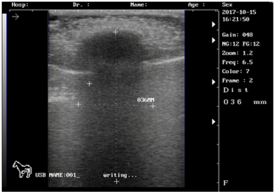
Figure 1: Ultrasound images of the testis of 3 months lamb showing: moderate echogenicity of the testicular parenchyma; mediastinum testicular highly echogenic
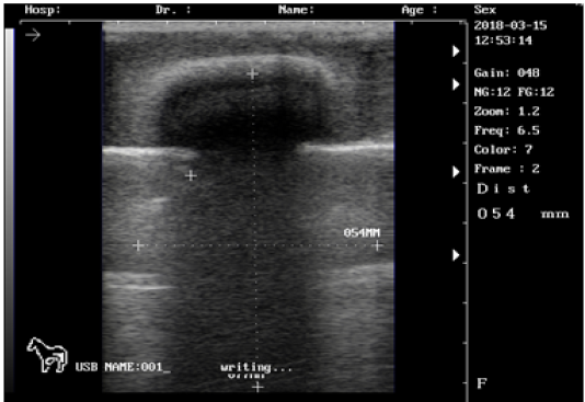
Figure 2: Ultrasound images of the testis of 8 months lamb showing: high echogenicity of the testicular parenchyma; mediastinum testicular moderately echogenic
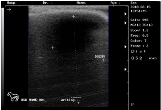
Figure 3: Ultrasound images of the testis of 9 months lamb showing: high echogenicity of the testicular parenchyma; mediastinum testicular lowly echogenic
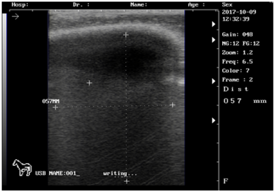
Figure 4: Ultrasound images of the testis of mature ram showing: high echogenicity of the testicular parenchyma; mediastinum testicular lowly echogenic
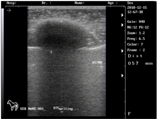
Figure 5: Ultrasound images of the testis of 6 months lamb showing: moderate echogenicity of the testicular parenchyma; mediastinum testicular moderately echogenic
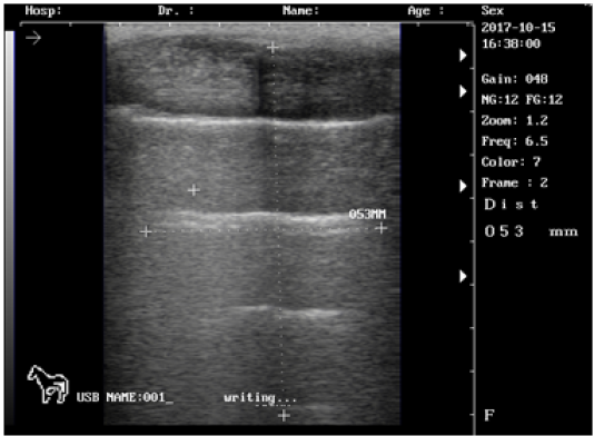
Figure 6: Ultrasound images of the testis of 4 months lamb showing: moderate echogenicity of the testicular parenchyma; mediastinum testicular highly echogenic
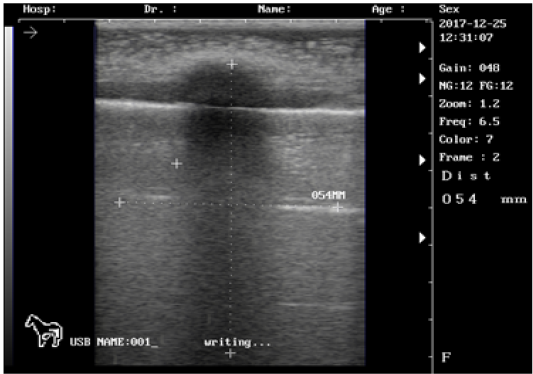
Figure 7: Ultrasound images of the testis of 5 months lamb showing: moderate echogenicity of the testicular parenchyma; mediastinum testicular diffusely echogenic
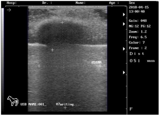
Figure 8: Ultrasound images of the testis of 7 months lamb showing: high echogenicity of the testicular parenchyma; mediastinum testicular diffusly echogenic
Table 1: Testis length (mm), testis width (mm) and testis thickness (mm) in pre-pubertal lambs and mature rams.
| Ages | Testis length* | Testis width* | Testis thickness* |
| 3 months | 48.2±0.2 c | 19.2±0.1 c | 16±01 b |
| 4 months | 55.5±0.3 bc | 35.9±0.1 bc | 31.4±0.1 ab |
| 5 months | 71.1±0.5 abc | 43.2±0.2 abc | 39±0.1 ab |
| 6 months | 87.2±0.5 abc | 51.1±0.1 abc | 42±0.2 ab |
| 7 months | 95.7±0.5 abc | 52.3±0.2 abc | 48.6±0.2 ab |
| 8 months | 116.2±0.4 abc | 55.8±0.1 abc | 48.9±0.2 ab |
| 9 months | 123.1±1 ab | 62.2±0.2 ab | 49.8±0.1 ab |
| Mature | 131.9±0.9 a | 76.8±0.3 a | 66±0.3 a |
| Correlations | +0.67** | +0.76** |
+0.75** |
The numbers represent mean ± standard error.
The similar small letters represent no significant differences.
The different small letters represent significant differences at level of *(P<0.05) or **(P<0.01).
Discussions
This study was the first of it kinds which applied in Iraq to study the testicular structures using sonar techniques. There was predominance of testis parenchyma images with moderate echogenicity in pre-pubertal lambs’ animals and high echogenicity in those that had reached near puberty age or mature rams. Although, there was decreased echogenicity in testis mediastinum with the animal age. This finding was corroborating by Gouletsou et al. (2003) and (Andrade et al., 2014). Furthermore, when comparing the testes of pre-pubertal lambs and mature rams, there was predominance of images with low echogenicity in pre-pubertal animals and moderate echogenicity in those that had reached puberty (Andrade et al., 2014). Although some authors have studied the ultrasonographic appearance of the testes and epididymes of clinically healthy sheep (Gouletsou et al., 2003; Andrade Moura et al., 2008; Juca et al., 2009). In this experiment of Gouletsou et al. (2003) and Juca et al. (2009), the testicular parenchyma in the lambs demonstrated homogeneity, with echogenicity ranging from low to moderate in both testes. Andrade Moura et al.
(2008), in their study that aimed to describe the testicular parenchyma echotexture of Santa Ines sheep at different ages, found a variation of hypoechoic images of low and high intensity in all groups, with predominance of hypoechoic images of low intensity. Andrade et al. (2014) stated that the mediastinum testis was visualized in 100% of the evaluated animals, presenting as a hyperechoic line of variable thickness at the center of the testicular parenchyma when visualized in the frontal plane and as a hyperechoic point in the center of this parenchyma when visualized in the transverse plane. Moreover, Andrade Moura et al. (2008), using cattle and sheep, respectively, concluded that the mediastinum thickness and echogenicity increased with age. Thus, this increase in the testicular mediastinum thickness may be explained by the important and considerable anatomical changes in the seminiferous tubules, which develop with age as they become longer, increase in diameter, twisted, the lumen forms and the basement membrane thickens. According to Tapping and Cast (2008), this structure can often be seen as a hypoechoic area with striated appearance. Ultrasound examination may also be useful in monitoring the progressive changes that occur in the testes.
Conclusions
These studies conclude that the use of sonar in ram lamb was useful in detecting the pubertal age in addition to the abnormal cases during evaluation of breeding soundness.
Acknowledgments
The author would like to thank Mrs. Najlaa Sami Ibrahim, Ph. D. In Surgery and Obstetrics Department - College of Veterinary Medicine –University of Baghdad, for reviewing and editing this paper.
Conflict of Interest
None of the authors have any conflict of interest to declare.
Authors Contribution
All authors contributed equally.
References


