Advances in Animal and Veterinary Sciences
Research Article
Trace Elements Status During Different Production Stages and Parities in Holstein Dairy Cows
Shimaa G. Yehia1, Eman S. Ramadan1*, Eissa A. Megahed2, Noha Y. Salem1
1Department of Internal Medicine and Infectious Diseases, Faculty of Veterinary Medicine, Cairo University, Giza, Egypt; 2Veterinary Medicine Directorate - Giza, El Haram, Giza, Egypt.
Abstract | Macro and microminerals are essential for dairy cattle health. The principal purpose of the current investigation was to explore the modifications of the blood levels of Copper (Cu), Zinc (Zn), Iron (Fe), Calcium (Ca), Phosphorus (P), and Magnesium (Mg) alterations during different production stages of clinically healthy dairy primiparous and multiparous cows. This study enrolled 20 healthy Holstein primiparous and multiparous dairy cattle (Ten primiparous and ten multiparous). Three blood samples were taken from each cow in close-up, early lactation, and mid-lactation phases. Samples were analyzed for Cu, Zn, Fe, Ca, P, and Mg levels. In the primiparous group, significant elevation in Cu, Zn, Ca, P, and Mg was seen in lactating groups compared to the prepartum group. Significant elevation in both P and Cu were detected in early and mid-lactating multiparous cows respectively compared to the prepartum phase. Serum Iron levels did not differ throughout different phases in primiparous and multiparous groups. The production stage strongly modified the trace elements profile in different parities. The parity and stage of lactation should yield more attention during the ration formulation and minerals mix supplementation.
Keywords | Parity, Microminerals, Macrominerals, Transition, Lactation.
Received | July 12, 2021; Accepted | July 24, 2021; Published | November 15, 2021
*Correspondence | Eman S Ramadan, Department of Internal Medicine and Infectious Diseases, Faculty of Veterinary Medicine, Cairo University, Giza, Egypt; Email: [email protected]
Citation | Yehia SG. Ramadan ES, Megahed EA, Salem NY (2021). Trace elements status during different production stages and parities in holstein dairy cows. Adv. Anim. Vet. Sci. 9(12): 2266-2271.
DOI | http://dx.doi.org/10.17582/journal.aavs/2021/9.12.2266.2271
ISSN (Online) | 2307-8316; ISSN (Print) | 2309-3331
Copyright © 2021 Ramadan et al. This is an open access article distributed under the Creative Commons Attribution License, which permits unrestricted use, distribution, and reproduction in any medium, provided the original work is properly cited.
INTRODUCTION
In many countries, dairy cows are the main pillar of dairy production. The health status of dairy cows is jeopardized by many physiological factors (Hussein and Staufenbiel, 2011). Pregnancy, transition, and lactation periods are key stressors that can impact body metabolites (Salem, 2017; Yehia et al., 2020).
Trace minerals also play a significant role in the immune function of dairy cattle (Karatzia et al., 2016). Copper and Zinc participate in many antioxidant enzymes, particularly Cu-Zn Superoxide dismutase (SOD) (Evans and Halliwell, 2001; Kubesy et al., 2017; Elsayed et al., 2020). In addition to its role in antioxidant defense against free radicals, Zinc has an integral part in body immunity (Weiss and Spears, 2006). Maintaining these microminerals within normal levels is essential for safeguarding animal health status (Karatzia et al., 2016). Iron (Fe) is an integral part of cytochromes and Iron-dependent proteins involved in electron transport; it is also a constituent of several Iron-activated enzymes (Nocek et al., 2006).
Minerals are the main components of production and reproduction in animals (Panda et al., 2016). The term homeostasis is valuable in describing the alterations associated with mineral concentrations in the body (Kincaid, 2008). These minerals principally act as catalysts in many enzyme and hormone systems that impact bone growth, enzyme structure and function, and appetite (Stef and Gergen, 2012). The levels of Magnesium (Mg), Calcium (Ca), and phosphorous (P) during the transition and lactation phases mirror their supply and utilization (Djokovic et al., 2014). Calcium has a major impact on the physiological status and body metabolites; it decreases at the beginning of lactation due to milk production and relatively slower intestinal up-regulates absorption associated with this phase (Kincaid, 2008; Piccione, 2009). Magnesium plays a pivotal role in the metabolism of lipids, nucleic acids, protein carbohydrates, and the appropriate functioning of the nervous system (Panda et al., 2016).
Macro-and microminerals are essential for the health status of animals; disturbances in their levels could play havoc with animal performance, production, and reproduction (Djokovic et al., 2014). We hypothesized that levels of macro-and microminerals would be affected by the production stage and parity. The principal goal of the current investigation was to explore the modifications of the blood levels of Cu, Zn, Fe, Ca, P and Mg alterations during different production stages of clinically healthy dairy primiparous and multiparous animals for early detection of health problems during this critical period.
MATERIALS AND METHODS
Ethical Approval
This study was approved by the Institutional Animal Care and Use Committee and allotted number (vet CU28/04/2021), Faculty of Veterinary Medicine, Cairo University, Egypt.
Study Population and Design
Twenty Holstein dairy cattle were enrolled in this study. The cow’s age range was between 2-5 years, the weight range was 550-670 kg, and the average milk production was 25-37 kg/day. The cows were kept in loose barns with shades and free access to the yard. The barns were divided into sections according to the production stage and milk yield; cows were fed as a total mixed ratio (TMR) using conserved plants during the whole year. The physical description according to production and stage is summarized in Table 1.
Examined cows were divided according to parity into primiparous (n=10) and multiparous (n=10). Blood samples from the same cows were taken in the transition period, early lactation period, and mid-lactation period of each cow.
The study period was from January 2021-March 2021. Blood samples were collected from a dairy cow farm on the Misr-Alexandria desert road.
Blood Samples and Analysis
Three blood samples were taken from each cow. The sampling time was in the morning before morning milking. The first sample was collected three weeks before parturition, the second sample was taken in the early lactation phase (14-21 DIM), and the third sample was taken in the mid-lactation phase (60-90 DIM). Blood samples were collected from tail vein puncture and serum was separated.
Serum samples were used to analyze Copper (Cu), Zinc (Zn), Iron (Fe), Calcium (Ca), Phosphorus (P), and Magnesium (Mg) using automated biochemistry analyzer.
Statistical Analysis
Data are presented as mean ± SE. Repeated-measures ANOVA was used to analyze data of each group using a completely randomized one-way ANOVA by SPSS program version 16.00. The measured parameters were expressed as mean value ± SE. A probability “P” value of ≤ 0.05 was assumed as statistically significant.
RESULTS and DISCUSSION
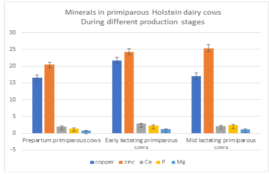
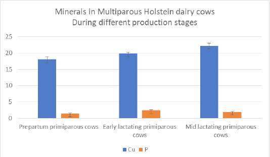
The mean concentrations of Cu, Zn, Fe P, Ca, and Mg in the blood serum of primiparous and multiparous cows are shown in Tables 2 -3 and Figure 1-2, respectively. They are essential for the healthy tissues of humans and animals (Wang et al., 2014). Several enzymes are copper-dependent (i:e Ceruloplasmin and SOD) (Djokovic et al., 2014). These enzymes are vital for collagen and elastin integrity, free radical detoxication, and energy metabolism (Djokovic et al., 2014). Zinc is an important element for over 70 enzymes in mammals. These enzymes participate in protein, nucleic acid, carbohydrate, and lipid metabolism (Kellogg et al., 2004).
Table 1: Physical composition of feed intake of dairy Holstein cow during different stages (amount/head/day)
| Feed ingredient (as feed base) | Dry pregnant cow | Medium milk production ≤ 28kg | Medium milk production < 28kg |
| Yellow corn | 2.65 | 4.9 | 5.5 |
| Soyabean meal (44%) | 1.60 | 2.6 | 4 |
| Soyabean meal plus | 0.5 | - | 1 |
| Gluten feed (16%) | 1.2 | 2 | 2 |
| Protected fat | 0.05 | - | 0.52 |
| Corn silage | 17 | 19 | 22 |
| Alfalfa hay | 2 | 4 | 4 |
| Minerals | 0.03 | 0.036 | 0.044 |
| Vitamins | 0.015 | 0.018 | 0.022 |
| Limestone | 0.205 | 0.150 | 0.08 |
| Dicalcium phosphate | 0.042 | - | 0.06 |
| Organic antioxidant | 0.010 | 0.01 | 0.015 |
| Sodium bicarbonate | - | 0.140 | 0.240 |
| Magnesium oxide | - | 0.05 | 0.06 |
| Inorganic antioxidant | - | 0.03 | 0.04 |
| Salt | 0.08 |
0.088 |
Table 2: Microminerals and Macrominerals status in primiparous Holstein dairy cows During different production stages.
| Parameters/Unit | Prepartum primiparous cows | Early lactating primiparous cows | Mid lactating primiparous cows |
| Mean ± SE | Mean ± SE | Mean ± SE | |
| Copper (µmol/l) |
16.62 ± 0.72a |
21.8 ± 0.81b |
17.03 ± 0.95a |
| Zinc (µmol/l) |
20.51 ± 0.53a |
24.31 ± 0.85b |
25.30 ± 1.078b |
| Iron (µmol/l) |
26.263 ± 2.60 a |
32.27 ± 3.057 a |
31.84 ± 2.85 a |
| Ca (mmol/l) |
2.004 ± 0.252a |
2.78 ± 0.097b |
2.182 ± 0.139ab |
| P (mmol/l) |
1.496 ± 0.115a |
2.335± 0.124 b |
2.4 ± 0.25b |
| Mg (mmol/l) |
0.74 ± 0.034 a |
1.233 ± 0.12 b |
1.18 ± 0.13 ab |
a and b: Mean on the same row of different superscripts are significantly different (P ≤ 0.05)
Data are presented as mean ± SE.
Table 3: Microminerals and Macrominerals status in Multiparous Holstein dairy cows during different production stages.
| Parameters/Unit | Prepartum multiparous cows | Early lactating multiparous cows | Mid lactating multiparous cows |
| Mean ± SE | Mean ± SE | Mean ± SE | |
| Copper (µmol/l) |
18.06 ±0.81a |
19.82± 0.54ab |
22.28 ± 0.81b |
| Zinc (µmol/l) |
22.82 ± 1.83a |
24.28 ± 2.26a |
27.9 ± 2.83a |
| Iron (µmol/l) |
25.40 ± 3.41 a |
30.45 ± 2.21 a |
29.03 ± 2.88 a |
| Ca (mmol/l) |
2.29 ± 0.24 a |
2.472 ± 0.227 a |
2.170 ± 0.218 a |
| P (mmol/l) |
1.449 ± 0.143a |
2.41 ± 0.167 b |
1.89 ± 0.119 ab |
| Mg (mmol/l) |
1.1 ± 0.15 a |
1.15 ± 0.119 a |
1.222 ± 0.085 a |
a and b: Mean on the same row of different superscripts are significantly different (P ≤ 0.05)
Data are presented as mean ± SE.
showed significant elevation in Cu level in early lactating group compared with prepartum and mid- lactating groups. Zinc, P showed significant elevations in both lactation phases compared to Prepartum phase. Ca , Mg levels showed showed significant elevations in early lactation phases compared to Prepartum stage (Table 2, Figure 1). Serum Copper concentration was significantly higher in early lactating primiparous cows compared to the prepartum and mid - lactating groups. ‘Hussein and Staufenbiel (2011) reported higher Cu activity in the fresh lactation group, which agreed with our findings in primiparous cows. Zinc was significantly elevated with lactation compared to the prepartum stage. Contrary, a lower Zinc value in puerperium cows compared with close-up, and full lactation cows were also reported (Djokovic et al., 2014). The lower Copper and Zinc concentrations in pregnant cattle in the current study could be explained by additional fetal demands and consumption of maternal copper and Zinc for the growth of the fetal nervous system (Elnageeb and Abdelatif, 2010; Slavik et al., 2006). Increased concentration of Copper and Zinc in fresh lactating cows may occur secondary to stressful stages and oxidative stress around calving (Castillo et al., 2005).
No significant variations were detected in Iron concentration in primiparous cows throughout different phases, as recorded by other reports (Noaman et al., 2012). Iron is an integral part of cytochromes and Iron-dependent proteins involved in electron transport; it is also a constituent of several Iron-activated enzymes (Nocek et al., 2006). Calcium was slightly higher in early lactating primiparous cows compared to the prepartum primiparous cows. This result was supported by Giuseppe et al. (2012), who reported elevated Calcium in the lactation period than in the dry period. A previous study recorded higher values of Calcium in lactating sheep (Yokus et al., 2004). On the contrary, DAS et al. (2016) reported a drop in Calcium levels during the early stage of lactation. The increase in Ca concentration in primiparous early lactating cows may be attributed to more Ca mobilization associated with osteoclast proportion with high levels of PTH, primiparous cows tend to have lower milk yield and a higher portion of osteoblast because the bone structure is still growing (Timaran, 2020).
The Phosphorus level was higher in lactation than in the dry period (Giuseppe et al,. 2012). Contrariwise, Sarker et al. (2015) stated that Phosphorus level was higher in the dry cows than lactating ones. All animals need minerals such as Calcium (Ca), Phosphorus (P), and Magnesium (Mg) for growth, reproduction, and lactation (Samardzija et al., 2011). Mg level was significantly higher in primiparous early lactating cows compared to pregnant primiparous cows, but no significant variations were detected between different lactation stages in primiparous and multiparous cows, as previously reported (Djokovic et al., 2014; Abd-EL Naser, 2014). This reduction may be attributed to an additional requirement for bone formation in the growing fetus (Zofkova and Kancheva, 1995).
P and Cu showed significant elevation in multiparous cows during early and mid-lactating stages respectively compared to prepartum phase. Zn, Fe, Ca, and Mg levels did not differ throughout the three stages (Table 3, Figure 2). Serum Copper concentration was significantly higher in mid-lactating multiparous cows compared to prepartum multiparous cows, as previously suggested (Noaman et al., 2012; Cortinhas et al., 2014). No significant variations were detected in Iron concentration in multiparous cows. However, Das et al. (2016) reported lower Iron concentrations in pregnant cows.
Ca level did not increase in lactating multiparous cows in the current study. The absence of sufficient Ca levels during lactation may increase the risk of developing postpartum diseases such as milk fever (Van Saun et al., 2010). Phosphorus level was found to be slightly higher in early lactating multiparous cows. During milk production, additional Phosphorus from the ingested ration is transferred to milk and less is excreted in feces (Valk et al., 2002). So, a higher serum phosphorus level is expected. No significant variations were detected between different lactation phases in multiparous cows. This finding agreed with previous reports (Djokovic et al., 2014; Abd-EL Naser, 2014).
This study investigated trace minerals profiles in different stages of production. The limitations of this study are the small sample size and the absence of seasonal comparison.
CONCLUSION
The production stage strongly modified the trace elements profile in different parities of Holstein dairy cows. The highest levels of micro and macro-minerals were exhibited between the different lactation stages during the early and mid-lactation. Trace element fluctuations were more pronounced in primiparous cows. The parity and stage of lactation should yield more attention during the formulation of rations and mineral mix supplementation. Trace elements monitoring is essential for early detection of health problems and enhancement of production.
CONFLICT OF INTEREST
The authors have declared no conflict of interest.
AUTHORS CONTRIBUTION
All authors contributed equally to the manuscript.
REFERENCES
during late pregnancy and early lactation in dairy cows. Vet. J.
169:286–292.
on serum mineral profile of banni buffalo (bubalus bubalis). Life sciences leaflets, 77:1-10. retrieved from https://petsd.org/ojs/index.php/lifesciencesleaflets/article/view/1071.
activity of dairy cows in relation to lactation stages with regard to ceruloplasmin to copper ratios. Biol. Trace Elem. Res., 146:47–52. https://doi.org/10.1007/s12011-011-9226-3
Gracner G, Dobranic V, Radisic B, Duricic D (2011). Comparison of blood
serum macromineral concentrations in meat and dairy goats during puerperium. Veterinarski Ahriv. T., 81: 1–11.
Estrada JM (2020). Parity and season affect hematological, biochemical, and milk parameters during the early postpartum period in grazing dairy cows from
high-tropics herds. Heliyon., 6(5):40-49.
Liu S, Dong S, Xia X, Li S (2014). Levels of Cu, Mn, Fe and Zn in Cow Serum and Cow Milk: Relationship with Trace Elements Contents and Chemical Composition in Milk. Acta Sci. Vet., 42: 1190.


