Advances in Animal and Veterinary Sciences
Case Report
Clinical Management of Elbow Hygroma in a Malayan Tigress (Panthera tigris jacksoni): A Case Report
Zubaidah Kamaruddin6, Chiu Nie Chong1, Idris Umar Hambali4, Faez Firdaus Abdullah Jesse1, 3, Fitri Wan-Nor1, Eric Lim Teik Chung2, 3, Yusuf Abba4, Asinamai Athliamai Bitrus5, Innocent Damudu Peter4, Bura Thlama Paul1, Siti Suzana Selamat6, Hasdi Hassan6, Azlan Che-Amat1*
1Department of Veterinary Clinical Studies, Faculty of Veterinary Medicine, Universiti Putra Malaysia, 43400 Serdang, Selangor, Malaysia, 2Department of Animal Science, Faculty of Agriculture, Universiti Putra Malaysia, 43400 Serdang, Selangor, Malaysia, 3Institute of Tropical Agriculture and Food Security, Universiti Putra Malaysia, 43400 Serdang, Selangor, Malaysia, 4Faculty of Veterinary Medicine, University of Maiduguri. P.M.B 1069 Maiduguri, Borno Nigeria, 5Department of Veterinary Microbiology and Pathology, Faculty of Veterinary Science, University of Jos, P.M.B 2084 Jos, Plateau Nigeria, 6National Wildlife Rescue Centre (NWRC), Department of Wildlife and National Parks (PERHILITAN), 35600 Sungkai, Perak.
Abstract | This case reports detail the clinical management of an elbow hygroma in a Malayan Tigress (Panthera tigris jacksoni). A twelve (12) years old Malayan Tigress weighing 112 kg with body condition score of 3/5 kept in captivity was reported by rangers in the National Wildlife Rescue Centre, Sungkai, Perak, Malaysia with a primary complaint of a lump at the right elbow of the forelimb. Physical and clinical examinations showed normal pulse and respiratory rates, additionally, an 8 cm x 9 cm well demarcated, soft lump caudal to the right forelimb elbow joint was palpated. Based on the physical examination and clinical signs, a diagnosis of elbow hygroma was made on that point of time. The tigress was managed by surgical lancing and wound cleaning. During the intra-operative session, the tigress was premedicated with an anti-cholinergic agent, an antibiotic, an anti-inflammatory agent and a supplement of vitamin D. During the post-operative medical management, antibiotic was administered to prevent secondary bacterial infection, papase as anti-inflammatory and iodine spray for wound care management. In conclusion, hygroma on the elbow was managed non-invasive surgical procedure and proper management by avoiding the overwhelming effects of possible risk factors can be a preventive measure for this case.
Keywords | Tiger, Elbow hygroma, Panthera tigris jacksoni, Clinical, Management
Received | November 05, 2019; Accepted | February 17, 2020; Published | February 20, 2020
*Correspondence | Azlan Che Amat, Department of Veterinary Clinical Studies, Faculty of Veterinary Medicine, Universiti Putra Malaysia, 43400 Serdang, Selangor, Malaysia; Email: [email protected]
Citation | Kamaruddin Z, Chong CN, Hambali IU, Jesse FFA, Wan-Nor F, Chung ELT, Abba Y, Bitrus AA, Peter ID, Paul BT, Selamat SS, Hassan H, Che-Amat A (2020). Clinical management of elbow hygroma in a Malayan tigress (Panthera tigris jacksoni): A case report. Adv. Anim. Vet. Sci. 8(2): 213-216.
DOI | http://dx.doi.org/10.17582/journal.aavs/2020/8.2.213.216
ISSN (Online) | 2307-8316; ISSN (Print) | 2309-3331
Copyright © 2020 Che-Amat et al. This is an open access article distributed under the Creative Commons Attribution License, which permits unrestricted use, distribution, and reproduction in any medium, provided the original work is properly cited.
INTRODUCTION
Tigers are regarded as national animals in Malaysia, India, Bangladesh and South Korea (Sethi and Choudhary, 2019; Sanderson et al., 2019). They are a group of highly adaptable felid species inhabiting a wide range of forest categories, climatic systems, and transformed landscapes (Upadhyaya, 2019). In most recent times, tigers are facing extinction across their range states, primarily due to territory loss and poaching (Ashraf, 2019).
Censuring territorial figures of Tiger had been of paramount interest of concerned nations. In Malaysia, a reliable population estimate of the Malayan Tiger (Panthera tigris jacksoni) is yet to be ascertained. But an estimate of Malayan tigers is between 250 and 340 individuals (Kawanishi, 2015), which was considered a signal to extinction. Globally, there are fewer than 3,500 tigers (Joshi et al., 2016; Ashraf, 2019).
A connective tissue sac lined by synovial membrane and contains synovial fluid is called a true bursa (Johnston, 1985; Sawyer and Varacallo, 2019). It is situated between tendons, ligaments and muscles and many bony prominences of the body. The function of a bursa is to reduce friction for the moving parts over the body such as tendons, ligaments and muscles (Johnston, 1985; Sawyer and Varacallo, 2019).
Furthermore, a hygroma is known as a false bursa which develops over bony prominences and pressure points of the body (Karen, 2018). Hygroma is characterized by soft, fluid-filled and painless swelling which develops due to repeated trauma on soft tissue surrounding the bony prominence (Fossum et al., 2013; Nath et al., 2014). According to Pavlectic and Brum (2015), it was reported that the common site of development of hygroma is olecranon or known as the point of elbow. Other sites such as the tuber calcanei, the trochanter major of the femur and the tuber coxae of the ilium are also prone to development of hygroma. In essence, this clinical case report describes the management of hygroma in a tiger.
CASE REPORT
History
A 12 years old, Malayan Tigress (Panthera tigris jacksoni) weighing 112 kg body weight with body condition score of 3/5 kept captive in the National Wildlife Rescue Centre was noticed to have a lump on the right elbow of the forelimb with no lameness observed by the rangers.
Physical examination
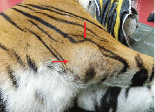
To achieve a thorough physical examination, the tiger was administered with Ketamine 2.0 mg/kg and Detomidine 0.05 mg/kg through a blow pipe intramuscularly at the semitendinosus in order to achieve anaesthesia. Upon physical examination, pulse and respiration rates were within the normal range. An 8 cm x 9 cm well demarcated, soft lump caudal to the right forelimb elbow joint was palpated (Figure 1). Thus, the differential diagnoses at that time were cyst or hygroma, haematoma and abscess.
Diagnostic work-up
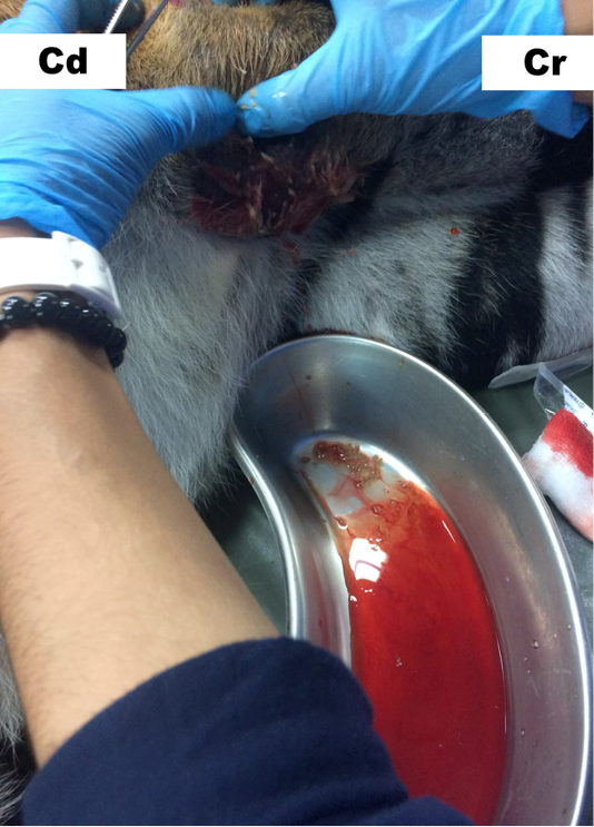
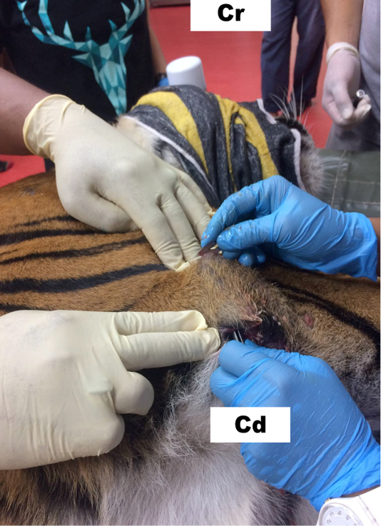
Based on the clinical signs which was a soft and fluid filled lump coupled with the location of the lump which was at the elbow region, the case was tentatively diagnose as elbow hygroma. The treatment regimen planned for this case was surgical lancing and wound cleaning. The tigress was then placed on a left lateral recumbency followed by shaving the site of incision and application of a preparation of chlorhexidine, 70% alcohol and povidone iodine. About 1.5 cm incision was made at the caudo-ventral part of the hygroma to drain the accumulated serosanguineous fluid with fresh blood that was approximately 15 ml (Figure 2). Another incision was made at the cranial part of the hygroma and this to ensure the entire sac was fully drained and flushed (Figure 3).

Figure 2: Incision (1.5 cm) at caudo-ventral part of elbow and drained 15 ml of serosanguineous fluid with blood.

Figure 3: Second incision was made at the cranial part of the hygroma to ensure the entire sac is fully drained and easier to flush to remove all debris accumulated inside the cavity.
Treatment
Intra-operative
During the intra-operative medical management, the tigress was subjected 0.02 mg/kg of Atropine, administered intramuscularly (IM) as an anti-cholinergic agent, 5 mg/kg of Enrofloxacin was given IM as a prophylactic antibiotic to prevent secondary bacterial infection, 0.1 mg/kg of Flunixin Meglumine was also administered IM to reduce inflammation, while 1mL/10kg, IM of Catosal® as vitamin supplementation.
Post-operative
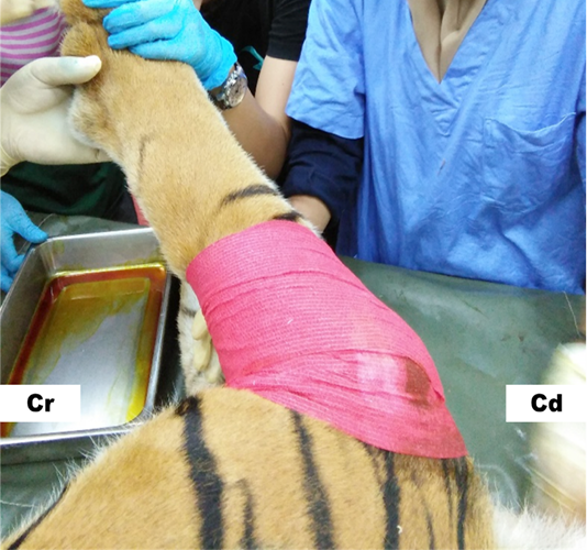
During the post-operative management, 10 mg/kg of Amoxycillin (PO), SID was given for 10 days as a prophylactic antibiotic to prevent secondary bacterial infection, iodine spray BID was administered as anti-septic solution for wound care and finally Papase (PO), SID for 7 days as NSAID to reduce further inflammation. The elbow joint was then bandaged with gauze, soaked with povidone iodine, cotton and water resistant compression wrap (Figure 4). The problem has resolved and there is no further recurrence of hygroma in this case.

Figure 4: The elbow joint was bandaged with gauze soaked with povidone iodine, cotton and water resistant compressing bandage.
DISCUSSION
Hard surfaces have been reported to be a crucial risk factor for the formation of hygroma (Kousi et al., 2017). Additionally, non-padded flooring coupled with heavy animal weight could also contribute to the formation of hygroma due to the direct pressure it exerts on the tissue underlying the olecranon. In the present clinical case report, the tigress was admitted being kept captive on a hard surface which might be a risk factor associated with the observed condition. The tigress in the current report weights about 112 kg body weights, such a body weight could contribute to the formation of hygroma as more pressure is exerted on the elbows when the tiger is sitting down.
Senescence is usually accompanied by a process of cellular (tissue) deterioration (Curtis, 2019). The current clinical case report indicated that the tigress was about 12 years old and this age is enough to suggest a higher tendency of getting injury and trauma such as osteoarthritis as well as social withdrawal and lazing behaviour syndrome, which may eventually cause a prolonged sitting down posture leading to hygroma formation (Downing, 2016).
Tigers are carnivorous in nature where their feeding habit suggests that they feed mostly on red meat which is high in zinc but deficit in required mineral and vitamin contents. This can cause mineral imbalances that can result in muscle weakness and recumbency thus increasing the risk of developing elbow hygroma condition (Kaiser et al., 2014).
To achieve preventive measures, impending risk factors must be checked where soft beddings such as wooden platforms and rubber mats can be improvised to pad the elbow and delineate pressure to prevent trauma. Stimulatory enriching activities can be promoted to minimize prolonged sitting behaviour on the floor. Lastly, adequate diet and proper supplements containing requisite vitamins, minerals and zinc are needed in order to maintain the general health of the captive wildlife and prevent metabolic bone disease.
The therapeutic plan and management of this case is in accord with Meena et al. (2014) where have stated that the clinical management of bilateral elbow hygroma in their case indicated the use of anti-cholinergic, antibiotics and anti-inflammatory agents coupled with the surgical approach and was followed by post-operative care using antibiotics and anti-inflammatory agents. Johnston (1975) also stated that conservative medical management of hygromas had always included the surgical drainage of hygromas and Swenberg et al. (1974) indicated that surgical approach requires partial elliptical incision of the hygroma, and overlying skin be the approach in managing such conditions. Therefore, lancing of hygromas is considered as the best reserved for such clinical conditions and has been adopted in the clinical management of this case.
CONCLUSION
In conclusion, this clinical case report on elbow hygroma in tigress was managed effectively surgically through lancing and wound management. The condition can be prevented by understanding and avoiding the risk factors associated with occurrence of hygroma.
ACKNOWLEDGEMENT
The authors wish to appreciate the management and staff of the National Wildlife Rescue Centre, Sungkai, Perak, PERHILITAN and Department of Veterinary Clinical Studies and University Veterinary Hospital, Faculty of Veterinary Medicine, Universiti Putra Malaysia (UPM).
Authors Contribution
ZK, SSS, HH, CNC were involved in the medical management and all authors were actively involved in designing and commencement of the manuscript.
Conflict of interest
The authors declares that there is no conflict of interest.
REFERENCES
- • Ashraf M (2019). Global tiger day: July 29, 2019: Five Essays.
- • Curtis A (2019). Targeting senescence within the Alzheimer’s plaque. Sci. Transl. Med., 11(488): eaax4869. https://doi.org/10.1126/scitranslmed.aax4869
- • Downing R (2016). Hygroma in dogs. Retrieved from https://vcahospitals.com/know-your-pet/hygroma-in-dogs.
- • Fossum TW (2013). Small animal surgery (4th ed). Elsevier Mosby, St Louis.pp:1619.
- • Johnston DE (1975). Hygroma of the elbow in dogs. J. Am. Vet. Med. Assoc. 167: 213-219.
- • Johnston DE (1985). Bursitis/Tendinitis. Textbook of small animal orthopaedics. Int. Vet. Inf. Ser., Ithaca, New York, USA.
- • Joshi AR, Dinerstein E, Wikramanayake E, Anderson ML, Olson D, Jones BS, Seidensticker J, Lumpkin S, Hansen MC, Sizer NC, Davis CL, Palminteri S, Hahn NR (2016). Tracking changes and preventing loss in critical tiger habitat. Sci. Adv., 2(4): e1501675. https://doi.org/10.1126/sciadv.1501675
- • Kaiser C, Wernery U, Kinne J, Marker L, Liesegang A (2014). The role of copper and vitamin A deficiencies leading to neurological signs in captive cheetahs (Acinonyx jubatus) and lions (Panthera leo) in the United Arab Emirates. Food Nutr. Sci., 05(20): 1978-1990. https://doi.org/10.4236/fns.2014.520209
- • Karen AM (2018). Overview of hygroma. MSD veterinary manual. Retrieved from https://www.msdvetmanual.com/integumentary-system/hygroma/overview-of-hygroma.
- • Kawanishi K (2015). Panthera tigris ssp. jacksoni. The IUCN red list of threatened species 2015. e.T136893A50665029.
- • Kousi T, Angellou V, Psalla D, Papazoglou LG (2017). Elbow hygroma in the dog. Which treatment works better? Hellenic J. Companion Anim. Med. 6(1): 22-28.
- • Meena A, Kumaresan A, Madheswaran R (2014). Surgical management of bilateral elbow hygroma in a boxer dog. 6th Int. Clin. Case Conf. Next Gener. Vet. Face Challenges Clin. Pract., 6(1): 47.
- • Pavletic MM, Brum DE (2015). Successful closed suction drain management of a canine elbow hygroma (2015). J. Small Anim. Pract., 56: 476-479. Nath I, Singh J, Sidhartha SB, Prasant KS, Dwibedy BK (2014). Bilateral hygroma in a great dane dog and its surgical management.
- • Sawyer E, Varacallo M (2019). Anatomy, Shoulder and upper limb, hand ulnar bursa. In Stat. Pearls [Internet]. Stat Pearls Publishing.
- • Sanderson EW, Moy J, Rose C, Fisher K, Jones B, Balk D, Walston J (2019). Implications of the shared socioeconomic pathways for tiger (Panthera tigris) conservation. Biol. Conserv., 231: 13-23. https://doi.org/10.1016/j.biocon.2018.12.017
- • Sethi S, Goyal SP, Choudhary AN (2019). Poaching, illegal wildlife trade and bush meat hunting in India and South Asia. Int. Wildl. Manage. Conserv. Challenges Changing World. pp. 157.
- • Swenberg JA (1974). Surgical closure of elbow hygroma in a dog. J. Am. Vet. Med. Assoc. 164: 147-149.
- • Upadhyaya SK (2019). Human-wildlife interactions in the Western Terai of Nepal. An analysis of factors influencing conflicts between sympatric tigers (Panthera tigris tigris) and leopards (Panthera pardus fusca) and local communities around Bardia National Park, Nepal (Doctoral dissertation).


