Advances in Animal and Veterinary Sciences
Research Article
Incidence and Management of Obstructive Urolithiasis in Buffalo Calves and Goats
Ajit Kumar Singh, Anil Kumar Gangwar*, Khangembam Sangeeta Devi, Harnam Singh
Department of veterinary surgery and radiology, College of Veterinary Science and Animal Husbandry, NarendraDeva University of Agriculture and Technology, Kumarganj, Faizabad-224 229, Uttar Pradesh, India.
Abstract | A total of 125 animals including male buffalo calves (n=83) and buck (n=42) with obstructive urolithiasis were prospectively studied. Most often males aged between 2 and 8 months (mean age 5 months) were affected, with young ones being more commonly affected. Prevalence was maximum in extreme winter and summer. Cystorrhexis was observed in 16.45% of buffalo calves and 5.56% in case of goats. All the animals were treated surgically by tube cystostomy. Simultaneously urinary acidifiers and calculolytic drugs were given to dissolve the calculi. Majority of the cases showed uneventful recovery.
Keywords | Buffalo calves; Goats; Obstructive urolithiasis; Tube cystostomy
Editor | Kuldeep Dhama, Indian Veterinary Research Institute, Uttar Pradesh, India.
Received | September 12, 2014; Revised | October 02, 2014; Accepted | October 03, 2014; Published | October 08, 2014
*Correspondence | Anil Kumar Gangwar, NarendraDeva University of Agriculture and Technology, Kumarganj, Faizabad-224 229, Uttar Pradesh, India; Email: [email protected]
Citation | Singh AK, Gangwar AK, Devi KS, Singh H (2014). Incidence and management of obstructive urolithiasis in buffalo calves and goats. Adv. Anim. Vet. Sci. 2 (9): 503-507
DOI | http://dx.doi.org/10.14737/journal.aavs/2014/2.9.503.507
ISSN (Online) | 2307-8316; ISSN (Print) | 2309-3331
Copyright © 2014 Singh et al. This is an open access article distributed under the Creative Commons Attribution License, which permits unrestricted use, distribution, and reproduction in any medium, provided the original work is properly cited.
Introduction
Obstructive urolithiasis is an economically important disease of bovine and caprine. Factors such as diet, age, sex, breed, genetic makeup, season, soil, water, hormone, mineral, and urinary tract infections play an important role in the genesis of urolithiasis (Udall and Chow, 1969). Young goats and calves are frequently affected with this frustrating condition (Amarpal et al., 2013). After formation of calculi in urinary tract, they may lodge anywhere within the urinary tract, causing urine retention. Surgery is the primary treatment of obstructive urolithiasis (Larson, 1996). Surgical procedures like urethrostomy (Stone et al., 1997), tube cystostomy (Williams and White, 1991), bladder marsupialization (May et al., 1998), penile catheterization and amputation were tried with little practical value in treating obstructive urolithiasis. Tube cystostomy together with medical dissolution of calculi is considered an effective technique for resolution of obstructive urolithiasis in small ruminants (Ewoldt et al., 2006). Animals with prolonged obstruction have high morbidity due to subsequent uraemia. Surgical management of such patients should be done very cautiously. In this study incidence of obstructive urolithiasis and its surgical as well as medical management in 125 cases including male buffalo calves and buck was reported.
Materials and methods
A total of 125 animals including buffalo calves (n=83) and goats (n=42) aged between 2 and 8 months with obstructive urolithiasis were prospectively studied. These cases were presented to TVCC, College of Veterinary Science and Animal Husbandry, Kumarganj, Faizabad (UP) from the adjoining villages between the years 2011-2013. Physical examination was done to check the status of the urethra and urinary bladder. Further, abdomenocentesis was performed in the cases showing water belly appearance to confirm cystorrhexis, if any. All the animals were treated surgically by tube cystostomy technique on the day of admission.
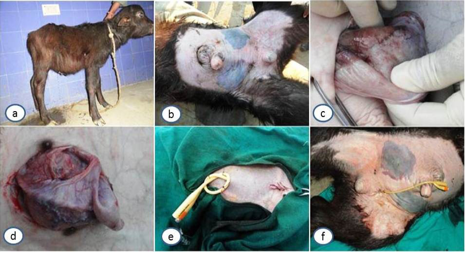
Local infiltration and lumbosacral epidural analgesia using 2% lignocaine hydrochloride was achieved in all the cases to desensitize the proposed surgical site. Patients with high morbidity were administered dexamethasone (4-8 mg) along with 0.5-2.0 litres normal saline intravenously, based on the condition of animal. Animals were placed in right lateral recumbency. Left side of the abdomen near the rudimentary teat was prepared for aseptic surgery. A linear skin incision was given anterior to the rudimentary teat. After incising the skin, fascia, muscles and the peritoneum, bladder was identified. The status of bladder was checked whether intact or ruptured. If bladder was intact, a subcutaneous tunnel parallel to the prepuce was made through which the Foleys catheter was passed (Figure 1e) with pointed end towards the incision. Foley’s catheter was passed from outside to abdominal cavity where the catheter tip was held in stilette and directly stabbed the bladder at an avascular area and its bulb was inflated with sterile normal saline to fix the tube within the bladder. Further the peritoneum, muscles and skin were closed routinely. In cases of ruptured bladder (Figure 1d), urine drainage was done slowly to prevent the animal from shock. Cystorrhaphy was done with catgut number 1 followed by catheter placement after necessary debridement. The bladder was irrigated with normal saline to remove concretions and cystic calculi. In cases where skin became necrosed and delicate to make subcutaneous tunnel, Foley’s catheter was secured at multiple sites on the ventral abdomen (Figure 1f).
Postoperatively amoxicillin-cloxacillin antibiotic combination (500 mg total dose in buffalo calves and 250 mg total dose in goats) was administered by intramuscular route for 5 days and analgesic meloxicam (0.5 mg/Kg) for 3 days. Owners were advised to give ammonium chloride at 100 mg/Kg body weight, twice daily, orally for 30 days. Local antiseptic dressing with dilute liquid povidone iodine was done for a week. The catheter was allowed to drain freely until normal urination resumed, after which it was clamped on every alternate day with infusion set flow regulating clamp to determine the urethral patency. Catheters were removed after normal urination resumed through urethra.
a. Fluid flowing out on abdominocentesis and showing water belly condition; b. Severe inflammation and necrosis on ventral abdomen; c. Ischemic and devitalized bladder; d. Ruptured bladder; e. Foleys catheter passed through a subcutaneous tunnel parallel to prepuce and flow of urine through catheter; f. In cases with skin necrosis, Foley’s catheter secured at multiple sites on the ventral abdomen.
Results
Cases of obstructive urolithiasis were more prevalent in the extreme winter and summer. 91.56% affected buffalo calves were of the age of 4-6 months and the remaining 8.44% calves were above 6 months of age. In case of goats, 87.43% were between age group of 1-5 months and rest 12.57% were more than 5 months of age. Duration of urine retention was less than 3 days in 95.05% of goats. In buffalo calves, 66.53% were reported during first 3-4 days of retention. Cystorrhexis was observed in 16.45% of buffalo calves and 5.56% in case of goats. 2.95% buffalo calves and 97.72% goats had a history of castration at the time of obstruction. Dribbling of urine was observed in 9.03% of buffalo calves and 11.63% in goats, indicating partial obstruction. Males were exclusively affected in case of both buffalo calves and goats.
Catheterization of urinary bladder and positioning of tube was achieved without difficulties. Following tube placement, flow of urine through the tube was observed in all the cases (Figure 1e). The signs of acute pain and distress reduced immediately after surgery and animals started to feed normally after 3-5 hours. Out of 125 animals affected with urolithiasis during study period, 33.60% (42) were goats and 66.40% (83) were buffalo calves. Follow up of 88.80% (111) of total cases (buffalo calves and goats) could be made. Catheter blockage was reported in 12% (15) animals (both goats and buffalo calves). Complication of urethral rupture was seen in 6.40% (8) of buffalo calves. Blocked catheter was cleared by flushing normal saline into the catheter. 12.08% (16) animals were found to have pus at the time of removal of the catheter which was cleared and then antiseptic dressing was done. Catheter was removed at an average period of 13-17 days, though in some cases especially in 12% buffalo calves (15), catheter was removed almost after a month due to delayed normal urination. Majority of the cases showed uneventful recovery.
Discussion
Obstructive urolithiasis was more prevalent in the extreme winter and summer. Occurrence of urolithiasis in peak winter may be due to the decreased water intake and deficiency of vitamin- A, arising from lesser availability of green fodder (Radostits et al., 2000). Desquamated epithelial cells may be due to deficiency of vitamin A and infections (Jones, 2009). Excess sunlight and vitamin D may play an important role in urolithiasis in summer. This may be related to water balance of animals, during winter animals will not take much water and produce concentrated urine (Kushwaha et al., 2011). Conversely, during summer, urine may be more concentrated due to increased water loss in heat. Incidence of urethral obstruction has been reported 49.83% in goats (Amarpal et al., 2004) and 12.66% in buffaloes (Singh et al., 2008). However, in this study much higher incidence (91.56%) was seen in case of male buffalo calves compared to goats (8.44%). The difference in occurrence of urolithiasis may arise as a result of the factors like particular species population, season and management practices in that area. Also the period for which buffalo calves are being maintained is much larger compared to goats. The age of the buffalo calves ranged from 2 to 8 months (mean age 5 months) with maximum calves aged between 2–4 and 4–6 months. Sharma et al. (2007) recorded about 60% urethral obstruction occurs at an early age in ruminants. Gugjoo et al. (2013) reported that 84.61 per cent affected buffalo calves were of the age of 4–7 months. The average duration of illness was 4.5 days. Amarpal et al. (2004, 2005 and 2013) also reported similar findings. All the buffalo calves Singh were male. Although, urolithiasis equally affects male and female animals but obstruction occurs mainly in males (Tamilmahan et al., 2014) due to presence of long and narrow urethra. When urinary bladder ruptures (Figure 1d), initial pain relief occurs, and clinical signs may not appear for 1 to 2 days (Kushwaha et al., 2014). However, over the course of the next few days, uraemia becomes severe, and the animal progresses to severe depression, anorexia and dehydration. Ventral abdominal distention may be noticed as water belly and fluid may be detected by ballottement and abdominocentesis (Figure 1a). In case of high degree of abdominal distension calves were reluctant to move forward and preferred to maintain standing posture to avoid the pressure over the diaphragm and lungs. Severe dehydration in these cases could be due to shifting of water from the interstitial and intravascular compartment into the peritoneal cavity (Radostits et al., 2000). Diet given (concentrate) and the changes brought about by weaning may be contributing factors for development of obstructive urolithiasis in young ruminants (Sharma et al., 2007). Early reporting of goats for urinary retention than buffalo calves could be due to the vocalization shown by goats even with slightest of discomfort (lesser threshold for pain) and also due to the delayed discomfort shown by buffalo calves and their difficult transportation. Delayed reporting in buffalo calves could also be related with the higher occurrence of cystorrhexis than in goats. Diuretic treatment given by the local veterinarian leading to more urine formation may increase the chances of cystorrhexis (Adams, 1995). The increasing pressure and distended stretching of bladder wall results in inflammation, pressure ischaemia, devitalization (Figure 1c), thinning, trabeculae formation and herniation of mucosa through the musculature of the urinary bladder leading to seepage of urine into the peritoneal cavity resulting in uroperitoneum (Makhdoomi and Ghazi, 2013). Uropretoneum cause severe inflammation of abdominal muscles and skin which may lead to necrosis of the affected part (Figure 1b). Singh et al. (2008) reported that complete urethral obstruction also predisposes younger animals to its rupture. Castrated male goats were more affected with urolithiasis. Belknap and Pugh, (2002) reported that castration at an early age may deprive animal from testosterone which is required for the normal development of urethra. In the absence of testosterone hydrophilic colloids decrease in urine thus increasing the incidence of urolithiasis (Rakestraw et al., 1995). However, in our study we did not find such correlation of castration with the incidence of urolithiasis in buffalo calves.
Urolithiasis was seen predominantly in males in both goats and buffalo calves. Though it occurs in both the sexes however, its more occurrences in males might arise due to the smaller diameter and more length of urethra (Thilagar et al., 1996). High phosphorous and low calcium are commonly used as concentrate rations which predispose the animal to phosphate uroliths (Funaba et al., 2001). It was found that feeding had more significant effect on urolith formation than castration, especially in buffalo calves.
The treatment of obstructive urolithiasis is primarily surgical (Van Metre et al., 1996). Before surgical procedure, animal must be stabilized by giving normal saline as metabolic derangements like hyperkalemia, hyponatremia and hypocalcemia exists (Makhdoomi and Ghazi, 2013). Tube cystostomy provides an alternative to number of the surgical techniques available for management of urolithiasis. Urethrostomy and urethrotomy have been used to relieve the obstruction. However, postoperative leakage of urine from the site of obstruction leads to necrosis of urethra and subcutaneous tissues (Gugjoo et al., 2013). Further, postoperative urethral constriction, loss in breeding potential and recurrent urolithiasis is potential factors that results in unfavourable outcome after urethrotomy (Türk et al., 2012). The tube cystostomy gives passage for removal of urine and prevents its accumulation which might lead to the rupture of bladder or the urethra (Dubey et al., 2006). Medical dissolution of the calculi is achieved by giving urine acidifiers and calculolytic drugs. The average duration of removal of catheter was about 15-20 days. Though in some previous reports, 14 days of hospitalization has been reported in goats (Ewoldt et al., 2006). Regular flushing of the catheter with the normal saline was done to prevent the blockade. The tube cystostomy has advantages like improved potential for preservation of breeding function of the animal and urinary continence, and the opportunity for removal of cystic calculi (May et al., 1998). In addition to surgical management, it is also important to advice the owner regarding importance of water consumption by the animal and restriction of salt in the diet.
Conclusion
Our findings suggested that tube cystostomy is a quick, practicable and field applicable method for the management of obstructive urolithiasis buffalo calves and goats.
Acknowledgement
Authors are thankful to Dean College of Veterinary science and Animal Husbandry for providing all the necessary facilities to carry out present study.
References


