Clinical abnormalities suggesting enteritis were recorded in 14 (30.43%) calves of different ages (1-4 weeks) that showed fever, diarrhea, dehydration, pneumonia recumbence and death of two calves. Similar clinical picture was reported by Al-Alo et al. (2018), Cho et al. (2010) and Singh et al. (2013). Hossain et al. (2013) reported that the symptoms of calf enteritis included diarrhea, a rise in body temperature, general weakness, dehydration and lack of appetite followed by coma and death within a few hours. The high morbidity 30.43% recorded in this study among new born calves may be attributed to several factors including the calf less mature immune system than in adults, agammaglobulinemia and the high level of hydrocortisone that probably due to transplacental transfer, as well as the fetus produces large quantity of corticosteroids beginning 8–10 days before birth that lead to immunosuppression as discussed by Hosein (2018).
Identification of 13 (92.85 %) isolates of E. coli out of the 14 collected samples of diarrheic calves was confirmed on the basis of their morphology, cultural and biochemical tests using standard bacteriological procedures. The 13 isolates were visualized as blue-black colonies (2-3mm diameter) with green metallic sheen on Eosin Methylene Blue Agar and bright pink colonies on MacConkey agar. Such findings indicated a presumptive identification of E. coli that was confirmed using standard bacteriological procedures.
The overall prevalence rate of colibacillosis in the present study among 46 calves was (28.26 %). This may be regarded to the fact that E. coli is a normal inhabitant in the intestine of warm-blooded animals as mentioned by Achá et al. (2004). The present result agreed and disagreed in respect of the prevalence of infection with the findings of different authors; Bashir et al. (2015), (35%); Cookson et al. (2006), (88.7%); Pourtaghi et al. (2013), (86.7%) and Shahrani et al. (2014), (76.45%). The difference in the rates of E. coli infection may be attributed to differences in preventive and hygienic measures and exposure to stress factors allowing the opportunistic E. coli to flourish and express its virulence genes causing pathogenic effect on calves as mentioned by Cho and Yoon (2014).
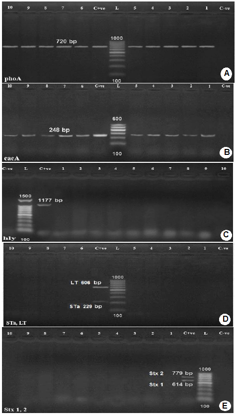
A monoplex PCR detecting alkaline phosphatase gene (phoAgene) specific for E. coli was performed on E. coli isolates. The results of the amplification of phoA gene using PCR, (Figure 1) revealed that all E. coli isolates showed positive result for the presence of phoAgene, thus confirming their identity as E. coli and proving that phoA gene is a housekeeping gene present in all E. coli strains as reported by Chang et al. (1986) and Kong et al. (1999). In order to categorize the different pathotypes of pathogenic E. coli, multiplex PCR was used for detection of virulence genes encoding virulence factors including shiga toxin1 (stx1), shiga toxin 2 (stx2), intimin (eae), hemolysin (hylA), and E. coli enterotoxin genes such as heat stable enterotoxin (st) and heat labile enterotoxin (lt). All tested E. coli isolates were positive for intimin (eae) A, attaching and effacing gene (gene species specific) for E. coli (100%). No isolate had shiga toxin1 (stx1), shiga toxin 2 (stx2), hemolysin (hylA), and E. coli enterotoxin genes heat stable enterotoxin (st) and heat labile enterotoxin (lt), photos (2-5). Intimin is a virulence factor of EPEC (e.g. E. coli O127:H6) and EHEC (e.g. E. coli O157:H7) E. coli strains. It is an attaching and effacing (A/E) protein, which is responsible for enteropathogenic and enterohemorrhagic diarrhea, (Stevens et al., 2006). The intimin gene detection is mainly linked to the EPEC pathotype (Beraldo et al., 2014) and eaeA-positive strains are considered to be more virulent to human than the eaeA-negative ones. This indicates a possible participation of calves in the zoonotic transmission of pathogenic E. coli as supported by the findings of Thiry et al. (2017). Nataro and Kaper (1998) reported that EPEC pathoype strains had a characteristic attaching and effacing (A/E) effect on the gut mucosa due to intimate bacterial adhesion to the enterocytes and effacement of the brush border microvilli. The formation of A/E lesions is governed by the locus of enterocyte effacement (LEE). This locus contained the eaegene, which encoded an outer protein called intimin, necessary for intimate attachment to epithelial cells. EPEC is a cause of diarrhea in ruminants and human and their pathogenicity is attributed to the production of protein intimin which was encoded by the eaegene as mentioned by Horcajo et al. (2012).
EPEC represented 100% of the tested E. coli strains. All isolates were obtained from diarrheic calves which indicated that EPEC is a calf pathogen as mentioned by Moxley and Smith (2010).
The rate of EPEC among pathogenic E. coli was investigated by many authors, Badouei et al. (2016), 1.33%, Nguyen et al. (2011), 2.6%, Arya et al. (2008), 7.7%, Orden et al. (1998), 8.1%, Wani et al. (2003), 9.73%, Islam et al. (2015) 12.5% and Güler et al. (2008) 13.3%.
In general, the high prevalence of EPEC among calves may be attributed to bad management system, lack of hygiene, overcrowding, insufficient attention to the newborn calves with insufficient intake of colostrum and lack of effective preventive measures as well as stress factors that allow the opportunistic E. coli to flourish and express virulence genes causing pathogenic effect on calves as mentioned by Cho and yoon (2014).