
Diarrhea is one of the leading public health problems in under developed countries. In Pakistan, diarrheal cases are treated empirically and, rarely, specific investigations are made. This study was designed to identify the prevalent strains of Escherichia coli among children of less than five years of age in Quetta, Balochistan, Pakistan. A total of 200 samples were collected from children suffering with diarrhea. E. coli was isolated using standard biological procedures and confirmed by colonial characteristics, biochemical profile and specie specific PCR. Multiplex PCR was applied to differentiate among the five main diarrheagenic E. coli pathotypes (enteropathogenic E. coli (EPEC), enteroaggregative E. coli (EAEC), enterotoxigenic E. coli (ETEC), enteroinvasive E. coli (EIEC), and enterohemorrhagic E. coli (EHEC). Results show that the virulent EAEC strain of E. coli was found to be the most prevalent (35%), while EPEC was identified in 15%, followed by EHEC (8%). Notably, none of the samples was found positive for EIEC. Moreover, ETEC was identified in 11% cases of co-infection. Results showed that children with less than two years of age were affected significantly. However, gender had no significant effect on acquiring diarrhea. In conclusion, pathogen spectrum of pathogenic E. coli is EAEC strain. Children under one year of age were affected significantly, and no significant effect was observed when data was stratified on gender base. Further studies are required to investigate the other pathological agents and control parameters should be strengthened to minimize the risks.
Research Article · Volume 51, Issue 6, 7 August 2019 · Open Access
Incidence of Diarrheagenic Escherichia coli Pathotypes in Children Suffering from Diarrhea in Tertiary Care Hospitals, Quetta, Pakistan
· · · · · · · ·
Affiliations & Notes ⌄
- 1Department of Zoology, Sardar Bahadur Khan Women’s University, Quetta 2Department of Zoology, University of Balochistan, Quetta 3Department of Microbiology, University of Balochistan, Quetta 4Centre of Advanced Studies in Vaccinology and Biotechnology (CASVAB), University of Balochistan, Quetta 5Institue of Basic Medical Sciences, Khyber Medical University, Peshawar 6Collge of Veterinary Sciences and Animal Husbandry, Abdul Wali Khan University, Mardan, KP , * Corresponding author: sadeeq@awkum.
Article Info ⌄
- Journal:
- Pakistan Journal of Zoology
- Published:
- 7 August 2019

From the article
- To evaluate the prevalence of DEC in children of less than five years of age, 200 rectal swab samples from children suffering with diarrhea were collected for analysis.
- The samples were analyzed by conventional and advanced diagnostic methods to document the prevalence of DEC in children suffering with diarrhea.
- Samples confirmed as E. coli with species specific PCR were further investigated by multiplex PCR to identify the specific type of E. coli by amplifying their most common virulent gene (Figure 1C).
- In conclusion, EAEC is an important pathogen associated with diarrhea was the most prevalent in children from Quetta, Pakistan.
Selected from the article's own text.
Abstract
INTRODUCTION
Diarrhea accounts for about 10% of all deaths globally in children of less than five years of age and ranked at second position after pneumonia. In Pakistan, 20-30% of total deaths in children of less than 5 years of age are caused by diarrhea . Based on specific virulence factors and other phenotypic characteristics diarrheagenic E. coli (DEC) has been categorized into six different pathotypes including enteropathogenic E. coli (EPEC), enteroaggregative E. coli (EAEC), enterotoxigenic E. coli (ETEC), enteroinvasive E. coli (EIEC), diffusely adherent E. coli (DAEC) and enterohemorrhagic E. coli (EHEC), and shiga-like toxin (STEC). The different DEC has been reported to share 40% of cases of acute diarrhea in children below 5 years ; ; . Although DEC pathotypes are of considerable cause of diarrhea in Pakistan, however, strains are not generally sought, particularly in developing countries like Pakistan, and thus prevalence of diarrhea caused by DEC generally remains unknown.
DEC has been reported involved in outbreaks of gastrointestinal infections in many developing countries. For instance, DEC was found in 23.3% of cases of diarrhea in Mexico . Besides developing countries, other developed countries have also witnessed the epidemics or random cases of gastrointestinal infections, which were mostly food borne associated such as the epidemics of STEC like E. coli in Germany in 2011 . Similarly, a report published about the contamination of raw vegetables with STEC E. coli in Pakistan report that 34% of vegetables were found positive indicating widespread presence of pathogenic E. coli presenting public health threats. Besides human, animals have also been reported to be infected by DEC, which were found multidrug resistant and leading to severe diarrhea .
Despite random reports of E. coli recovered from diarrheic patients in Pakistan, molecular characterization and pathotyping of clinical isolates is totally lacking. Such analysis are cruial to understand pathogenicity of E. coli isolates and can further help policy makers to devise effective strategy for its control. This study is thus aimed to determine the relative prevalence of five major DEC types in children younger than 5 years of age in Quetta, Pakistan and its virulence.
MATERIALS AND METHODS
Ethical approval
The study was approved by the Ethical Committee of University of Balochistan and all procedures described here were performed as per local institutional ethical guidelines. Informed consent was taken from donors.
Sample collection and microbiology
The samples were collected from Bolan Medical Complex Hospital and Children Hospital Quetta for a period of seven months. Children under the age of five years suffering with diarrhea were included in this study. Patients diagnosed with other infectious disease in addition to diarrhea were excluded from the study. Samples were obtained from rectums of children suffering with diarrhea with help of a sterile cotton swabs. The collected samples were then placed in Cary Blair Transport Medium and transferred to the laboratory for culturing and isolation of E. coli. The samples were inoculated and streaked on eosin methylene blue (EMB) and MacConkey agars (Oxoid, UK) for colony formation. After overnight incubation at 37°C, the colonies were observed for characteristic of E. coli. The presumable E. coli colonies were then streaked onto fresh sterilized nutrient agar (Oxoid, UK) to perform conventional biochemical tests for E. coli identification. E. coli was identified by positive methyl red and indole test, negative Voges proskauer, citrate and urease tests. All isolates of E. coli from each rectal swab showing typical characteristics of colony morphology and biochemical properties were maintained in the laboratory in nutrient broth at 4o C as well as were stocked at -70 °C until further use.
Genomic DNA extraction and differentiation of DEC
Extraction of DNA was carried out by cetyletrimethyle ammonium bromide (CTAB) method as described by Minas et al. (2011). The extracted DNA was kept at -70oC until used as a template for PCR. PCR was carried out to confirm E. coli by using universal primer of E. coli (uidA). Conditions for PCR were: 94oC for 5 min for initial template denaturation, 35 cycles at 98oC for 10 seconds for final denaturation, primer annealing at 68oC for 35 seconds and primer extension at 72oC for 45 seconds and finally at 72ºC for 7 min (final extension). Multiplex PCR was used to investigate and categorize isolates into their respective pathotypes/diarrheagenic groups . Total reaction volume of PCRs performed was 90µl, containing 45µl of PCR Master mix (Gene All), 1µl of each set of 11 primers (Macrogen),10µl of DNA template extracted from samples and 15µl molecular grade water. Using multiplex PCR, 11 specific primers (Macrogen) were used to identify the particular E. coli pathotypes as shown in Table I. Amplification conditions for multiplex PCR include 95oC (initial template denaturation), 35 cycles at 94oC for 50s (final denaturation), primer annealing starting from 45ºC for 45s with a regular interval increase of 0.5oC after each cycle, till the temperature becomes 60oC, primer extension at 72oC for 1 min and finally at 72ºC for 7 min (final extension). Amplicons were examined by electrophoresis on agarose gel (2% w⁄v) followed by staining with ethidium bromide
RESULTS
To evaluate the prevalence of DEC in children of less than five years of age, 200 rectal swab samples from children suffering with diarrhea were collected for analysis. The samples were analyzed by conventional and advanced diagnostic methods to document the prevalence of DEC in children suffering with diarrhea. Out of 200 collected samples, 100 samples were found positive for E. coli by cultivating on selective EMB agar media. Results demonstrated that positive samples of E. coli presented typical colonies with greenish metallic sheen in reflected light with blue black center as shown in Figure 1A. Purified and biochemically confirmed E. coli isolates were confirmed by specieS specific PCR (Fig. 1B).
Samples confirmed as E. coli with species specific PCR were further investigated by multiplex PCR to identify the specific type of E. coli by amplifying their most common virulent gene (Figure 1C). The multiplex PCR results demonstrated that 35 % of the isolates have the virulent gene AggR (430bp) of EAEC as shown in Table II. Importantly, AggR showed co-infection with EHEC (12%) and EAEC and ETEC (11%). The eae virulent gene (229bp) expressed by EPEC was 15% of the samples positive for E. coli. The EHEC pathotype expressing Shiga-like toxin 2 (stx2) gene (779bp) was amplified in 8% of the total positive samples for E. coli and Shiga-like-toxin 1 (stx1) gene (614bp) of EHEC was only detected in co-infected samples with AggR (EAEC) or combined with AggR (EAEC) and elt (ETEC).
Table I.- Primer sequences of E. coli virulent genes.
| Patho-type | Gene | amplicon size(bp) | Primer sequence | Reference |
| EHEC | Stx 1(F) | 614 | ACA CTG GAT GAT CTC AGT GG | |
| Stx 1(R) | CTG AAT CCC CCT CCA TTA TG | |||
| Stx 2(F) | 779 | CCATGA CAA CGG ACA GCA GTT | ||
| Stx 2(R) | CCT GTC AAC TGA GCA CTT TG | |||
| HylA(F) | 534 | GCA TCA TCA AGC GTA CGT TCC | ||
| HylA(R) | AAT GAG CCA AGCTGG TTA AGCT | |||
| PcvD(F) | 630 | CTG GCG AAA GAC TGT ATC AT | ||
| PcvD(R) | CAA TGT ATA GAA ATC CGC TGTT | |||
| EPEC | bfpA(F) | 450 | CAC CGT TAC CGC AGG TGT GA | |
| bfpA(R) | GTT GCC GCT TCA GCA GGA GT | |||
| eae(F) | 229 | CTG AAC CAG ATC GTA ACG GC | ||
| eae(R) | TGA TAA GCT GCA GTC GAA TCC | |||
| ETEC | elt(F) | 322 | TCT CTA TGT GCA TAC GGA GC | |
| elt(R) | CCA TAC TGA TTG CCG CAA T | |||
| estl(F) | 170 | TCT TTC CCC TCT TTT AGT CAGTC | ||
| estl(R) | CAG CAC AGG CAG GAT TAC | |||
| EIEC | ipaH(F) | 619 | GTT CCT TGA CCG CCT TTC CGA TAC CGT C | |
| ipaH(R) | GCC GGT CAG CCA CCC TCT GAG AGT AC | |||
| EAEC | AggR(F) | 430 | AGA CGC CTA AAG GAT GCC C | ; ; |
| AggR(R) | GAG TTA TCA AGC AAC AGC AAT GC | |||
| E.coli | uidA(F) | 619 | CCA AAA GCC AGA CAG AGT | |
| uidA(R) | GCA CAG CAC ATC AAA GAG |
Table II.- Prevalence of Escherichia coli (DEC) strains in children suffering with diarrhea.
| Escherichia coli (DEC) strains | Total samples | Positive (n) |
| EAEC | 100 | 35 |
| EPEC | 100 | 15 |
| EHEC | 100 | 08 |
| EAEC+ETEC+EHEC | 100 | 11 |
| EAEC+EHEC | 100 | 12 |
| EPEC+EHEC+EAEC | 100 | 13 |
| EPEC+EHEC | 100 | 06 |
| EIEC | 100 | 00 |
Similarly, ETEC pathotype expressing elt gene (322bp) was only found in samples infected with EHEC and EAEC. Such co-infections were present in 11% of the positive E. coli samples. Moreover, stx2 of EHEC and AggR of EAEC were found together in 12% of the positive samples. EPEC co-infection with EHEC and EAEC was found 13%, while EPEC and EHEC co-infection was 6% in all samples analyzed.
To estimate the prevalence of EIEC, specific primers against the virulent gene ipaH, (619bp) was employed however no sample was found positive for the said strain (Fig. 1C).
The results of multiplex PCR were further analyzed to dissect the prevalence of DEC in children on basis of gender and age. Of the 100 samples positive for DEC, 49% of them were boys while rests were girls (51%). Majority of the children were less than 12 months (37%) followed by age between 1-2 years (30%), 2-3 years (19%), 3-4 years (6%) and 4-5 years (3%) as shown in Table III. These results indicate that children under five years of age suffering with diarrhea carry high burden of DEC. Importantly, EAEC strain proved to be the most prevalent in area under study.
Table III.- Escherichia coli (DEC) strains identified by multiplex PCR and their prevalence sub-divided according to the gender and age of children suffering with diarrhea.
| Escherichia coli (DEC) strains | Gender | Age (Years) | |||||
| Male Female | <1 | 1-2 | 2-3 | 3-4 | 4-5 | ||
| EAEC | 15 | 25 | 12 | 14 | 8 | 1 | 0 |
| EPEC | 9 | 6 | 7 | 4 | 3 | 0 | 1 |
| EHEC | 5 | 3 | 3 | 4 | 1 | 0 | 0 |
| EAEC+ETEC+ EAEC | 5 | 6 | 3 | 3 | 2 | 2 | 1 |
| EAEC+EHEC | 5 | 7 | 4 | 4 | 3 | 1 | 0 |
| EPEC+EHEC+ EAEC | 7 | 6 | 5 | 4 | 1 | 2 | 1 |
| EPEC+EHEC | 3 | 3 | 3 | 2 | 1 | 0 | 0 |
| EIEC | 0 | 0 | 0 | 0 | 0 | 0 | 0 |
DISCUSSION
In developing countries, the DEC strains are not regularly sought as stool pathogens but treated as per symptoms. Since the infections and complications caused by DEC are critical, especially in infants, proper investigations should be practiced before prescription of any drug. In this study, the low percentage (50%) of E. coli obtained from children with diarrhea may be due to the fact that antimicrobial therapy prior to sample collection might have been administered in some cases and it is a known fact that this can reduce the percentage of bacterial enteropathogens isolation . Our results are in agreement with previous studies and reporting important DEC, which were found in samples obtained from children suffering with diarrhea . In this study four categories of DEC were identified. One of the major emerging pathogens that has been associated with diarrhea is EAEC and it is known to cause persistent diarrhea in children in developing nations. Furthermore, EAEC has been identified in patients infected by human immunodeficiency virus and specially children and travelers in the developing world ; ; . EAEC are a heterogeneous group of bacteria that display a wide range of virulence factors . The diarrhea caused by EAEC is watery diarrhea with no blood and manifested by occasional abdominal cramps, but with no fever, and has been documented globally as described earlier consistent with our findings . EPEC was found to be the second most prevalent DEC (15%) in this study. Diarrhea caused by EPEC is usually watery that may contain mucus but normally does not necessarily have blood in it. Symptoms include fever, vomiting and dehydration. In infants, typical EPEC is quite common cause of gastroenteritis. ETEC produce heat-labile (LT) and/or heat-stable (STa and STb) toxins that cause diarrhea in humans and animals. Traveler’s diarrhea is commonly caused by the exposure of ETEC in these areas . The occurrence of ETEC (11% in the diarrhea group as a mix infection) was lower in our study as reported by studies conducted in other countries .
Very less EHEC strains were identified in this study. It has been observed that there is low prevalence of EHEC in developing countries ; ; which is in agreement with result of our study. EHEC produce verotoxins 1 and 2 (stx1 and stx2) and these strains are related with food-borne diseases predominantly due to consumption of raw shredded meat, uncooked milk or by intake of unclean foods polluted with faeces .
EIEC is a source of broad range human’s infections. Several studies around the globe have reported the low frequencies of EIEC which is also in agreement with our studies ; . No work has been done previously in Quetta, Pakistan regarding the use of multiplex PCR to detect DEC. There has been much similarity and sensitivity to our studies concerning the multiplex PCR as assays conducted earlier ; . In this study, we used only one specific and sensitive multiplex PCR assay that has reduced the number of several assays for gene detection so the time and effort have been saved . There is a possibility of detecting mixed infections by these assays which involve more than one DEC strain. It is very important to identify DEC accurately so that the spectrum of the disease, the infection source and transmission routs can be understood clearly. By identifying this way the clinician will be able to properly manage and treat the disease. Common antibiotics usually face resistance by strains of DEC. It is therefore very important to rapidly detect the specific pathogen and accomplish the assay within 24 hours. If multiplex PCR assay is used in diagnostic laboratories it will definitely provide a rapid and practical method to identify DEC .
CONCLUSION
In conclusion, EAEC is an important pathogen associated with diarrhea was the most prevalent in children from Quetta, Pakistan. Multiplex PCR proved to be specific, sensitive and much rapid method of diagnosis therefore it can be used in diagnostic laboratories to overcome the limitations of traditional techniques.
Acknowledgments
No funding was available for this study.
Statement of conflict of interest
The authors declare no conflict of interest.
Figures
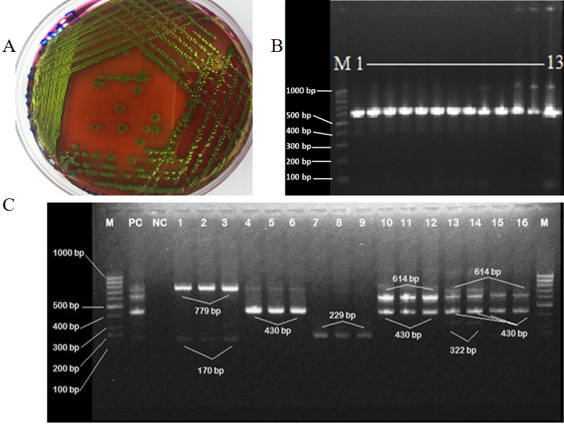
Isolation and identification of various E. coli pathotypes based on virulent gene profile. A, growth of purified E. coli isolate on EMB agar; B, representative results of amplification of uidA gene of E.coli from collected samples: Lanes 1-13 positive amplification of uidA gene (630bp). M is molecular weight marker, size of which is demarcated on the left; C, multiplex PCR results showing amplification fragments of different DEC. Lanes 1-3 positive amplification of stx2 (779bp). 4-6 shows positive amplification of Agg (430bp), 7-9 for eae gene (229bp), 10-12 shows amplification of stx1 (614bp) and Agg (430bp). Lanes 13-14 denotes the amplification of stx1, Agg and elt genes (614bp, 430bp and 322bp respectively). Lanes 15-16 show amplification of stx1 (614bp) and Agg genes (430bp). M, PC and NC denotes molecular weight marker, positive control and negative control, respectively.
References
- Adachi, J.A., Ericsson, C.D., Jiang, Z.-D., DuPont, M.W., Pallegar, S.R. and DuPont, H.L., 2002. Natural history of enteroaggregative and enterotoxigenic Escherichia coli infection among US travelers to Guadalajara, Mexico. J. Infect. Dis., 185: 1681-1683. https://doi.org/10.1086/340419[CrossRef]
- Adnan, M., Khan, H., Kashif, J., Ahmad, S., Gohar, A., Ali, A., Khan, M.A., Shah, S.S.A., Hassan, M.F. and Irshad, M., 2017. Clonal expansion of sulfonamide resistant Escherichia coli isolates recovered from diarrheic calves. Pakistan Vet. J., 37: 230-232.
- Bern, C., Martines, J., De Zoysa, I. and Glass, R., 1992. The magnitude of the global problem of diarrhoeal disease: a ten-year update. Bull. Wld Hlth Organ., 70: 705.
- Beutin, L., 2006. Emerging enterohaemorrhagic Escherichia coli, causes and effects of the rise of a human pathogen. Zoon. Publ. Hlth., 53: 299-305 https://doi.org/10.1111/j.1439-0450.2006.00968.x[CrossRef]
- Canizalez-Roman, A., Flores-Villaseñor, H.M., Gonzalez-Nuñez, E., Velazquez-Roman, J., Vidal, J.E., Muro-Amador, S., Alapizco-Castro, G., Díaz-Quiñonez, J.A. and León-Sicairos, N., 2016. Surveillance of Diarrheagenic Escherichia coli strains isolated from diarrhea cases from children, adults and elderly at Northwest of Mexico. Front. Microbiol., 7: 1924 https://doi.org/10.3389/fmicb.2016.01924[CrossRef]
- Chase-Topping, M.E., Rosser, T., Allison, L.J., Courcier, E., Evans, J., McKendrick, I.J., Pearce, M.C., Handel, I., Caprioli, A. and Karch, H., 2012. Pathogenic potential to humans of bovine Escherichia coli O26, Scotland. Emerg. Infect. Dis., 18: 439-448. https://doi.org/10.3201/eid1803.111236[CrossRef]
- de Sousa, C.P. and Dubreuil, J.D., 2001. Distribution and expression of the astA gene (EAST1 toxin) in Escherichia coli and Salmonella. Int. J. med. Microbiol., 291: 15-20. https://doi.org/10.1078/1438-4221-00097[CrossRef]
- Frank, C., Werber, D., Cramer, J.P., Askar, M., Faber, M., an der Heiden, M., Bernard, H., Fruth, A., Prager, R., Spode, A., Wadl, M., Zoufaly, A., Jordan, S., Kemper, M.J., Follin, P., Müller, L., King, L.A., Rosner, B., Buchholz, U., Stark, K. and Krause, G., 2011. Epidemic profile of shiga-toxin–producing Escherichia coli O104:H4 outbreak in Germany. N. Engl. J. Med., 365: 1771-1780. https://doi.org/10.1056/NEJMoa1106483[CrossRef]
- Franzolin, M.R., Alves, R.C.B., Keller, R., Gomes, T.A.T., Beutin, L., Barreto, M.L., Milroy, C., Strina, A., Ribeiro, H. and Trabulsi, L.R., 2005. Prevalence of diarrheagenic Escherichia coli in children with diarrhea in Salvador, Bahia, Brazil. Mem. Inst. Oswaldo Cruz, 100: 359-363. https://doi.org/10.1590/S0074-02762005000400004[CrossRef]
- Habib, M.A., Soofi, S., Sadiq, K., Samejo, T., Hussain, M., Mirani, M., Rehmatullah, A., Ahmed, I. and Bhutta, Z.A., 2013. A study to evaluate the acceptability, feasibility and impact of packaged interventions (“Diarrhea Pack”) for prevention and treatment of childhood diarrhea in rural Pakistan. BMC Publ. Hlth., 13: 922-932. e?. https://doi.org/10.1186/1471-2458-13-922[CrossRef]
- Jenkins, C., van Ijperen, C., Dudley, E.G., Chart, H., Willshaw, G.A., Cheasty, T., Smith, H.R. and Nataro, J.P., 2005. Use of a microarray to assess the distribution of plasmid and chromosomal virulence genes in strains of enteroaggregative Escherichia coli. FEMS Microbiol Lett., 253: 119-124. https://doi.org/10.1016/j.femsle.2005.09.040[CrossRef]
- Kaper, J.B., Nataro, J.P. and Mobley, H.L., 2004. Pathogenic Escherichia coli. Nature Rev. Microbiol., 2: 123-140. https://doi.org/10.1038/nrmicro818[CrossRef]
- Kimata, K., Shima, T., Shimizu, M., Tanaka, D., Isobe, J., Gyobu, Y., Watahiki, M. and Nagai, Y., 2005. Rapid categorization of pathogenic Escherichia coli by multiplex PCR. Microbiol Immunol., 49: 485-492. https://doi.org/10.1111/j.1348-0421.2005.tb03752.x[CrossRef]
- Kotloff, K.L., Nataro, J.P., Blackwelder, W.C., Nasrin, D., Farag, T.H., Panchalingam, S., Wu, Y., Sow, S.O., Sur, D. and Breiman, R.F., 2013. Burden and aetiology of diarrhoeal disease in infants and young children in developing countries (the Global Enteric Multicenter Study, GEMS): a prospective, case-control study. The Lancet, 382: 209-222. https://doi.org/10.1016/S0140-6736(13)60844-2[CrossRef]
- McDaniels, A., Rice, E., Reyes, A., Johnson, C., Haugland, R. and Stelma, G., 1996. Confirmational identification of Escherichia coli, a comparison of genotypic and phenotypic assays for glutamate decarboxylase and beta-D-glucuronidase. Appl. environ. Microbiol., 62: 3350-3354.
- Minas, K., McEwan, N.R., Newbold, C.J. and Scott, K.P., 2011. Optimization of a high-throughput CTAB-based protocol for the extraction of qPCR-grade DNA from rumen fluid, plant and bacterial pure cultures. FEMS Microbiol. Lett., 325: 162-169. https://doi.org/10.1111/j.1574-6968.2011.02424.x[CrossRef]
- Moyo, S.J., Maselle, S.Y., Matee, M.I., Langeland, N. and Mylvaganam, H., 2007. Identification of diarrheagenic Escherichia coli isolated from infants and children in Dar es Salaam, Tanzania. BMC Infect. Dis., 7: 92-98. https://doi.org/10.1186/1471-2334-7-92[CrossRef]
- Nataro, J.P., 2005. Enteroaggregative Escherichia coli pathogenesis. Curr. Opin. Gastroenterol., 21: 4-8.
- Nessa, K., Ahmed, D., Islam, J., Kabir, F.L. and Hossain, M.A., 2016. Usefulness of a multiplex PCR for detection of diarrheagenic Escherichia coli in a diagnostic microbiology laboratory setting. Bangladesh J. med. Microbiol., 1: 38-42. https://doi.org/10.3329/bjmm.v1i2.21506[CrossRef]
- Nguyen, T.V., Le Van, P., Le Huy, C., Gia, K.N. and Weintraub, A., 2005a. Detection and characterization of diarrheagenic Escherichia coli from young children in Hanoi, Vietnam. J. Clin. Microbiol., 43: 755-760. https://doi.org/10.1128/JCM.43.2.755-760.2005[CrossRef]
- Ogura, Y., Ooka, T., Terajima, J., Nougayrède, J.-P., Kurokawa, K., Tashiro, K., Tobe, T., Nakayama, K., Kuhara, S. and Oswald, E., 2007. Extensive genomic diversity and selective conservation of virulence-determinants in enterohemorrhagic Escherichia coli strains of O157 and non-O157 serotypes. Genome Biol., 8: R138. https://doi.org/10.1186/gb-2007-8-7-r138[CrossRef]
- Pereira, A.L., Ferraz, L.R., Silva, R.S. and Giugliano, L.G., 2007. Enteroaggregative Escherichia coli virulence markers: positive association with distinct clinical characteristics and segregation into 3 enteropathogenic E. coli serogroups. J. Infect. Dis., 195: 366-374. https://doi.org/10.1086/510538[CrossRef]
- Persson, S., Olsen, K., Scheutz, F., Krogfelt, K. and Gerner-Smidt, P., 2007. A method for fast and simple detection of major diarrhoeagenic Escherichia coli in the routine diagnostic laboratory. Clin. Microbiol. Infect., 13: 516-524. https://doi.org/10.1111/j.1469-0691.2007.01692.x[CrossRef]
- Pickering, L.K., Obrig, T.G. and Stapleton, B.F., 1994. Hemolytic-uremic syndrome and enterohemorrhagic Escherichia coli. Pediat. Infect. Dis. J., 13: 459-475. https://doi.org/10.1097/00006454-199406000-00001[CrossRef]
- Rajendran, P., Ajjampur, S.S.R., Chidambaram, D., Chandrabose, G., Thangaraj, B., Sarkar, R., Samuel, P., Rajan, D.P. and Kang, G., 2010. Pathotypes of diarrheagenic Escherichia coli in children attending a tertiary care hospital in South India. Diagn. Microbiol. Infect. Dis., 68: 117-122. https://doi.org/10.1016/j.diagmicrobio.2010.06.003[CrossRef]
- Rappelli, P., Folgosa, E., Solinas, M.L., DaCosta, J.L., Pisanu, C., Sidat, M., Melo, J., Cappuccinelli, P. and Colombo, M.M., 2005. Pathogenic enteric Escherichia coli in children with and without diarrhea in Maputo, Mozambique. Pathog. Dis., 43: 67-72. https://doi.org/10.1016/j.femsim.2004.07.006[CrossRef]
- Rappelli, P., Maddau, G., Mannu, F., Colombo, M., Fiori, P. and Cappuccinelli, P., 2001. Development of a set of multiplex PCR assays for the simultaneous identification of enterotoxigenic, enteropathogenic, enterohemorrhagic and enteroinvasive Escherichia coli. New Microbiol., 24: 77-83.
- Rohner, P., Pittet, D., Pepey, B., Nije-Kinge, T. and Auckenthaler, R., 1997. Etiological agents of infectious diarrhea: implications for requests for microbial culture. J. clin. Microbiol., 35: 1427-1432.
- Samad, A., Abbas, F., Ahmad, Z., Pokryrshko, O. and Asmat, T.M., 2018. Prevalence of foodborne pathogens in food items in Quetta, Pakistan. Pakistan J. Zool., 50: 1597-1600. http://dx.doi.org/10.17582/journal.pjz/2018.50.4.sc17[Full text][CrossRef]
- Sethabutr, O., Venkatesan, M., Murphy, G.S., Eampokalap, B., Hoge, C.W. and Echeverria, P., 1993. Detection of Shigellae and enteroinvasive Escherichia coli by amplification of the invasion plasmid antigen H DNA sequence in patients with dysentery. J. Infect. Dis., 167: 458-461. https://doi.org/10.1093/infdis/167.2.458[CrossRef]
- Sheikh, J., Czeczulin, J.R., Harrington, S., Hicks, S., Henderson, I.R., Le Bouguénec, C., Gounon, P., Phillips, A. and Nataro, J.P., 2002. A novel dispersin protein in enteroaggregative Escherichia coli. J. clin. Investig., 110: 1329-1337. https://doi.org/10.1172/JCI200216172[CrossRef]
- Sobel, J.D., Wiesenfeld, H.C., Martens, M., Danna, P., Hooton, T.M., Rompalo, A., Sperling, M., Livengood III, C., Horowitz, B. and Von Thron, J., 2004. Maintenance fluconazole therapy for recurrent vulvovaginal candidiasis. N. Engl. J. Med., 351: 876-883. https://doi.org/10.1056/NEJMoa033114[CrossRef]
- Stritt, A., Tschumi, S., Kottanattu, L., Bucher, B.S., Steinmann, M., von Steiger, N., Stephan, R., Hächler, H. and Simonetti, G.D., 2012. Neonatal hemolytic uremic syndrome after mother-to-child transmission of a low-pathogenic stx 2b harboring Shiga toxin–producing Escherichia coli. Clin. Infect. Dis., 56: 114-116. https://doi.org/10.1093/cid/cis851[CrossRef]
Services
Article usage statistics
Loading…