
The disease that caused reproductive and respiratory problems in pigs was once known as “mystery swine disease” before being renamed “porcine reproductive and respiratory syndrome, (PRRS)” in the United States and Europe. In the early 1990s, the PRRS virus (PRRSV), an arterivirus, was discovered to be the disease’s causative agent. Numerous studies have been carried out since then. Since spreading to Vietnam and Cambodia, “porcine high fever disease”, PRRSV continues to cause severe illness in pigs in China as of 2010. This illness was first reported in 2006, when PRRSV was recognized as a critical virus linked to a high rate of mortality and morbidity (20%). The complete method of infection transmission is still unknown despite a great deal of research on epidemiology regarding the PRRS. This paper provides a brief historical summary of PRRS and the associated PRRSV. It lists the current objectives for eliminating or reducing PRRS as well as the areas whereby research is currently missing and preventing the creation of effective vaccines. It is hoped that this discussion will stimulate additional collaboration between researchers and swine veterinarians around the world to develop solutions that advance our knowledge of PRRS and PRRSV in an effort to eradicate this economically important disease.
Review · Volume 44, 22 May 2026 · Open Access
Understanding PRRS: Epidemiology, Pathogenesis, and Histopathology in the Context of Global Swine Health
· · · ·
Affiliations & Notes ⌄
- 1Faculty of Veterinary Science, University of Agriculture, Faisalabad, Pakistan. , * Corresponding author: 2022ag5900@uaf.edu.pk
Article Info ⌄
- Journal:
- South Asian Journal of Zoology
- Published:
- 22 May 2026
From the article
- Respiratory illness: Pneumonia brought on by a PRRS virus infection in piglets and growing pigs can lower growth rates and raise mortality.
- Immunological system suppression: The PRRS virus has the ability to lower immunological function, which increases pigs’ susceptibility to recurrent infections.
- Enhanced vulnerability to other illnesses: Porcine circovirus-associated disease (PCVAD) is more common in pigs infected with PRRS.
- Porcine reproductive and respiratory syndrome virus (PRRSV) in pig meat.
Selected from the article's own text.
Abstract
Introduction
A well-known and economically important swine disease, porcine reproductive and respiratory syndrome (PRRS) is characterized by reproductive failure in pregnant sows or respiratory tract distress in nursing pigs ; . In the mid-1980s, the condition was first recognized as “mystery swine disease” or “blue ear disease” in the United States . The PRRS virus (PRRSV), the causative agent, was first discovered in the US in 1987 ; ; . Later, in the early 1990s, it was discovered throughout Asia and Europe ; . PRRS causes significant financial losses to the world’s pig output, especially in enormous manufacturing systems . In the USA alone, PRRS is thought to cost the pork sector roughly US$ 560 million in damages every year. A disease known as “porcine high fever syndrome (PHFS)” first appeared in the People’s Republic of China in 2006 and quickly spread throughout the nation, inflicting extremely serious illness in pigs with severe fever (40–42 oC). The disease’s characteristics included substantial death rates in suckling piglets, infants, and growers, as well as miscarriages in sows across all age groups. Pigs with PHFS were used to isolate PRRSVs in several Chinese facilities. The illness was linked to an unusually virulent strain of the PRRSVs, according to a later genomic and pathogenicity examination of those viruses. Highly pathogenic PRRS is the new term for the illness brought on by this novel variant strain ; ; ; , 2009; . Thus, in 2006, highly pathogenic PRRS (HP-PRRS) first appeared in China and has subsequently expanded to Southeast Asian nations. It has seriously harmed the region’s pig productivity and put a strain on pig farmers . To lower the financial losses brought on by this illness and stop it from spreading to other parts of the world, suitable control measures must be developed and put into place. This requires a thorough understanding of the disease’s characteristics, the virus, and its epidemiology. In order to lay the groundwork for the creation of practical and effective disease management strategies, the goal of this study is to review the data currently available on HP-PRRS.
Characteristics of the HP-PRRSV
Parkinson’s disease is caused by an RNA virus that is a member of the genera Arterivirus, family Arteriviridae, order Nidovirales Genotype I, which represents the prototype Lelystad virus, which is the type of virus that is most common in Europe, and genotype II, represented by VR 2332, the prototype strain that was initially identified in North America, are two closely related but genetically and antigenically different strains. The genetic and antigenic makeup of these early isolates differed greatly from one another. Their genetic makeups are less than 70% similar. Disease control is constantly threatened by genetic and antigenic variability among isolates, even within a country. There are at least eight open-reading frames (ORFs) in the virus’s 15–15.5 kb genome, which collectively encode over 20 complete proteins. The HP-PRRSV is an altered strain of the PRRSV linked to genotype II (North American genotype) of the virus, according to a genetic sequence study . Phylogenetic examination of the ORF5 gene suggests that the HP-PRRSV may have descended from two of the PRRSVs that were previously discovered in China. Further analysis of a nonstructural protein (NSP2) revealed a trait unique to the HP-PRRSV: The discontinuous deletion of 30 protein amino acids (at locations 482 and 534-562) . This loss is proposed as a genetic feature of the HP-PRRSV, even though it has been shown that the deletions are not the sources of the virus’s increased virulence.
Epidemiology of HP-PRRS virus
Up to 70% of animals in Southeast Asia and China are still kept in traditional backyard and small-scale settings . This results in the proximity of production systems with varying sizes and levels of biosecurity, which are crucial factors that can influence the potential for disease spread and the impact that diseases may have on pig populations. Given that over 70% of pig farmers are smallholders, one of the main issues that persists is livestock producers’ lack of awareness and comprehension of the advantages of disease control. Commercial pig farmers are aware of the advantages of disease control, but their understanding of the prerequisites for a cooperative national disease monitoring program is limited. The conventional farrow-to-finish methods are typically used by medium- and small-scale pig producers, frequently with very close age group mixing. Figure 1 shows that the direction of spread is more significant and more intense from sow farms to other farms.
Replacement stock frequently originates from a range of places, is of unknown health, and is not properly quarantined before arrival. When it comes to interactions between farm workers and pigs outside of their farms, or between strangers and pigs housed on the farms, hygiene application is typically very inadequate or nonexistent. Vaccination is typically used to reduce disease in small-scale hog farming. Without proper supervision or veterinary advice, the use of large concentrations of antibiotic and antibacterial agent combinations is rather prevalent. Pork, which has been consumed at the highest rates about the entire amount of meat produced in various countries (Vietnam, China, Cambodia, as well as the Philippines), is one of the most important agricultural goods in the region. Commercial pig production rises in response to rising pork consumption. According to statistics, the main places where HP-PRRS has been spreading are those with larger populations of pigs. The Red River Delta in Vietnam is the primary region for heavy pig production in the country’s north and is also a major hub for pig diseases, such as the rise of highly PRRS.
Live pigs, including piglets, fatteners, and finishers, are traded from this location to the southern region of Vietnam. The quick dissemination of PRRSV is likewise consistent with and probably explains pig migrations in these value chains. The illness may potentially be spreading between neighboring nations due to the transportation of HP-PRRS-infected pigs, including deceased pigs . The trans-boundary spread of HP-PRRSV from southern China to South-East Asia strongly implies the existence of bio-security lapses, such as the inability to regulate animal movements and border trade between neighboring nations. The spread of viruses within a nation also demonstrates the inadequacy of biosecurity measures, mostly due to unchecked human mobility in highly polluted regions, particularly at loading zones and slaughterhouses. Sharing such contaminated places with other cars could lead to a more community-wide spread of the virus.
Age of the pigs at the time of the infection
Klinge et al. (2009) showed that viraemias in 3-week-old pigs were noticeably longer than those in finishers or grownup pigs, regardless of the PRRSV isolate that was utilized as the inoculum. Similarly, 2-month-old pigs had much higher viral burdens within their lymph nodes the lungs, alongside tracheobronchial swabs over 6-month-old animals, regardless of the challenge strain’s pathogenicity ; . Additionally, Thanawongnuwech et al. (1998) showed that pulmonary macrophages from 4-week-old pigs generated higher viral titers than those from 4-months old pigs. All of the prior data provide indirect evidence that piglets are more pathogenic than finishers or older pigs, notwithstanding the lack of a precise evaluation. In farrow-to-finish farms, this can be important for controlling the illness, particularly in light of the potential for sows or newborn pigs from nurseries to become infected again.
HISTOPATHOLOGY OF PRRS
The porcine reproductive and respiratory syndrome virus is the root cause of PRRS. The indications of PRRS include interstitial pneumonia, thickened alveolar septa, and suppurative bronchopneumonia, with suppurative bronchopneumonia and proliferative and necrotizing pneumonia also observed, especially with virulent strains.
Lung lesions
Interlobular setae become thickened due to edema and proteinaceous exudates. Necrosis, degeneration can be seen in the lumen of bronchioles. Hyperplasia and hypertrophy can be seen. Interstitial pneumonia with vasculitis is’s consistent finding . Figure 2 shows interstitial pneumonia and hyperplasia.
Lymphoid organs
Severe lymphoid depletion results in the formation of cavities in the germinal centers of lymphoid follicles. Figure 3 shows thrombus formation, congestion, and hemorrhages. There may also be fibrin deposition also ; .
Vascular lesions
Skin lesions consist of small round macules and papules and irregular patches; distributed to hindquarters, thorax, abdomen and margins of ears. Necrotizing and leukocytoclastic vasculitis of small blood vessels was observed within the dermis. Figure 4 shows microscopically, lymphocytic infiltration and lymphadenopathy can be seen ; .
Reproductive lesions
Gross lesions in the umbilical cords varied, ranging from 1 to 2 cm segmental hemorrhagic regions to a full cord involvement that was enlarged and bled profusely. Gross cord lesions in virally infected fetuses were associated with necrotizing umbilical arteritis with per arterial bleeding, as demonstrated by a histopathologic study. In some experimentally infected sows, several foci of lymphoplasmacytic inflammation have been seen in the myometrium. Myometritis, endometritis, and placentitis of the mother placenta were noted in sows that were infected both naturally and in an experiment . Figure 5A, B show moderate multifocal and perivascular lymphoplasmacytic inflammation along with interstitial edema .
Cellular infiltrates
A characteristic lesion of interstitial pneumonia is mononuclear cell infiltration of alveolar walls with normal airway epithelium, particularly in newborn pigs experiencing respiratory distress. Comparably, lymphoplasmacytic inflammation and edema development are evident .
Comparison between PRRS strains
In this investigation, three strains of porcine reproductive and respiratory syndrome virus-1 suffered segregation from pigs that tested negative for PRRS using porcine alveolar macrophages (PAM) cells. In 1993, strain 18794 was isolated from a pig in Denmark. In 2009 saw the isolation of strain ILI6 from Russian weaner pig lung tissue. Strain BOR59 was identified in 2009 from the lung tissue of a Belarusian swine that had died from respiratory disease symptoms.
In the current investigation, the virulence of two distinct strains of PRRSV-1 subtype 2 and an example of PRRSV-1 subtype 1 strain were evaluated. Four groups of eight-week-old pathogen-free pigs were set up and given fake inoculations, subtype 1 strain 18794, subtype 2 strain ILI6, or subtype 2 strain BOR59.
Overall, the outcomes revealed that the BOR59 strain is extremely pathogenic, although the severity of infection of the other subtype 2 strain, ILI6, was in the middle zone of that of the BOR59 and subtype 1 strains ; .
Impact of immunopathology
The immunopathology of PRRS is complex and involves an overactive immune response, cytokine imbalance, and tissue damage, leading to the various disease manifestations . The immunopathology of PRRS significantly impacts both the pig industry and animal health. Some of the impacts are given below.
- Reproductive failure: When pregnant sows are infected with the PRRS virus, it can cause abortion, stillbirth, and underweight piglets.
- Respiratory illness: Pneumonia brought on by a PRRS virus infection in piglets and growing pigs can lower growth rates and raise mortality.
- Immunological system suppression: The PRRS virus has the ability to lower immunological function, which increases pigs’ susceptibility to recurrent infections.
- Enhanced vulnerability to other illnesses: Porcine circovirus-associated disease (PCVAD) is more common in pigs infected with PRRS.
Histopathological findings in vaccinated animals
- The testis and epididymis’ adluminal compartments contain both mononuclear and multinuclear giant cells (Fig. 6A, B).
- Damage and inflammation in the epididymis and testes (Fig. 6C, D).
- Seminiferous epithelial cells are desquamated.
- Apoptosis of bystander cells.
- Lung alveolar edema and interstitial pneumonia.
- Histiocytic proliferation and lymphoid depletion in the lymph nodes.
- Multinucleated giant cells seen in lymph nodes and lungs.
- Damage and inflammation in other organs, like the spleen and liver.
- A histological investigation revealed luteinizing cysts formed in the ovaries instead of lesions of endometritis or myometritis. The vaccinated animals had a decreased prevalence of these cysts. The development of an ovarian cyst impairs the ability to reproduce, leading to prolonged anestrus and complete infertility .
Diagnostic utility of histopathology in PRRS
- Verification of infection: By detecting distinctive lesions and viral antigens in tissues, histopathology can verify the presence of PRRS virus infection.
- Evaluation of disease severity: Histopathology can assess the degree of lesions linked to PRRS.
- Distinguishing PRRS from other diseases: Histopathology can differentiate PRRS from other reproductive and respiratory conditions, allowing for a precise diagnosis.
- Identification of secondary infections: Histopathology can detect secondary bacterial or viral infections, which often complicate PRRS cases.
- Monitoring vaccine efficacy: Histopathology can evaluate the effectiveness of PRRS vaccines by examining lesions and viral antigens in vaccinated animals .
Future directions in PRRS histopathology research
Immunohistochemically staining: Enhancing detection of PRRSV antigen and immune cell characterization in tissues.
Digital pathology: Implementing digital tools for image analysis and automated scoring of histopathological lesions.
Comparative pathology: Investigating PRRS lesions in different animal models and species.
Vaccine efficacy evaluation: Assessing the impact of various vaccines on PRRS lesions and immune responses .
Pathogenesis and transmission of PRRSV
The PRRS, commonly known as blue ear pig disease. It is a condition that can affect pigs and is caused by the virus betaarterivirus suid 1. This economically significant panzootic disease results in respiratory tract sickness in young pigs. Betaarterivirus sud 1 is an unclassified virus within the realm Riboviria, belonging to the kingdom Orthornavirae, phylum Pisuviricota, and class Pisoniviricetes. It is part of the order Nidovirales and falls under the family Arteriviridae. At the genus level, it is classified as Betaarterivirus, with the subgenus Eurpobartervirus. The full species name is Betaarterivirus sud 1 .
According to a 2021 study by Derald Holtkamp of Iowa State University, PRRS harms the US pork sector by $664 million annually. Based on data recently compiled by Pipestone Management Company, PRRS costs their system roughly $200 per sow year.
Transmission of PRRSV
Zimmerman et al. (1997) reported that transmission of PRRSV occurred in pigs through direct or indirect contact. It can also be transmitted through inhalation.
The experiment was performed to detect PRRSV in muscle (longissimus dorsi), lymphoid tissue, and serum. 135 pigs (49 negative controls and 89 PRRSV-injected pigs). Thirteen out of eighty-nine (14.6%) muscle samples tested positive for qRT-PCR between 28 and 202 days following inoculation. Within these 13, PRRSV was recovered from three out of thirteen lymphoid tissue samples and four out of thirteen suited serum samples. Experiments were also performed to check the transmissibility of PRRSV through meat. No meat transmission was observed .
Target cells and tissues
Many experiments were performed to detect the target cells and tissues of PRRSV, the virus was inoculated and detected at 3, 14, 21, 35 days of post-inoculation (DPI). On day 3 post inoculation, PRRSV replication was found in retropharyngeal lymph nodes, bronchial lymph nodes, tonsils, spleen, alveolar macrophages, and thoracic aortic lymph nodes. On day 14 post inoculation, the same tissues were PRRSV positive, except for the thoracic aortic and retropharyngeal lymph nodes. On 35 DPI, lung and alveolar macrophage PRRSV positivity was seen. The heart, bone marrow cells, and peripheral blood mononuclear cells did not contain PRRSV. During the acute stage of infection, PRRSV primarily replicates in lymphoid tissue and lung macrophages and remains in lung macrophages .
Impact on reproduction and respiratory system
PRRSV can be transmitted horizontally or vertically. Horizontally, it is transmitted through body secretions such as semen. Abortion can occur at any stage of gestation . The virus has a direct effect on the conceptus and follows transplacental infection. Sometimes, gestation becomes prolonged and fetuses are infected transplacentally and often die in utero. Fetal circulation is also affected. Segmental hemorrhages in the umbilical cord can be seen , along with cellular infiltration and cellular changes in the antibodies produced.
Lungs fail to collapse, and they exhibit distinct dark, mottled patches of pneumonia . Acute pneumonia is brought on by the acute cytocidal replication of PRRSV in alveolar lung macrophages. Expiration was more influential than inspiration. Lowering the compliance of the airways, decreased lung CO-transfer factor, and peripheral airway blockage were discovered while rebreathing test gases (He, CO) and using impulse oscillometry for non-invasive pulmonary function testing ; .
Viral replication
Basic research focuses on the process by which the virus enters its host cell, which is a critical first stage in the infection. Porcine alveolar macrophages (PAMs) are the primary target cells of PRRSV, which has a limited cell tropism. Target cell’s restricted cell tropism is caused by the presence of particular entry mediators in the target cell. The virus’s DNA is bound, internalized, and released by a number of cellular components, including heparin sulphate, CD163, porcine sialoadhesin (pSn), non-muscle myosin heavy chain 9 (MYH9), vitremin, CD151, and CD209 (DC-SIGN) (Van Breedam et al., 2010; .
Mechanisms
PRRSV enters macrophages through receptor-mediated mechanisms, first binding to heparin sulfate glycosaminoglycans on the cell surface (Fig. 7). Then there is an increase in integration with pSn. Clathrin-mediated endocytosis identifies pSn as an internalization receptor. After that, the genome is released into the cytoplasm. CD163, a scavenger receptor cysteine rich (SRCR) for Hb clearance, is the most selective receptor for invasion and infection development. The viral glycoproteins GP2 and GP4 also bind to CD163. CD163 is essential for viral removal of coating and genetic release in conjunction with cellular proteases, such as trypsin-like serine proteases, cathepsin E, and aspartic proteases ; . The genome is used as a template that is translated to pp1 and pp1ab. Proteases hydrolyze polyproteins into mature non-structural proteins. The two essential components of nsps; nsp9 and nsp10 are assembled by the the transcription and replication complex of viruses. Making use of an interrupted transcription method, the RTC first produces both full-length and subgenome (sg)-length minus strands. The latter serve as templates for the production of plus-strand sg mRNAs, which are required for the expression of the structural protein genes found in the genome’s 3′-proximal quarter .
Assembling and releasing the virion is the final step. Novel RNA genomes are bundled into nucleocapsids, which after budding from smooth internal membranes converted into enveloped virions by structural proteins. The new virions are then exocytosed from the cell ; .
Systemic effects
Most specifically, reproductive and respiratory systems are affected but other lymph organs and tissues also get disrupted. There are lesions present along with cellular infiltrates. PRRSV was also found in retropharyngeal lymph nodes, bronchial lymph nodes, tonsils, spleen, alveolar macrophages, and thoracic aortic lymph nodes.
Host factors influencing pathogenesis
Age
Nursery pigs are more susceptible to PRRSV than adults. Pulmonary intravascular macrophages in young pigs are more virulent to infection than those of adult pigs . Age has no bearing on the presence of antibody responses to PRRSV. Infection is restricted to cells of monocytic lineage, and fully developed pig alveolar macrophages are the primary target of spontaneous infection .
Breed
Duroc, Hampshire pigs are susceptible to VR2385, a strain with high pathogenicity of PRRSV . The degree of resistance to PRRSV infection fluctuates
among sow breeds and strains. Contrary to other breeds, including the Large White (LW), Meishan and Tongcheng (TC) are less prone to disease because of their enhanced ability to resist PRRSV ; .
Other factors
The vulnerability of various pig breeds to PRRSV is influenced by environmental factors, pigs’ nutritional and health conditions, the virulence of PRRSV, and other factors .
Chronic infection and persistence
When PRRSV enters the thymus, it depletes T-cell precursors and modifies the TCR repertoire. Affected thymocytes are developed. The cytotoxic and helper αβ-T cells both exhibit repertoire diversification restriction. As a consequence, infection becomes prolonged and vital viral epitopes are sustained. Developed antibodies recognize virus but can’t neutralize it.
Studies have shown that high levels of virus are detected in serum than the swab. Virus concentration decreases but is not eliminated from blood bloodstream and mucosal areas till three weeks have passed.
PRRSV diminishes αβ-T cell clones necessary for neutralizing epitope identification and impairs the thymus’s developing αβ-T cell repertoire. As an outcome, the repertoire lacks certain T helpers needed for particular B cells to generate high-affinity antibodies capable of neutralizing PRRSV. It will lead the virus to persist longer (Fig. 8) ; .
Overall pathogenesis
Primary replication takes place in lymphoid tissues when the virus has been transmitted to the tonsils or upper respiratory system. The next step is viremia, which might last for a few weeks. The spleen, thymus, tonsils, lymph nodes, and Peyer’s patches are among the lymphoid tissues that the virus likes to infect. It spreads to pulmonary alveolar and intravascular macrophages and causes interstitial pneumonia. It causes abortion in late gestation by crossing the placenta.
Control and prevention of PRRS
These are the keys to managing PRRS (a) Create and rigorously implement biosecurity measures to stop PRRSV from entering or reentering, (b) Prevent the virus from spreading among or infecting sows in breeding herds. Currently, biosecurity, testing and removal, immunization, and management techniques like whole-herd depopulation/repopulation and herd closure are employed to combat PRRS.
Although there are vaccines available, their efficacy varies. This may be partly caused by the many viral strains that are out there, the viral load that infects the animal after vaccination, and vaccination-related processes or circumstances that influence immune response. As a result, the duration of immunity is unknown. Pig vaccination may be beneficial in herds with PRRS issues or herds at high risk of contracting PRRSV infection, even though it does not prevent PRRSV infection. Generally speaking, the goal of vaccination is to lower clinical losses. A vaccine technique is more affordable for pig farmers than other PRRS management methods, and it is workable for all sizes of pig producers (small, medium, and large). Commercially accessible PRRS vaccines come in two varieties: a live vaccination and a killed viral vaccine. The live vaccine is widely acknowledged for its ability to provide protection against genetically similar PRRS viruses. However, there are issues with its safety, cross-protective effectiveness, and immunogenicity. On the other hand, the PRRS killed vaccination is widely recognized for its safety, albeit its protection is restricted.
Declarations
Acknowledgement
The authors express their sincere gratitude to their colleagues and mentors for their valuable guidance, constructive discussions, and continuous encouragement throughout the preparation of this review.
Funding
This review article received no specific grant from any funding agency in the public, commercial, or not-for-profit sectors.
Generative AI and AI-assisted technology statement
The authors declare that no generative AI and AI assisted technology was used in the creation of this manuscript.
Statement of conflict of interest
The authors have declared that they have no known competing financial interests or personal relationships that could have appeared to influence the work reported in this paper.
Figures
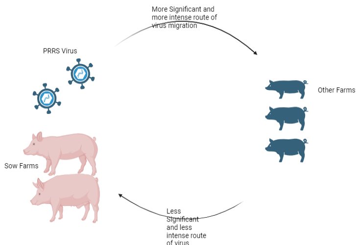
Illustration of PRRSV transmission dynamics between sow farms and other pig farms. The figure highlights that virus migration from sow farms to other farms is a more significant and intense route compared to the reverse direction, which is less significant and less intense.
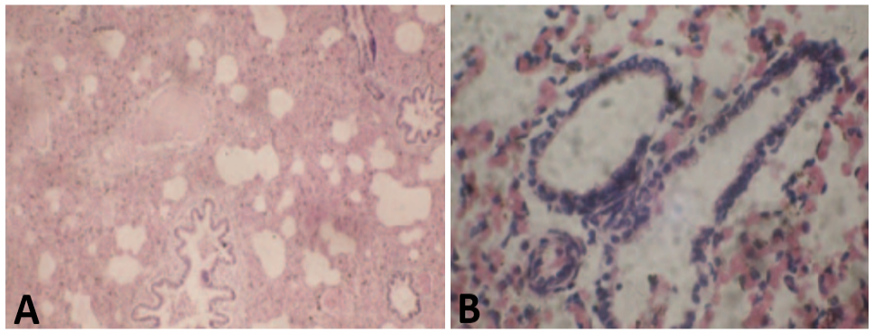
A, Histopathological section of pig lung showing multifocal interstitial pneumonia with alveolar septal thickening and infiltration of inflammatory cells, characteristic of PRRSV infection. B, Higher magnification of affected lung tissue illustrating peribronchiolar lymphoid hyperplasia and bronchiolar epithelial damage, commonly observed in PRRSV-infected pigs. Stain: H and E stain. Magnification: A, 10x; B, 40x).
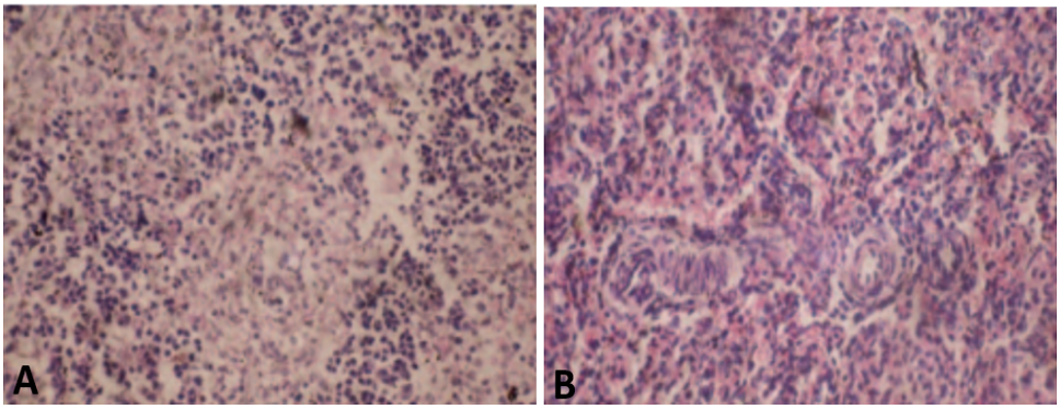
A, Microscopic view of lymphoid tissue reveals prominent interfollicular vascular congestion. B, diffuse monocyte infiltration, indicating an active inflammatory response. Stain: H and E, Magnification: 40x.
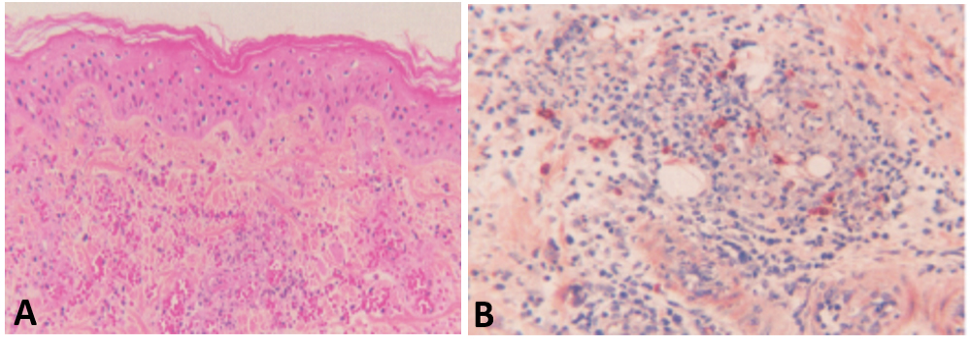
A, Microscopic image reveals multifocal hemorrhages within the dermis along with perivascular. B, interstitial infiltration of lymphocytes, indicating an ongoing inflammatory process. Stain: H and E, Magnification: 40x.
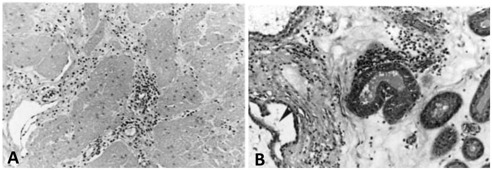
A, Histopathological section showing moderate, multifocal inflammatory infiltrates scattered throughout the tissue. B, Perivascular accumulation of lymphocytes and plasma cells (lymphoplasmacytic inflammation), suggestive of a chronic immune response. Stain: Hematoxylin and Eosin. Magnification:
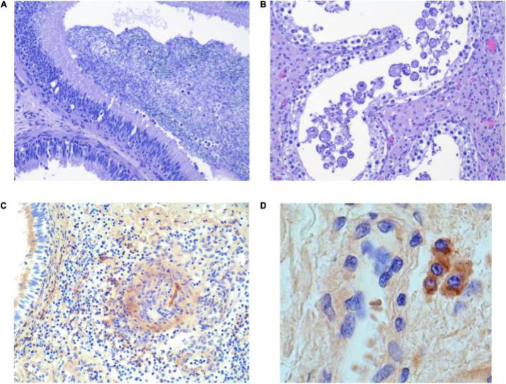
A, Histopathological section showing the invasion of the epithelium by intraluminal mononuclear cells, indicating an inflammatory response. B, Enlarged and swollen cells present within the lumen, suggesting a pathological alteration in cellular morphology. C, Focally scattered macrophages within the testicular interstitium, reflecting localized immune activity. D, Macrophages dispersed in the interstitium of the epididymis, indicative of chronic inflammation. Stain: H and E. Magnification: 40x.
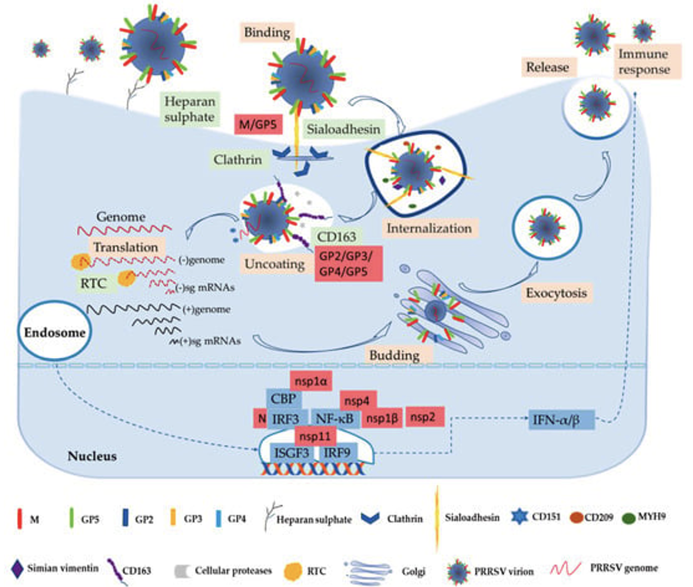
Schematic representation of the viral entry pathway into the host cell. The diagram illustrates the various steps involved, including virus attachment to the host cell receptor, fusion with the host membrane, and subsequent entry via endocytosis or direct fusion. These processes are essential for viral infection and replication within the host cell (Nan et al., 2017).
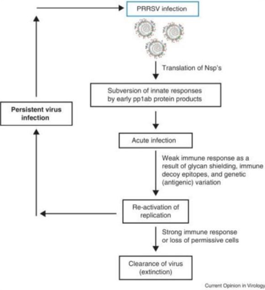
Flow sheet diagram showing the persistence of virus in host cell (Ostrowski et al., 2002).
References
- Albina, E., 1997. Epidemiology of porcine reproductive and respiratory syndrome (PRRS): An overview. Vet. Microbiol., 55: 309-316. https://doi.org/10.1016/S0378-1135(96)01322-3[CrossRef]
- Albina, E., Carrat, C. and Charley, B., 1998. Interferon-alpha response to swine arterivirus (PoAV), the porcine reproductive and respiratory syndrome virus. J. Interferon Cytok. Res., 18: 485-490. https://doi.org/10.1089/jir.1998.18.485[CrossRef]
- An, T.Q., Li, J.N., Su, C.M. and Yoo, D., 2020. Molecular and Cellular Mechanisms for PRRSV pathogenesis and host response to infection. Virus Res., 286: 197980. https://doi.org/10.1016/j.virusres.2020.197980[CrossRef]
- Cho, J.G., Dee, S.A., Deen, J., Trincado, C., Fano, E., Jiang, Y., Faaberg, K., Murtaugh, M.P., Guedes, A., Collins, J.E. and Joo, H.S., 2006. The impact of animal age, bacterial coinfection, and isolate pathogenicity on the shedding of porcine reproductive and respiratory syndrome virus in aerosols from experimentally infected pigs. Can. J. Vet. Res., 70: 297-301.
- Cho, J., Laschinger, H.K. and Wong, C., 2006. Workplace empowerment, work engagement and organizational commitment of new graduate nurses. Nurs Leadersh (Tor Ont), 19: 43-60. https://doi.org/10.12927/cjnl.2006.18368[CrossRef]
- Christianson, W.T., Choi, C.S., Collins, J.E., Molitor, T.W., Morrison, R.B. and Joo, H.S., 1993. Pathogenesis of porcine reproductive and respiratory syndrome virus infection in mid-gestation sows and fetuses. Can. J. Vet. Res., 57: 262-268.
- Christianson, W.T., Collins, J.E., Benfield, D.A., Harris, L., Gorcyca, D.E., Chladek, D.W., Morrison, R.B. and Joo, H.S., 1992. Experimental reproduction of swine infertility and respiratory syndrome in pregnant sows. Am. J. Vet. Res., 53: 485-488. https://doi.org/10.2460/ajvr.1991.53.04.485[CrossRef]
- Darith, S., 2017. Optimizing livestock structure: The case of cattle, pig and chicken in Cambodia.
- Duinhof, T.F., van Schaik, G., van Esch, E.J. and Wellenberg, G.J., 2011. Detection of PRRSV circulation in herds without clinical signs of PRRS: Comparison of five age groups to assess the preferred age group and sample size. Vet. Microbiol., 150: 180-184. https://doi.org/10.1016/j.vetmic.2011.01.001[CrossRef]
- Gao, P., Chai, Y., Song, J., Liu, T., Chen, P., Zhou, L., Ge, X., Guo, X., Han, J. and Yang, H., 2019. Reprogramming the unfolded protein response for replication by porcine reproductive and respiratory syndrome virus. PLoS Pathog., 15: e1008169. https://doi.org/10.1371/journal.ppat.1008169[CrossRef]
- Grebennikova, T.V., Clouser, D.F., Vorwald, A.C., Musienko, M.I., Mengeling, W.L., Lager, K.M., Wesley, R.D., Biketov, S.F., Zaberezhny, A.D., Aliper, T.I. and Nepoklonov, E.A., 2004. Genomic characterization of virulent, attenuated, and revertant passages of a North American porcine reproductive and respiratory syndrome virus strain. Virology, 321: 383-390. https://doi.org/10.1016/j.virol.2004.01.001[CrossRef]
- Guo, R., Katz, B.B., Tomich, J.M., Gallagher, T. and Fang, Y., 2016. Porcine reproductive and respiratory syndrome virus utilizes nanotubes for intercellular spread. J. Virol., 90: 5163-5175. https://doi.org/10.1128/JVI.00036-16[CrossRef]
- Han, D., Hu, Y., Li, L., Tian, H., Chen, Z., Wang, L., Ma, H., Yang, H. and Teng, K., 2014. Highly pathogenic porcine reproductive and respiratory syndrome virus infection results in acute lung injury of the infected pigs. Vet. Microbiol., 169: 135-146. https://doi.org/10.1016/j.vetmic.2013.12.022[CrossRef]
- Hoff, E.J. and Vandevelde, M., 1981. Case report: Necrotizing vasculitis in the central nervous systems of two dogs. Vet. Pathol., 18: 219-223. https://doi.org/10.1177/030098588101800209[CrossRef]
- Hopper, S.A., White, M.E. and Twiddy, N., 1992. An outbreak of blue-eared pig disease (porcine reproductive and respiratory syndrome) in four pig herds in Great Britain. Vet. Rec., 131: 140-144. https://doi.org/10.1136/vr.131.7.140[CrossRef]
- Kelly, R.I., 1995. Cutaneous vasculitis and cutaneous vasculopathies. Australas. J. Dermatol., 36: 109-117; quiz 118-109. https://doi.org/10.1111/j.1440-0960.1995.tb00952.x[CrossRef]
- King, S.J., Ooi, P.T., Phang, L.Y., Allaudin, Z.N., Loh, W.H., Tee, C.Y., How, S.P., Yip, L.S., Choo, P.Y. and Lim, B.K., 2017. Phylogenetic characterization of genes encoding for viral envelope glycoprotein (ORF5) and nucleocapsid protein (ORF7) of porcine reproductive and respiratory syndrome virus found in Malaysia in 2013 and 2014. BMC Vet. Res., 13: 3. https://doi.org/10.1186/s12917-016-0933-z[CrossRef]
- Klein, C. and Reinhold, P., 2001. Analysis of respiratory mechanics by impulse oscillometry in non-sedated and diazepam-sedated swine. Res. Vet. Sci., 70: 181-189. https://doi.org/10.1053/rvsc.2001.0458[CrossRef]
- Klein, R., Nicholls, R. and Thomalla, F., 2004. Resilience to natural hazards: How useful is this concept? Environ. Hazard., 5: 35-45. https://doi.org/10.1016/j.hazards.2004.02.001[CrossRef]
- Klinge, K.L., Vaughn, E.M., Roof, M.B., Bautista, E.M. and Murtaugh, M.P., 2009. Age-dependent resistance to Porcine reproductive and respiratory syndrome virus replication in swine. Virol. J., 6: 177. https://doi.org/10.1186/1743-422X-6-177[CrossRef]
- Lager, K.M. and Halbur, P.G., 1996. Gross and microscopic lesions in porcine fetuses infected with porcine reproductive and respiratory syndrome virus. J. Vet. Diagn. Invest., 8: 275-282. https://doi.org/10.1177/104063879600800301[CrossRef]
- Lee, S.M., Schommer, S.K. and Kleiboeker, S.B., 2004. Porcine reproductive and respiratory syndrome virus field isolates differ in in vitro interferon phenotypes. Vet. Immunol. Immunopathol., 102: 217-231. https://doi.org/10.1016/j.vetimm.2004.09.009[CrossRef]
- Li, Y., Wang, X., Bo, K., Wang, X., Tang, B., Yang, B., Jiang, W. and Jiang, P., 2007. Emergence of a highly pathogenic porcine reproductive and respiratory syndrome virus in the Mid-Eastern region of China. Vet. J., 174: 577-584. https://doi.org/10.1016/j.tvjl.2007.07.032[CrossRef]
- Lunney, J.K. and Chen, H., 2010. Genetic control of host resistance to porcine reproductive and respiratory syndrome virus (PRRSV) infection. Virus Res., 154: 161-169. https://doi.org/10.1016/j.virusres.2010.08.004[CrossRef]
- Lunney, J.K., Fang, Y., Ladinig, A., Chen, N., Li, Y., Rowland, B. and Renukaradhya, G.J., 2016. Porcine reproductive and respiratory syndrome virus (PRRSV): Pathogenesis and interaction with the immune system. Annu. Rev. Anim. Biosci., 4: 129-154. https://doi.org/10.1146/annurev-animal-022114-111025[CrossRef]
- Ma, Z., Yang, L. and Zhang, Y.J., 2018. Porcine reproductive and respiratory syndrome virus: propagation and quantification. Curr. Protoc. Microbiol., 48: 15m.11.11-15m.11.14. https://doi.org/10.1002/cpmc.51[CrossRef]
- Murakami, Y., Kato, A., Tsuda, T., Morozumi, T., Miura, Y. and Sugimura, T., 1994. Isolation and serological characterization of porcine reproductive and respiratory syndrome (PRRS) viruses from pigs with reproductive and respiratory disorders in Japan. J. Vet. med. Sci., 56: 891-894. https://doi.org/10.1292/jvms.56.891[CrossRef]
- Nan, Y., Wu, C., Gu, G., Sun, W., Zhang, Y.J. and Zhou, E.M., 2017. Improved vaccine against PRRSV: current progress and future perspective. Front. Microbiol., 8: 1635. https://doi.org/10.3389/fmicb.2017.01635[CrossRef]
- Neumann, E.J., Kliebenstein, J.B., Johnson, C.D., Mabry, J.W., Bush, E.J., Seitzinger, A.H., Green, A.L. and Zimmerman, J.J., 2005. Assessment of the economic impact of porcine reproductive and respiratory syndrome on swine production in the United States. J. Am. Vet. med. Assoc., 227: 385-392. https://doi.org/10.2460/javma.2005.227.385[CrossRef]
- Nilubol, D., Tripipat, T., Hoonsuwan, T. and Kortheerakul, K., 2012. Porcine reproductive and respiratory syndrome virus, Thailand, 2010-2011. Emerg. Infect. Dis., 18: 2039-2043. https://doi.org/10.3201/eid1812.111105[CrossRef]
- Oleksiewicz, M.B., Bøtner, A., Madsen, K.G. and Storgaard, T., 1998. Sensitive detection and typing of porcine reproductive and respiratory syndrome virus by RT-PCR amplification of whole viral genes. Vet. Microbiol., 64: 7-22. https://doi.org/10.1016/S0378-1135(98)00254-5[CrossRef]
- Ostrowski, M., Galeota, J.A., Jar, A.M., Platt, K.B., Osorio, F.A. and Lopez, O.J., 2002. Identification of neutralizing and nonneutralizing epitopes in the porcine reproductive and respiratory syndrome virus GP5 ectodomain. J. Virol., 76: 4241-4250. https://doi.org/10.1128/JVI.76.9.4241-4250.2002[CrossRef]
- Pallarés, F.J., Halbur, P.G., Opriessnig, T., Sorden, S.D., Villar, D., Janke, B.H., Yaeger, M.J., Larson, D.J., Schwartz, K.J., Yoon, K.J. and Hoffman, L.J., 2002. Porcine circovirus type 2 (PCV-2) coinfections in US field cases of postweaning multisystemic wasting syndrome (PMWS). J. Vet. Diagn. Invest., 14: 515-519. https://doi.org/10.1177/104063870201400614[CrossRef]
- Raymond, P., Bellehumeur, C., Nagarajan, M., Longtin, D., Ferland, A., Müller, P., Bissonnette, R. and Simard, C., 2017. Porcine reproductive and respiratory syndrome virus (PRRSV) in pig meat. Can. J. Vet. Res., 81: 162-170.
- Renukaradhya, G.J., Meng, X.J., Calvert, J.G., Roof, M. and Lager, K.M., 2015. Live porcine reproductive and respiratory syndrome virus vaccines: Current status and future direction. Vaccine, 33: 4069-4080. https://doi.org/10.1016/j.vaccine.2015.06.092[CrossRef]
- Robinson, S.R., Rahe, M.C., Gray, D.K., Martins, K.V. and Murtaugh, M.P., 2018. Porcine reproductive and respiratory syndrome virus neutralizing antibodies provide in vivo cross-protection to PRRSV1 and PRRSV2 viral challenge. Virus Res., 248: 13-23. https://doi.org/10.1016/j.virusres.2018.01.015[CrossRef]
- Sarli, G., D’Annunzio, G., Gobbo, F., Benazzi, C. and Ostanello, F., 2021. The role of pathology in the diagnosis of swine respiratory disease. Vet. Sci., 8. https://doi.org/10.3390/vetsci8110256[CrossRef]
- Snijder, E.J., Kikkert, M. and Fang, Y., 2013. Arterivirus molecular biology and pathogenesis. J. Gen. Virol., 94: 2141-2163. https://doi.org/10.1099/vir.0.056341-0[CrossRef]
- Stadejek, T., Stankevicius, A., Storgaard, T., Oleksiewicz, M.B., Belák, S., Drew, T.W. and Pejsak, Z., 2002. Identification of radically different variants of porcine reproductive and respiratory syndrome virus in Eastern Europe: Towards a common ancestor for European and American viruses. J. Gen. Virol., 83: 1861-1873. https://doi.org/10.1099/0022-1317-83-8-1861[CrossRef]
- Stadejek, T., Larsen, L.E., Podgórska, K., Bøtner, A., Botti, S., Dolka, I., Fabisiak, M., Heegaard, P.M.H., Hjulsager, C.K., Huć, T., Kvisgaard, L.K., Sapierzyński, R. and Nielsen, J., 2017. Pathogenicity of three genetically diverse strains of PRRSV Type 1 in specific pathogen free pigs. Vet. Microbiol., 209: 13-19. https://doi.org/10.1016/j.vetmic.2017.05.011[CrossRef]
- Stockhofe-Zurwieden, N., Navarro-Camarro J.A., Grosse-Beilage, E., Chavez, J. and Pohlenz, J., 1993. Uterine and placental alterations in pregnant sows associated with the porcine epidemic abortion and respiratory syndrome (PEARS). Zentralbl Vet. B, 40: 261-271. https://doi.org/10.1111/j.1439-0450.1993.tb00137.x[CrossRef]
- Thanawongnuwech, R., Thacker, E.L. and Halbur, P.G., 1998. Influence of pig age on virus titer and bactericidal activity of porcine reproductive and respiratory syndrome virus (PRRSV)-infected pulmonary intravascular macrophages (PIMs). Vet. Microbiol., 63: 177-187. https://doi.org/10.1016/S0378-1135(98)00245-4[CrossRef]
- Tian, K., Yu, X., Zhao, T., Feng, Y., Cao, Z., Wang, C., Hu, Y., Chen, X., Hu, D., Tian, X., Liu, D., Zhang, S., Deng, X., Ding, Y., Yang, L., Zhang, Y., Xiao, H., Qiao, M., Wang, B., Hou, L., Wang, X., Yang, X., Kang, L., Sun, M., Jin, P., Wang, S., Kitamura, Y., Yan, J. and Gao, G.F., 2007. Emergence of fatal PRRSV variants: Unparalleled outbreaks of atypical PRRS in China and molecular dissection of the unique hallmark. PLoS One, 2: e526. https://doi.org/10.1371/journal.pone.0000526[CrossRef]
- Van-Breedam, W., Delputte, P.L., Van Gorp H., Misinzo, G., Vanderheijden, N., Duan, X. and Nauwynck, H.J., 2010. Porcine reproductive and respiratory syndrome virus entry into the porcine macrophage. J. Gen. Virol., 91: 1659-1667. https://doi.org/10.1099/vir.0.020503-0[CrossRef]
- Veit, M., Matczuk, A.K., Sinhadri, B.C., Krause, E. and Thaa, B., 2014. Membrane proteins of arterivirus particles: Structure, topology, processing and function. Virus Res., 194: 16-36. https://doi.org/10.1016/j.virusres.2014.09.010[CrossRef]
- Vincent, A.L., Thacker, B.J., Halbur, P.G., Rothschild, M.F. and Thacker, E.L., 2005. In vitro susceptibility of macrophages to porcine reproductive and respiratory syndrome virus varies between genetically diverse lines of pigs. Viral Immunol., 18: 506-512. https://doi.org/10.1089/vim.2005.18.506[CrossRef]
- Welch, S.K. and Calvert, J.G., 2010. A brief review of CD163 and its role in PRRSV infection. Virus Res., 154: 98-103. https://doi.org/10.1016/j.virusres.2010.07.018[CrossRef]
- Wensvoort, G., 1993. Lelystad virus and the porcine epidemic abortion and respiratory syndrome. Vet. Res., 24: 117-124.
- Wensvoort, G., Terpstra, C., Pol, J.M., Ter Laak, E.A., Bloemraad, M., de Kluyver, E.P., Kragten, C., van Buiten, L., den Besten, A. and Wagenaar, F., 1991. Mystery swine disease in The Netherlands: The isolation of Lelystad virus. Vet. Q., 13: 121-130. https://doi.org/10.1080/01652176.1991.9694296[CrossRef]
- Wills, R.W., Zimmerman, J.J., Yoon, K.J., Swenson, S.L., Hoffman, L.J., McGinley, M.J., Hill, H.T. and Platt, K.B., 1997. Porcine reproductive and respiratory syndrome virus: Routes of excretion. Vet. Microbiol., 57: 69-81. https://doi.org/10.1016/S0378-1135(97)00079-5[CrossRef]
- You, X., Qu, Y., Zhang, Y., Huang, J., Gao, X., Huang, C., Luo, G., Liu, Q., Liu, M. and Xu, D., 2020. Mir-331-3p inhibits PRRSV-2 replication and lung injury by targeting PRRSV-2 ORF1b and Porcine TNF-α. Front. Immunol., 11: 547144. https://doi.org/10.3389/fimmu.2020.547144[CrossRef]
- Young, J.E., Dvorak, C.M.T., Graham, S.P. and Murtaugh, M.P., 2021. Isolation of porcine reproductive and respiratory syndrome virus GP5-Specific, neutralizing monoclonal antibodies from hyperimmune sows. Front. Immunol., 12: 638493. https://doi.org/10.3389/fimmu.2021.638493[CrossRef]
- Yu, P., Wei, R., Dong, W., Zhu, Z., Zhang, X., Chen, Y., Liu, X. and Guo, C., 2019. CD163(ΔSRCR5) MARC-145 cells resist PRRSV-2 Infection via inhibiting virus uncoating, which requires the interaction of CD163 with calpain 1. Front. Microbiol., 10: 3115. https://doi.org/10.3389/fmicb.2019.03115[CrossRef]
- Zhou, L., Zhang, J., Zeng, J., Yin, S., Li, Y., Zheng, L., Guo, X., Ge, X. and Yang, H., 2009. The 30-amino-acid deletion in the Nsp2 of highly pathogenic porcine reproductive and respiratory syndrome virus emerging in China is not related to its virulence. J. Virol., 83: 5156-5167. https://doi.org/10.1128/JVI.02678-08[CrossRef]
- Zhou, Y.J., Hao, X.F., Tian, Z.J., Tong, G.Z., Yoo, D., An, T.Q., Zhou, T., Li, G.X., Qiu, H.J., Wei, T.C. and Yuan, X.F., 2008. Highly virulent porcine reproductive and respiratory syndrome virus emerged in China. Transb. Emerg. Dis., 55: 152-164. https://doi.org/10.1111/j.1865-1682.2008.01020.x[CrossRef]
- Ziecik, A.J., Likszo, P., Klos, J., Gromadzka-Hliwa, K., Knapczyk-Stwora, K., Peltoniemi, O., Gajewski, Z. and Kaczmarek, M.M., 2023. Atretic preovulatory follicles could be precursors of ovarian lutein cysts in the pig. Sci. Rep., 13: 7758. https://doi.org/10.1038/s41598-023-34563-4[CrossRef]
- Zimmerman, J.J., Yoon, K.J., Wills, R.W. and Swenson, S.L., 1997. General overview of PRRSV: A perspective from the United States. Vet. Microbiol., 55: 187-196. https://doi.org/10.1016/S0378-1135(96)01330-2[CrossRef]
Services
Article usage statistics
Loading…