
Given the economic consequences of infection with porcine reproductive and respiratory syndrome virus (PRRSV) for the global swine industry a rapid and practical early detection assay is required. This study established a reverse transcription-loop-mediated isothermal amplification (RT-LAMP) assay to detect PRRSV. Four primers were designed targeting the ORF5 gene of PRRSV were designed. Reverse transcription and amplification of the viral RNA using AMV reverse transcriptase was optimal at a constant temperature of 65 °C. The output of the RT-LAMP assay was visualized using 1% agarose gel electrophoresis or color change after the addition of the SYBR Green I dye. The RT-LAMP method was approximately 100-fold more sensitive than RT-PCR for PRRSV detection. The assay was also specific for PRRSV and did not cross react with CSFV, PCV-2, PPV or PRV. In clinical samples (n=5) RT-PCR and RT-LAMP both identified the same 2 PRRSV infected samples. Thus, the novel RT-LAMP assay described here is a rapid, simple, sensitive, specific test for PRRSV that can potentially be applied in clinical settings.
Research Article · Volume 50, Issue 5, 6 August 2018 · Open Access
Rapid and Simple Detection of Porcine Reproductive and Respiratory Syndrome Virus (PRRSV) by Reverse Transcription Loop-Mediated Isothermal Amplification
· · · · · · ·
Affiliations & Notes ⌄
- 1Institute of Applied Animal Technology, Mianyang Normal University, Mianyang 621000, China 2Research Center of Ecological Agriculture and Animal Husbandry in Northwest Sichuan, Mianyang, 621000, China 3College of Life Science, Southwest Forestry University, Kunming, 650224, China , * Corresponding author: wangxq193@163.com
Article Info ⌄
- Journal:
- Pakistan Journal of Zoology
- Published:
- 6 August 2018

From the article
- Given the limits of current diagnostic assays, it is necessary to develop a rapid, sensitive, specific, and simple method of detecting PRRSV.
- In addition, all five of the clinical samples tested yielded the same PRRSV results (+ or -) by RT-LAMP and RT-PCR.
- In conclusion, the PRRSV RT-LAMP assay was rapid (60 min) and required minimal equipment (heating source).
- Therefore, the RT-LAMP is a viable candidate for developing a rapid PRRSV detection kit for use in a clinical setting.
Selected from the article's own text.
Abstract
Introduction
Porcine reproductive and respiratory syndrome virus (PRRSV) is a member of the genus Arterivirus within the family Arteriviridae, which also includes equine arteritis virus (EAV), murine lactate dehydrogenase-elevating virus (LDV), and simian hemorrhagic fever virus (SHFV) ; . PRRSV is a positive-sense, single-stranded RNA virus that contains an approximately 15Kb genome with nine open reading frames (ORFs) termed ORF1a, ORF1b, ORF2a, ORF2b, ORF3, ORF4, ORF5, ORF6, and ORF7. PRRSV infection was first documented in the United States in acute virus infection in 1987 . Since that time PRRSV has been reported in other countries and has caused substantial economic losses in the global swine industry. In the United States alone the costs of PRRSV are estimated to reach $ 560 million ; . In China, highly pathogenic strains of PRRSV began to emerge in the spring of 2006 that have been costly to the Chinese swine industry ; ; . There are no effective treatments for PRRSV and control measures often fail. Therefore, early detection and identification of PRRSV plays an important role in controlling the spread of the disease.
Currently, some laboratory techniques such as serological, molecular biology and immunology are utilized to confirm or eliminate PRRSV infection. Because of the high specificity and sensitivity, reverse transcriptase polymerase chain reaction (RT-PCR) was widely utilized in laboratories. However, these methods require certain equipment, expensive reagents and highly trained operators . It is less economical and practical at the grassroots level. It is necessary to develop a rapid, sensitive, specific and simple detection method.
Loop-mediated isothermal amplification method (LAMP) is a rapid, highly sensitive, specific, simple, low cost assay that might be an alternative to traditional PRRSV detection assays . This method has already been widely used to detect numerous viruses, bacteria, parasites, and fungi ; ; . Specificity is ensured by four primers that recognize six specific regions of the target genes. The visual read out is apparent to the naked eye and is based on the SYBR Green I or visualized by agarose gel electrophoresis . The purpose of this study was to develop an RT-LAMP assay for rapid detection of PRRSV infection.
Materials and methods
DNA and RNA extraction
Total DNA and RNA were extracted from the feces and, internal organs of PRRSV infected and uninfected pigs using the MiniBEST Viral RNA/DNA Extraction Kit Ver. 5.0 (TaKaRa, Dalian, China) according to the manufacturer’s specification. DNA and RNA were stored at -20 °C until it was used for experiments.
RT-LAMP primer design
In order to ensure the specificity of the test, two set of primers were designed targeting the ORF5 gene. The sets forward and backward outer primers (F3 and B3) and forward and backward inner primers (FIP and BIP) were designed based on the sequences published in GenBank using the online program Primer Explorer version 4.0 software (http://primerexplorer.jp/e/index.html). The RT-LAMP primer sequences are shown in Table I. Primers were synthesized by Beijing Genomics Institute.
Empirical optimization of the RT-LAMP reaction
To ensure the most accurate assay, the time and temperature of the RT-LAMP reactions were empirically optimized. The RT-LAMP reactions were carried out on ice in sterile tubes. The 25μL reaction mixture contained 1 × ThermoPol buffer (New England Bio-labs, USA), 6mM MgSO4 (New England Bio-labs, USA), 1.4mM dNTPs (TaKaRa, Dalian, China), 0.2μM each outer primer (F3 and B3), 1.6μM each inner primer (FIP and BIP), 2.5U of AMV reverse transcriptase (TaKaRa, Dalian, China), ddH2O, and 500ng of RNA template. The reaction mixture was incubated in a 95°C water bath 5 min and then quickly cooled on ice before the enzyme mix was added. After adding the enzymes, the reactions were incubated in a water bath kept at 59, 61, 63, 65, or 67 °C for 10 min to determine the optimal reaction temperature. The reaction was then terminated by incubating at 80 °C for 2 min. To determine the optimal time, the reactions were allowed to proceed for 45, 50, 55, 60, or 65 min.
Specificity of LAMP detection
The specificity of the new assay was determined using RNA extracted from PRRSV and CSFV infected pigs and DNA extracted from PCV-2, PPV, and PRV infected pigs. The assay products were analyzed by 1% agarose gel electrophoresis and monitored by color change in the reaction tube when SYBR Green I dye was added to the reaction.
Sensitivity of LAMP detection
Total RNA was extracted from the feces and internal organs of PRRSV infected pigs. The RNA was prepared as described in section 2.1 and brought to a final concentration of 10 ng/μL. A 10-fold serial dilution series was mades frome the sample (final concentration range: 10-1-10-5 ng/μL) and used as the templates for the RT-LAMP assay. The sensitivity of the RT-LAMP assay was compared to RT-PCR.
Results
The RT-LAMP method successfully detected PRRSV
To determie whether the RT-LAMP reaction was detecting PRRSV, a positive control template was amplified using RT-LAMP and RT-PCR. The output of the RT-LAMP assay was visualized by both 1% gel electrophoresis under ultraviolet light and visual color change. By gel electrophoresis, RT-LAMP amplification of the positive control template produced a ladder of multiple hands, and there were no bands in the negative control (Fig. 1A). Similarly, only the positive control templated changed color from orange to green when the SYBR Green I dye was added (Fig. 1B). The presence of PRRSV in the positive control, and its absence in the negative control, was confirmed by RT-PCR (Fig. 1C).
Table I.- Primer sequences used for the RT-LAMP assay.
| Primer name | Sequences (5`-3`) | Nucleotide positiona |
| F3 | CCCGTGTTGACTCACATTGT | 217-236 |
| B3 | AGAGTAGCGCCAGGACAT | 400-417 |
| FIP (F1c+F2) | CCGTGATAATATCCGGCGGTGG-CTCACCACCAGCCATTTCC | F1c:296-317 F2:250-268 |
| BIP (B1c+B2) | TTACGCAGTCTGTGCTCTGGC-GCAGTTCTTCGCAAGCCTAA | B1c:339-359 B2:380-399 |
aSequence positions are for the TJM strain of PRRSV in the ORF5 gene (GenBank accession No. HQ679913).
Optimization of PRRSV RT-LAMP reaction conditions
A successful RT-LAMP reaction should produce a ladder of multiple bands which consist of several inverted repeat structures, while an RT-LAMP reaction lacking template should have no bands. To optimize the time of the reaction, RT-LAMP reactions were at 65 °C for 45, 50, 55, 60, or 65min, respectively. No product amplification occurred with a 45 min reaction time. While product amplification was detected 50-65min, the peak occurred at 60 min (Fig. 2). To optimize the RT-LAMP reaction temperature, reactions were carried out at 59, 61, 63, 65, or 67°C for 60 min. No products were amplified at 59 or 61°C. The peak product amplification was observed at 65°C (Fig. 3). Therefore, the final reaction conditions were a 60 min incubation period at 65 °C.
Assessing the specificity of the RT-LAMP assay
Having optimized the reaction conditions, the specificity of the RT-LAMP reaction was tested using primary samples containing PRRSV, CSFV, PCV-2, PPV, or PRV. PRRSV containing samples were the only positive samples (Fig. 4). Indicating that the RT-LAMP assay was specific for PRRSV.
Assessing the sensitivity of the RT-LAMP assay
To determine the sensitivity of PRRSV RT-LAMP, serial 10-fold dilutions of PRRSV RNA were assayed. PRRSV could be clearly detected by RT-LAMP at the 105 dilution of the stock template (10 ng/μL), but was only just above the limit of detection in the 103 dilution of the stock template (Fig. 5A, C). Detection with RT-LAMP was compared to detection by RT-PCR (Fig. 5B). RT-LAMP was approximately 100-fold more sensitive than RT-PCR.
Application of the RT-LAMP method for diagnosis of PRRSV in serum samples
The optimized RT-LAMP assay was then used to detect PRRSV infection in 5 unknown serum samples. PRRSV infection was confirmed using RT-PCR. Of these samples, 2 were found to contain PRRSV by both RT-PCR and RT-LAMP (Fig. 6), indicating that RT-LAMP is likely suitable for clinical use.
Discussion
Pork is historically one of the most economically important meat products in China, and infection with PRRSV has become a major concern due to its high infectivity and mortality is sows. However, at present, PRRSV infection is confirmed using techniques such as serology (antibody testing), molecular biology (PCR, in situ hybridization, etc.), and immunodetection assays (immunohistochemistry, immunofluorescence, etc.). These methods are dependent on levels of specific antibody, viral nucleic acids, or viral antigens being above background. For example, the immune peroxidase single-shell test (IPMA) is available to detect PRRSV specific serum antibodies using a virus neutralization test (Fernandez et al., 1991). However, the IPMA test is not sensitive enough to detect acute infection, and therefore unsuitable for early diagnosis. With the advent of PCR, it was feasible to use in situ hybridization to diagnosis PRRSV by detecting expression using probes for ORF1b, ORF6, and ORF7 ; . However, this method requires skilled, professional operators and expensive equipment. Finally, indirect immunofluorescence assays (IFA) were the first assays to detect PRRSV in pigs in China . Immunohistochemical techniques have also been established to detect PRRSV infection in laboratory infected piglets , but like in situ hybridization these assays are complex and require skilled staff. Thus, neither molecular nor immunodetection assays are suitable for rapid use on farms ; , 2010; . Given the limits of current diagnostic assays, it is necessary to develop a rapid, sensitive, specific, and simple method of detecting PRRSV.
The RT-LAMP method has been successfully used to test for numerous pathogens in humans, livestock, poultry and aquatic animals. However, in clinical detection of swine, did not see the related application of LAMP method for detecting PRRSV. In this study, the primers for the RT-LAMP assay was designed to target the ORF5 gene (TJM strain) that codes for the envelope glycoprotein E, which can induce cell apoptosis and is a target for neutralizing antibody in infected swine . As this assay requires all four primers to bind to the target DNA, the risk of non-specific amplification is very low. The RT-LAMP output could be visualized three ways; by agarose gel electrophoresis (laddered products), UV transillumination, or color change with the SYBR Green I dye (positive samples turned yellow-green). The multiple easy to read outputs products make RT-LAMP more convenient than RT-PCR. In this study, RT-LAMP was also approximately 100-fold more sensitive than RT-PCR and did not cross react with the other tested viruses. In addition, all five of the clinical samples tested yielded the same PRRSV results (+ or -) by RT-LAMP and RT-PCR. Given the sensitivity of the RT-LAMP assay, it is important to minimize potential contamination by separating the reaction and sample preparation areas.
Conclusion
In conclusion, the PRRSV RT-LAMP assay was rapid (60 min) and required minimal equipment (heating source). The assay was both faster and more sensitive than RT-PCR. Therefore, the RT-LAMP is a viable candidate for developing a rapid PRRSV detection kit for use in a clinical setting.
Acknowledgement
We acknowledge the financial supports of the Sichuan Province Basic Research Program 2013JY0127, Key Laboratory of Ecological Security and Protection of Sichuan Province project ESP1505, Department of Education of Sichuan Province Project (16ZA0321), and the Mianyang Normal University Project QD2014A004/2015A02.
Statement of conflict of interest
The authors declare that there is no conflict of interests regarding the publication of this article.
Figures
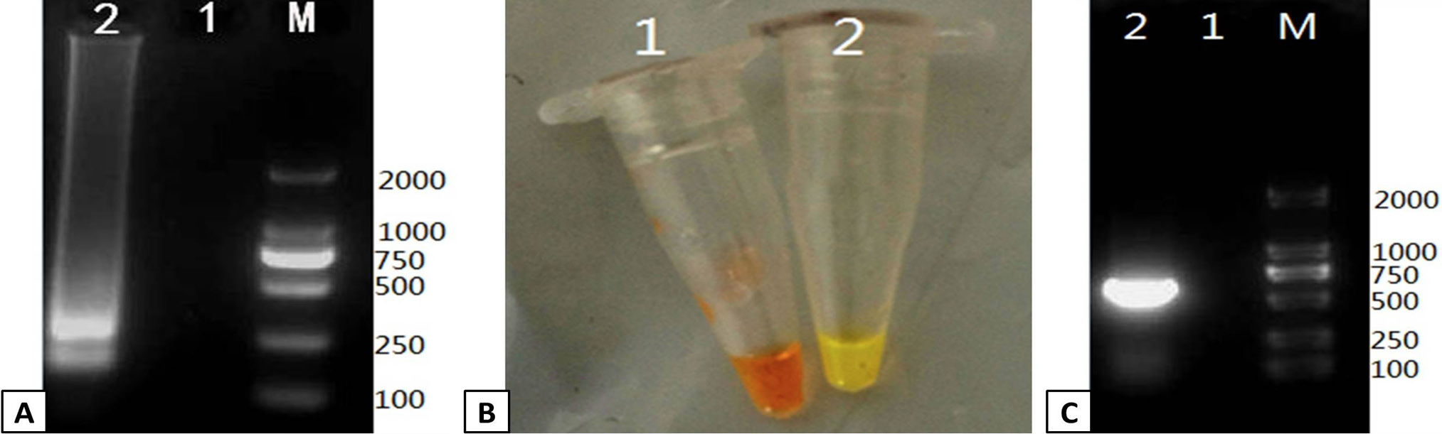
Verification of the PRRSV RT-LAMP assay. RT-LAMP was used to amplify a positive control template and compared to a negative control. The output of the RT-LAMP assay was monitored by gel electrophoresis and color change. (A) 1% agarose gel electrophoresis of the RT-LAMP products. Lanes: M; DL2000 Marker, 1; negative control, 2; positive control. (B) Color change in the RT-LAMP reaction was monitored followig addition of SYBR Green I dye. Tube 1: negative control, Tube 2: positive control. (C) The positive and negative controls were verified by RT-PCR 1% agarose gel electrophoresis of the RT-PCR products. Lanes: M; DL2000 Marker, 1; negative control, 2; positive control.
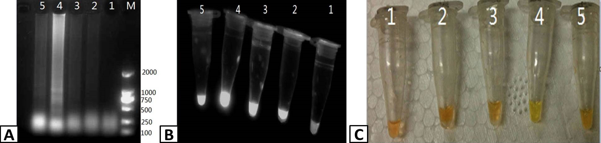
Optimizing the duration of the RT-LAMP reaction. RT-LAMP was used to amplify PRRSV containing samples. The products of the RT-LAMP reactions were visualized by (A) 1% agarose gel electrophoresis, (B) UV transillumination, and (C) color change using SYBR Green I dye. Lanes M; DL2000 Marker (A only), 1-5; 45, 50. 55, 60, and 65 min, respectively.
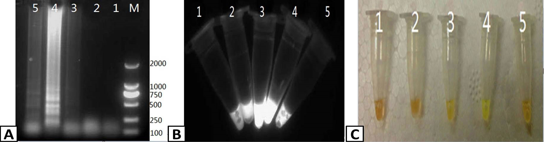
Optimizing the temperature of the RT-LAMP reaction. RT-LAMP was used to amplify PRRSV containing samples. The products of the RT-LAMP reactions were visualized by (A) 1% agarose gel electrophoresis. (B) UV transillumination, and (C) color change using SYBR Green I dye. Lanes: M; DL2000 Marker (A only), 1-5; 59, 61, 63, 65, and 67 C, respectively.
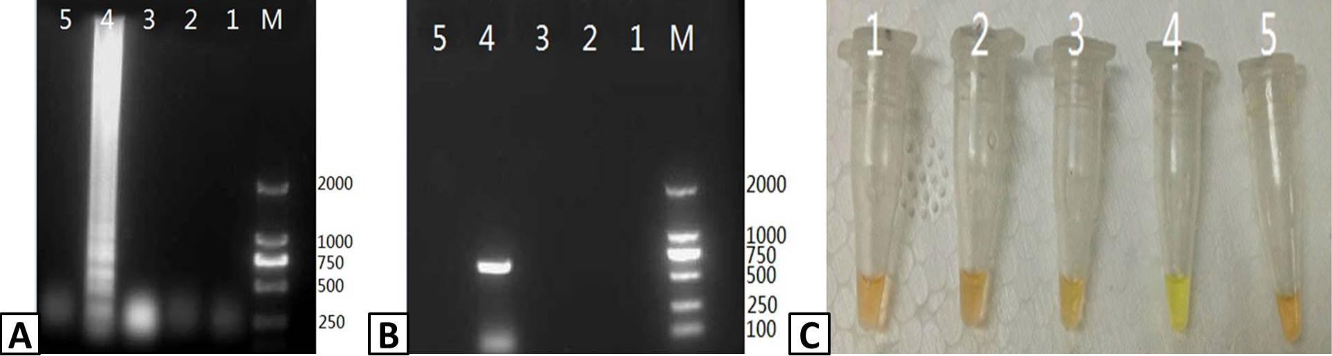
Assessing the specificity of RT-LAMP for PRRSV. Samples were isolated from pigs infected with PRRSV, CSFV, PCV-2, PPV, and PRV and tested usig the RT-LAMP assay. The output of the assay was visualized using (A) agarose gel electrophoresis and (C) color change with SYBR Green I dye. (B) The presence of PRRSV in the samples was confirmed by RT-PCR and visualized using agarose gel electrophoresis. Lanes: M; DL2000 Marker (A and B only). 1; CSFV, 2; PCV-2, 3; PPV, 4; PRRSV, 5; PRV.
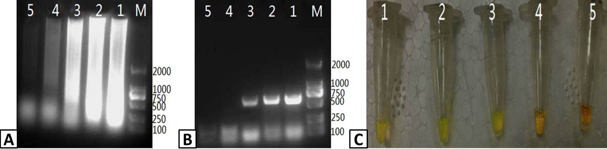
Assessing the sensitivity of the PRRSV RT-LAMP assay. 10-fold serial dilutions of a stock PRRSV RNA template (10ng/μL) were used to assess the sensitivity of the RT-LAMP assay compared to RT-PCR. Output from the RT-LAMP assay was visualized by (A) agarose gel electrophoresis and (C) color change with SYBR Green I dye. (B) RT-PCR amplification of PRRSV RNA was visualized by agarose gel electrophoresis. Lanes: M; DL2000 Marker (A and B only); 1-5: 10-1, 10-2, 10-3, 10-4, and 10-5 dilutions of the stocktemplate.
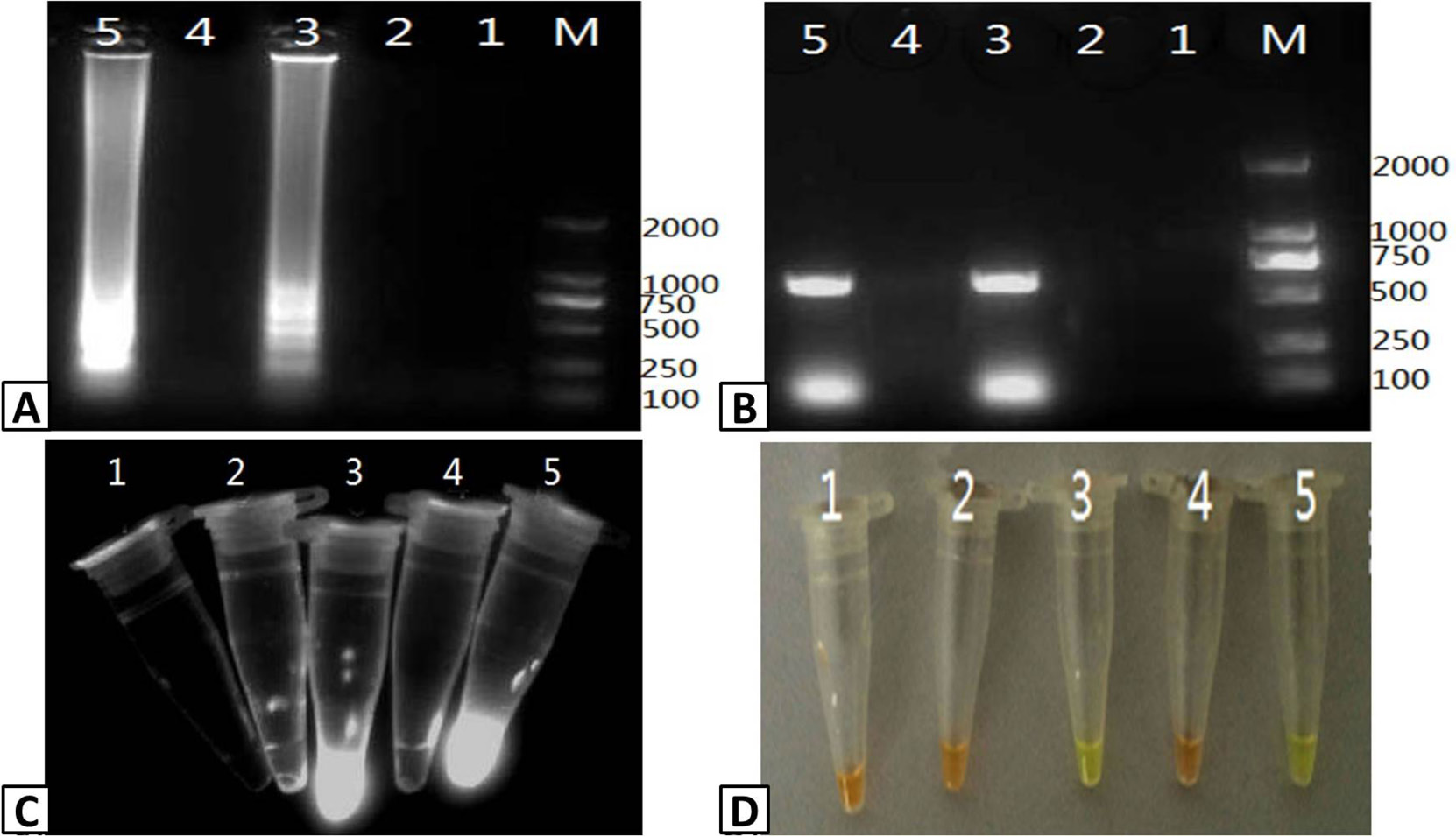
Detection of PRRSV infection in serum samples. Five (5) serum samples were tested for PRRSV infection using RT-LAMP and RT-PCR. The output of the RT-LAMP assay was visualized using (A) agarose gel electrophoresis, (C) UV transillumination, and (D) color change with SYBR Green I dye. (B) RT-PCR products were visualized using agarose gel electrophoresis of RT-PCR products. Lanes M; DL2000 Marker (A and B only); 1-5; serum samples.
References
- Akhter, H., Aslam, B., Shahzad, N., Farooq, T., Umer, M. and Rasool, M.H., 2017. Molecular and serological detection of avian influenza h9n2 virus in asymptomatic commercial layers in Faisalabad District, Punjab. Pakistan J. Zool., 49: 395-398. http://dx.doi.org/10.17582/journal.pjz/2017.49.1.sc6[CrossRef]
- Beura, L.K., Dinh, P.X., Osorio, F.A. and Pattnaik, A.K., 2011. Cellular poly(c) binding proteins 1 and 2 interact with porcine reproductive and respiratory syndrome virus nonstructural protein 1 beta and support viral replication. J. Virol., 85: 12939-12949. https://doi.org/10.1128/JVI.05177-11[CrossRef]
- Bilodeau, R., Dea, S., Sauvageau, R.A. and Martineau, G.P., 1991. Porcine reproductive and respiratory syndrome in Quebec. Vet. Rec., 129: 102-103. https://doi.org/10.1136/vr.129.5.102[CrossRef]
- Cavanagh, D., 1997. Nidovirales: A new order comprising Coronaviridae and Arteriviridae. Arch. Virol., 142: 629-633.
- Cho, J.G. and Dee, S.A., 2006. Porcine reproductive and respiratory syndrome virus. Theriogenology, 66: 655-662. https://doi.org/10.1016/j.theriogenology.2006.04.024[CrossRef]
- Fang, Y., Schneider, P., Zhang, W.P., Faaberg, K.S., Nelson, E.A. and Rowland, R.R., 2007. Diversity and evolution of a newly emerged North American Type 1 porcine arterivirus: Analysis of isolates collected between 1999 and 2004. Arch. Virol., 152: 1009-1017. https://doi.org/10.1007/s00705-007-0936-y[CrossRef]
- Fernandez, A., Suarez, P., Castro, J.M. and Tabares, E., 1999. Characterization of the domains implicated in apoptosis induction by the PRRSV GP5 protein. Proceedings of the International Symposium on PRRS and Aujeszky’s Disease, Ploufragan, France, June 21-24.
- Kwang, J., Zuckermann, F., Ross, G., Yang, S., Osorio, F., Liu, W. and Low, S., 1999. Antibody and cellular immune responses of swine following immunisation with plasmid DNA encoding the PRRS virus ORF’s 4, 5, 6 and 7. Res. Vet. Sci., 67: 199-201. https://doi.org/10.1053/rvsc.1998.0291[CrossRef]
- Li, Y., Wang, X., Bo, K., Wang, X., Tang, B., Yang, B., Jiang, W. and Jiang, P., 2007. Emergence of a highly pathogenic porcine reproductive and respiratory syndrome virus in the Mid-Eastern region of China. Vet. J., 174: 577-584. https://doi.org/10.1016/j.tvjl.2007.07.032[CrossRef]
- Liu, W.Q., Fan, Q.S., Jiang, Y., Xia, X.Z., Huang, G., Wang, J.G., Fang, J.B. and Wang, L., 2001. Establishment of a commonly used PCR technique for detection of carnivore parvoviruses. Chin. J. Vet. Sci., 21: 249-251.
- Malele, I.I., Ouma, J.O., Enyaru, J.C., Matovu, E., Alibu, V., Auma, J.E., Onyoyo, S.G., Bateta, R., Changasi, R.E., Mukiria, P.W., Ndung’u, K., Gitonga, P.K., Mwaniki, L.M., Nyingilili, H.S., Lyaruu, E.A., Kapange, L.A., Kamau, P.K. and Masiga, D.K., 2013. Comparative diagnostic and analytical performance of PCR and LAMP-based trypanosome detection methods estimated using pooled whole tsetse flies and midguts. Vet. Parasitol., 197: 549-556. https://doi.org/10.1016/j.vetpar.2013.05.022[CrossRef]
- Maruyama, F., Kenzaka, T., Yamaguchi, N., Tani, K. and Nasu, M., 2003. Detection of bacteria carrying the stx2 gene by in situ loop-mediated isothermal amplification. Appl. environ. Microbiol., 69: 5023-5028. https://doi.org/10.1128/AEM.69.8.5023-5028.2003[CrossRef]
- Meulenberg, J.J., Petersen den Besten, A., de Kluyver, E., van Nieuwstadt, A., Wensvoort, G. and Moormann, R.J., 1997. Molecular characterization of Lelystad virus. Vet. Microbiol., 55: 197-202. https://doi.org/10.1016/S0378-1135(96)01335-1[CrossRef]
- Njiru, Z.K., 2012. Loop-mediated isothermal amplification technology: Towards point of care diagnostics. PLoS Negl. Trop. Dis., 6: e1572. https://doi.org/10.1371/journal.pntd.0001572[CrossRef]
- Notomi, T., Okayama, H., Masubuchi, H., Yonekawa, T., Watanabe, K., Amino, N. and Hase, T., 2000. Loop-mediated isothermal amplification of DNA. Nucl. Acids Res., 28: E63. https://doi.org/10.1093/nar/28.12.e63[CrossRef]
- Pirzadeh, B. and Dea, S., 1998. Immune response in pigs vaccinated with plasmid DNA encoding ORF5 of porcine reproductive and respiratory syndrome virus. J. Gen. Virol., 79: 989-999. https://doi.org/10.1099/0022-1317-79-5-989[CrossRef]
- Snijder, E.J. and Meulenberg, J.J., 1998. The molecular biology of arteriviruses. J. Gen. Virol., 79: 961-979. https://doi.org/10.1099/0022-1317-79-5-961[CrossRef]
- Song, Q., Zhu, R., Sun, Y., Zhao, L., Wang, F., Deng, J. and Qian, Y., 2014. Identification of human metapneumovirus genotypes A and B from clinical specimens by reverse transcription loop-mediated isothermal amplification. J. Virol. Methods, 196: 133-138. https://doi.org/10.1016/j.jviromet.2013.10.037[CrossRef]
- Sur, J.H., Doster, A.R. and Osorio, F.A., 1998. Apoptosis induced in vivo during acute infection by porcine reproductive and respiratory syndrome virus. Vet. Pathol., 35: 506-514. https://doi.org/10.1177/030098589803500605[CrossRef]
- Tian, K., Yu, X., Zhao, T., Feng, Y., Cao, Z., Wang, C., Hu, Y., Chen, X., Hu, D., Tian, X., Liu, D., Zhang, S., Deng, X., Ding, Y., Yang, L., Zhang, Y., Xiao, H., Qiao, M., Wang, B., Hou, L., Wang, X., Yang, X., Kang, L., Sun, M., Jin, P., Wang, S., Kitamura, Y., Yan, J. and Gao, G.F., 2007. Emergence of fatal PRRSV variants: unparalleled outbreaks of atypical PRRS in China and molecular dissection of the unique hallmark. PLoS One, 2: e526. https://doi.org/10.1371/journal.pone.0000526[CrossRef]
- Van Nieuwstadt, A.P., Meulenberg, J.J., van Essen-Zanbergen, A., Petersen-den Besten, A., Bende, R.J., Moormann, R.J. and Wensvoort, G., 1996. Proteins encoded by open reading frames 3 and 4 of the genome of Lelystad virus (Arteriviridae) are structural proteins of the virion. J. Virol., 70: 4767-4772.
- Zhang, H.L., Yan, X.J., Chai, X.L. and Wu, W., 2007. Establishment and application of PCR for detection of mink enteritis virus. Spec. Wild Econ. Anim. Pl. Res., 34: 652-654.
- Zhang, X., Liao, M., Jiao, P., Luo, K., Zhang, H., Ren, T., Zhang, G., Xu, C., Xin, C. and Cao, W., 2010. Development of a loop-mediated isothermal amplification assay for rapid detection of subgroup J avian leukosis virus. J. clin. Microbiol., 48: 2116-2121. https://doi.org/10.1128/JCM.02530-09[CrossRef]
- Zhao, X., Li, Y., Wang, L., You, L., Xu, Z., Li, L., He, X., Liu, Y., Wang, J. and Yang, L., 2010. Development and application of a loop-mediated isothermal amplification method on rapid detection Escherichia coli O157 strains from food samples. Mol. Biol. Rep., 37: 2183-2188. https://doi.org/10.1007/s11033-009-9700-6[CrossRef]
Services
Article usage statistics
Loading…