Research Article · Volume 12, Issue 8, 25 June 2024 · Open Access
Toxicity Assessment Due to Chronic Exposure to Manganese in Male Wistar Rat
· · · · · ·
Affiliations & Notes ⌄
1Laboratory of Biology and Health, Department of Biology, Faculty of Science, University Ibn Tofail, Kenitra, Morocco. *Correspondence | Hala Harifi, Mouloud Lamtai, Laboratory of Biology and Health, Department of Biology, Faculty of Science, University Ibn Tofail, Kenitra, Morocco
Manganese (Mn) is present everywhere: in rocks, soil, water, food, and in various forms. Our bodies utilize it to contribute to energy metabolism and perform many other functions, but prolonged exposure can lead to toxicity in the organism. This study aimed to evaluate the effects of chronic exposure to Mn, specifically its impact on the general condition of male Wistar rats, by recording all clinical symptoms associated with this intoxication. Different doses ranging from the lowest to the highest (6 mg/kg, 25 mg/kg, 30 mg/kg, and 40 mg/kg i.p.) were administered intraperitoneally over a period of 12 weeks. The objective of the study was to identify the lethal dose and the tolerated toxic dose in order to establish a toxicity model. Weight gain was monitored weekly, and, at the end of the exposure period, the rats were euthanized and their organ weights (liver and kidneys) were recorded. Our results indicate signs of intoxication resulting from chronic exposure to Mn. Specifically, doses of 25 mg/kg and 30 mg/kg were found to be toxic and adversely affect the general condition of the rats. In contrast, the 6 mg/kg dose showed no adverse effects when compared to the 25 mg/kg and 30 mg/kg doses. Additionally, the 40 mg/kg dose proved to be lethal. Weight changes were observed in rats injected with the highest doses of Mn, along with alterations in organ weights. This study led us to conclude that chronic exposure to Mn induces dose-dependent toxic effects, as evidenced by the observed clinical signs of toxicity.
Keywords | Manganese, Chronic exposition, Toxicity, Clinical symptom, Dose-dependent, Rat
INTRODUCTION
Manganese (Mn) ranks as the 12th most abundant element and the 5th most abundant metal on Earth. It is widely distributed in the environment, and found in water, soil, and food sources. This metallic element is essential for cell survival ; , playing a crucial role in various physiological processes. Mn acts as a trace element vital for the normal functioning of numerous bodily processes, including the metabolism of sugars, amino acids, and lipids. Additionally, it plays pivotal roles in the nervous and immune systems, cellular energy regulation, the formation of bone and connective tissue, and the activation of specific enzymes . The primary pathway for Mn absorption is via the gastrointestinal tract. However, absorption can also occur through the lungs upon inhalation exposure ; . Prolonged exposure to Mn can be detrimental to health, particularly notable among mining and welding workers who experience chronic exposure to aerosols or dust containing Mn , as well as individuals with chronic oral exposure to contaminated water . Moreover, genetic disorders may impact or alter the distribution and function of Mn within the organism .
The primary target organ of Mn is the brain, as evidenced by previous studies indicating that exposure to Mn leads to its accumulation in both the brain and liver. Moreover, it has been observed that the longer the duration of exposure, the higher the concentration of Mn in these organs . Numerous studies have reported that exposure to high doses of Mn can lead to neurodegenerative diseases including Parkinson’s disease, Alzheimer’s disease, Huntington’s disease, and prion diseases . The neurotoxic impacts of this metal are similar to those provoked by other heavy metals ; ; , b). On the other hand, the liver serves as the primary organ responsible for regulating body Mn levels by facilitating endogenous Mn losses in the intestine . Liver cells express a variety of Mn transporters on their cell membranes , including Mn exporters, which regulate the efflux of excess Mn . Consequently, the liver plays a crucial role in storing, redistributing, and eliminating Mn from the body. However, accumulation of Mn in the liver can lead to liver damage by interfering with its excretory functions . In addition, Mn absorption in the kidney is regulated by several Mn transporters, including ZIP8, ZIP14, and DMT1, located in renal proximal tubule epithelial cells . It has also been reported that the kidneys were affected in rats exposed to Mn through oral ingestion . In a study, Mn-treated rats exhibited impaired kidney function, as evidenced by elevated serum urea and creatinine levels. This indicates that any increase in these levels in serum or plasma is a marker of kidney damage. The kidney tissue showed numerous atrophied and shrunken glomeruli, and the renal tubules were deformed with signs of cell apoptosis and necrosis . At high doses, this metal disrupts the overall health of the organism, manifesting visible signs of toxicity such as weight loss or slowed weight gain . Additionally, a study by Sárközi et al. (2009) reported similar effects of Mn on body weight . In vivo studies investigating Mn toxicity in rats have indicated that high doses of Mn can be lethal . Hence, determining the LD50 (dose at which 50% of the animals tested died) is crucial. This determination is typically the initial step in evaluating the toxic properties of a substance and is a critical aspect of any experiment.
This study aims to evaluate the dose-dependent effects of Mn on rats. Over 12 weeks, we will record the changes and alterations resulting from chronic Mn intoxication. Additionally, we aim to identify the lethal dose and establish a model dose that can be used to induce Mn toxicity.
MATERIALS AND METHODS
Conditioning and constitution of rat batches for manganese toxicity assessment
A total of 35 male Wistar rats from the breeding of the Faculty of Life Sciences at the University of Ibn Tofaïl were used in the current work. All rats were maintained under LD 12/12 (12 h light/12 h darkness) and at a standard temperature (21±1 °C), during which water and food were provided. The experimental procedures were carried out by the National Institutes of Health Guide for the Care and Use of Laboratory Animals and approved by the Animal Ethics Committee (Local Institutional Research Committee). All efforts were made to minimize the number of rats and their suffering.
The rats were divided into four groups of seven animals each (7 males). The different groups of rats are distributed as follows:
Control group: Rats were injected with physiological saline buffer (0.9% NaCl).
Mn-6 group: Injected with 6 mg/kg body weight .
Mn-25 group: Injected with 25 mg/kg body weight .
Mn-30 group: Injected with 30 mg/kg body weight.
Mn-40 group: Injected with 40 mg/kg body weight.
Mn doses were administered daily during the 3-month treatment period by intraperitoneal injection. The rats were weighed every week, and their weights were noted. At the end of the 12 weeks, the rats were sacrificed, and their livers and kidneys were collected and weighed in a precision balance. The relative organ weights were calculated using the formula: Relative Organ Weight = (Organ Weight (g)/ Body Weight (g)) × 100. This value is expressed as a percentage of the total body weight and allows for comparisons between treated and control groups, regardless of variations in overall body weight.
Manganese poisoning protocol
The Mn solution is prepared from a commercial form called manganese chloride tetrahydrate (MnCl24H2O). We selected four doses to determine the most toxic and lethal dose: 6 mg/kg and 25 mg/kg, as per the literature cited in the referenced articles, using a 0.9% NaCl solution.
The saline solution or MnCl24H2O (obtained from Sigma-Aldrich, St. Louis, MO, USA) used in this study was injected intraperitoneally and chronically, once daily for 12 weeks. The doses of 30 mg/kg and 40 mg/kg were tested for the first time in this study by our team.
Groups not treated with Mn (control) received intraperitoneal injections of 0.9% NaCl. The injection volume of Mn was 0.2 ml/100 g body weight each week. The control group was injected with an equal volume of physiological saline solution as the group treated with Mn.
Observation of symptomatic disorders
After treatment with Mn at different doses, the animals were returned to their cages, where they could access food and water again. They were observed immediately, and then daily for 12 weeks starting from day one, and thereafter once a day. Symptomatic disorders such as aggressiveness, lack of appetite, motor difficulties, changes in hair color, etc., were noted in animals injected with different doses. In addition, to assess the deterioration of olfaction, a piece of cheese was hidden in the cage, and the rat was allowed to search for it. The time it took for the rat to find the cheese was recorded and compared with the time taken by control rats.
Evaluation of toxicological parameters: Determination of DMT, LD100 and LD50
The number of deceased animals was recorded in each batch of this toxicity experiment, which aimed to determine the following toxicological parameters:
The 50% lethal dose (LD50), defined as a dose that kills 50% of the animals,
The 100% lethal dose (LD100), a dose that kills all animals
The maximum tolerated dose (MTD), representing the maximum dose that kills no animals when Mn is administered
In theory, the average lethal dose, or LD50, provides information on the amount of a substance required to cause adverse effects in the body. Moreover, there are various methods to assess effects, including physiological, biochemical, and behavioral measurements. However, toxicity remains one of the most extensively examined indicators , and one of the methods used to calculate the LD50 is the Behren-Karber method, which is a non-parametric approach. It involves observing equal spacing of dose intervals and an equal number of subjects at each dose from 0% to 100%.
It is calculated as follows:
LD50 = LD100 - Σ (a x b) / n
LD100: 100 % mortality dose
LD50: 50% mortality dose
n: Number of animals in each group
a: The difference between two consecutive doses
b: The arithmetic mean of the deaths from two consecutive doses.
Statistical analysis
IBM’s SPSS version 23 was used for all statistical analysis (IBM Corp., Armonk, NY, United States). Body weight and organ weight data were statistically analyzed using ANOVA (One-way). The values were expressed as mean ± SEM. A p < 0.05 was interpreted as a significant difference.
RESULTS and Discussion
Clinical signs and mortality
Clinical signs
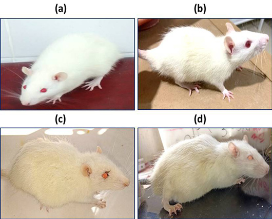
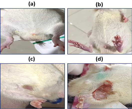
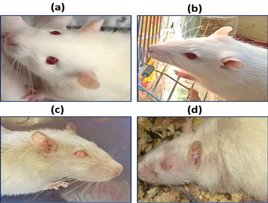
During the treatment period, rats exhibited symptoms following exposure to different doses of Mn. Rats treated with a single dose (25 mg/kg or 30 mg/kg) of Mn displayed signs of Mn intoxication as presented in Table 1. Additionally, rats treated with both doses showed changes in coat quality, characterized by yellowish hair, hair loss, and piloerection, as well as weight loss. Some rats in the 30 mg/kg group exhibited abdominal and testicular swelling, while abdominal lesions were observed in both groups. Asthenia, bradykinesia, weakness of muscle tone, deterioration of olfaction, and digestive tract diarrhea were also observed. Signs of toxicity in the 6 mg/kg group were minimal. Not all signs were observed in rats treated with the 40 mg/kg dose, as they succumbed within the first few weeks. These observations for the different groups are illustrated in Figures 1, 2, and 3.
Mortality
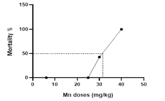
Following intraperitoneal injection of Mn at different doses, the mortality rate was recorded for each dose administered. An increase in animal mortality was observed with increasing doses, indicating a dose-dependent effect (Figure 4). The highest dose resulting in mortality for all animals was 40 mg/kg, while the MTD was determined to be 30 mg/kg. The LD50 of the metal was calculated to be approximately 32 mg/kg, as detailed in Table 2.
Table 1: The observed symptoms of toxicity associated with different doses treated by manganese and the melatonin effect in Rat at 1, 2, 3, 4, 5, 6,7, 8, 9, 10, 11, and 12 weeks after exposure, respectively.
Observations
Control
6 mg/kg Mn
25 mg/kg Mn
30 mg/kg Mn
40 mg/kg Mn
Weight fluctuation
____
____
+
+++
+++
Depression/anxiety
____
+
++
+++
+++
Hair color
White
White
Yellowish
Yellowish
Yellowish
Olfactory impairment (Hyposmia)
____
____
++
+++
+++
Hair loss
____
____
+
++
+++
Piloerection
____
____
+
+++
+++
anorexia
____
____
+
+++
+++
Diarrhea
____
+
+
++
+++
Aggressiveness
____
____
+
++
____
Normal gait
+++
+++
++
+
+
Abdominal ulcers
____
____
+
+++
+++
Testicular swelling
____
____
____
+
____
Abdominal swelling
____
____
____
+
____
Asthenia
____
____
++
+++
++
Stiffness
____
____
+++
+++
+++
Slow movement
____
+++
++
+++
+++
Sens of exploration
+++
+++
++
____
____
Eye color
Red
Red
Light red
Light red
Light red
Mortality
____
____
____
++
+++
Hunched back
____
____
+
+++
+++
Table 2: Determination of the LD50 according to arithmetic and Behrens and Kerber methods.
Mn doses (mg/kg)
Number of rats
Number of dead rats
Mortality %
a
b
a*b
6
7
0
0
19
0
0
25
7
0
0
5
1.5
7.5
30
7
3
42.85
10
5
50
40
7
7
100
Effect on body weight gain and organ weight of rats
Effect of Mn on body weight gain
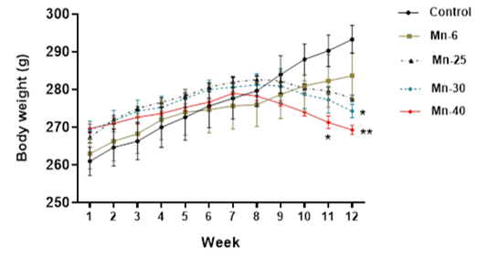
Figure 5 shows the evolution of the average body weight of rats throughout the Mn treatment period. Analysis ofdaily body weight measurements showed a slow variation in body weight compared with the other groups, with a significant decrease in the 30 and 40 mg/kg dose groups during the last weeks of treatment (from week 11 onwards).
In addition, compared with the control group, rats treated with 25 mg/kg showed slow weight growth and a decrease in body weight from week 9, and rats treated with 6 mg/kg showed almost similar weight growth to the controls.
Effect of Mn on the organ weight of rats
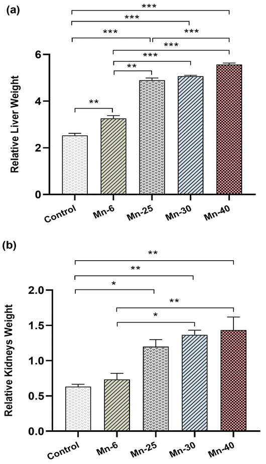
Statistical analysis has revealed that Mn-6, Mn-25, Mn-30, and Mn-40 significantly increased the relative liver weights in a dose-dependent fashion when compared to the control group (p < 0.01, p < 0.001, p < 0.001, and p < 0.001, respectively). In addition, the groups treated with Mn at the doses of 25, 30, and 40 mg/kg had increased relative kidney weights (in a dose-dependent manner) in comparison to the control group (p < 0.01), while Mn-0.6 non-significantly increased the relative kidneys weight (p > 0.05) (Figure 6).
Mn is essential for both humans and animals when consumed within the limits of their daily dietary requirements. However, exposure to high doses can be detrimental, and contamination of air and water sources poses a risk of Mn poisoning to the general population. Despite its necessity for various physiological functions, excessive accumulation of Mn in the human body can result in severe toxicity, impacting the entire organism. Our study aims to investigate the toxic effects of Mn at varying doses on the rat organism by documenting visible symptoms and monitoring weight fluctuations relative to the injected dose. Additionally, we aim to calculate the lethal dose and establish the appropriate dose for an animal model of Mn poisoning.
In our study, the intraperitoneal injection of Mn at various doses into rats and their observation for 12 weeks revealed differing effects depending on the injected dose. Chronic exposure to Mn resulted in notable consequences, manifested as visible alterations in the general condition of the rats. These alterations were characterized by the emergence of signs of intoxication stemming from toxic effects. It was observed that in rats treated with doses of 25 mg/kg and 30 mg/kg, symptoms of toxicity occurred at these levels and were characterized by external signs that increased in severity with the dose injected. These signs included yellowing and loss of hair, as well as bristling. Research suggests that environmental stress can alter skin structure, predisposing to stress-related skin diseases . In addition, it has been noted that hair growth can slow due to growth inhibition . Chronic stress related to strain modifies the active hair cycle . We also observed the appearance of ascites in some rats at the doses of 25 mg/kg and 30 mg/kg, which is in line with findings reported in another study . Additionally, abdominal lesions and diarrhea were noted, along with paleness of the mucous membranes of the eyes and pads of the extremities in the high-dose Mn group. Other symptoms, such as reduced exploratory behavior, increased fatigue, and decreased activity, were also observed. In this sense, research reporting the observation of some of these symptoms in rats after exposure to Mn includes the study conducted by O’Neal et al., which found that Mn treatment reduced exploratory behavior . The study also noted that with longer durations of exposure, rats exhibited reduced activity, hair loss, diarrhea, and other symptoms. Another study reported that muscle and joint weakness, hunched posture, and kinetic tremors were observed in Mn-intoxicated rats , which is consistent with our observations in rats treated with doses of 25 mg/kg and 30 mg/kg. Moreover, our results align with the study conducted by El-Fari et al. (2019) which affirmed that Mn intoxication decreases spontaneous locomotor behavior and muscle strength . In addition, in our study, the Mn dose of 6 mg/kg showed low toxicity compared to Mn doses of 25 mg/kg, 30 mg/kg, and 40 mg/kg, which exhibited signs of toxicity. The significant gap between the doses is chosen to establish a dose-response relationship, grounded in scientific rationale, supported by previous research, and aligned with regulatory requirements. The Estimated Safe and Adequate Daily Dietary Intake (ESADDI) for adults for manganese is 5 mg Mn/day. The LOAEL (lowest observable adverse effect level) for manganese is 4.2 mg Mn/day for a 70-kg individual .
Observing the mortality rate is crucial in toxicity studies . In addition to detecting abnormalities and behavioral signs caused by Mn, throughout the 90-day observation period, rats in this study exhibited mortality. The finding of our study showed that intraperitoneal administration of Mn induces toxicity in rats, with a dose-response effect evident as a dose of 40 mg/kg resulted in the mortality of all rats. In a study by Tian et al. (2018) the dose lethal to all rats was reported as 50 mg/kg . However, the toxicological parameters in our study were as follows: The MTD was 25 mg/kg (ip), and the LD50 of the rats was estimated to be 32 mg/kg ip. The LD50 was calculated using the Behren-Karber method. Based on the results of our study and their alignment with previous findings, we can conclude that 25 mg/kg serves as a suitable preliminary dose for establishing a model of Mn poisoning.
Body and organ weights were used to assess the general state of health and toxic effects on the rat organism following chronic exposure to Mn. The results showed a significant reduction in weight gain during the last weeks of exposure in the groups exposed to 25 mg/kg and 30 mg/kg Mn. Our results are consistent with those of ; , who reported a decrease in body weight in adult rats following chronic exposure to Mn ; . This decrease in body weight gain may result from a generalized disruption of normal physiological processes, such as a deficiency in energy metabolism and functional alterations in the hypothalamic nuclei that contribute to body weight control ; . Additionally, a study indicates that the reduction in body weight gain could be attributed to a lack of food intake caused by loss of appetite or gastrointestinal disorders provoked by this metal .
The relative organ weight in toxicology studies is also an important indicator of the harmful effects of the tested compound ; . Importantly, an increase in kidney and liver volume was observed in rats treated with Mn in a dose-dependent manner compared to the control group. Our results are consistent with a study reporting that Mn administration caused a significant increase in relative liver weight in treated rats ; another study also observed this increase . As known, the liver is the primary organ responsible for storing, redistributing, and eliminating Mn to maintain Mn homeostasis in the body. Consequently, exposure to excessive levels of Mn can lead to severe liver damage, resulting in various chronic liver diseases . Other works suggest that this increase in liver weight could be attributed to high-fat accumulation in hepatocytes. Additionally, the mechanisms by which Mn exposure affects kidney weight may involve oxidative stress, inflammation, and disruption of normal cellular functions. Mn-induced oxidative stress can damage renal tissue, leading to compensatory hypertrophy, where the kidney tissue increases in size in response to injury or stress . These alterations in the kidneys and liver may explain the changes in weight and volume observed in our study.
CONCLUSIONS AND RECOMMENDATIONS
This study revealed the toxic effects on the general health of rats due to prolonged exposure to high doses of Mn, including effects on weight, liver, and kidneys. In addition, it enabled us to select an assay for a toxicity model to be explored in our future studies, focusing specifically on the neurotoxic effects of Mn on neuroaffective, locomotor, and olfactory functions. These results underline the importance of continuing research into the mechanisms underlying Mn-induced toxicity. Continued analysis and research are crucial for gaining a comprehensive understanding of these symptoms and for the development of potential treatments.
ACKNOWLEDGMENTS
We extend our gratitude to the staff of the Biology Department at the Faculty of Science, Ibn Tofail University, for their invaluable technical support.
NOVELTY STATEMENT
This study aims to present a clear representation of the clinical symptoms resulting from chronic exposure to Mn and to determine the lethal dose, thus enabling researchers interested in studying Mn to visualize its effects at different doses in rats.
AUTHOR’S CONTRIBUTION
All authors contributed equally to the manuscript.
Data availability
The data sets analyzed in this study are available from the corresponding author on reasonable request.
Conflict of interest
The authors have declared no conflict of interest.
Figures
Figure 1:.
Hunched back with flattened limbs in control group (a), rat treated with 6 mg/kg (b), rat treated with 25 mg/kg (c), rat treated with 30 mg/kg (d).
Figure 2:.
Abdominal lesions in control group (a), rat treated with 6 mg/kg (b), rat treated with 25 mg/kg (c), rat treated with 30 mg/kg (d).
Figure 3:.
Hair color, piloerection, hair loss, and eyes color and paleness of the mucous membranes of the eyes in control group (a), rat treated with 6 mg/kg (b), rat treated with 25 mg/kg (c), and rat treated with 30 mg/kg (d).
Figure 4:.
Dose-lethality curve after intraperitoneal injection of Mn at different doses.
Figure 5:.
Mean body weight (Mean ± S.E.M) of male rats treated with Mn repeated intraperitoneal doses (6, 25, 30, and 40 mg/kg) for 12 weeks. Significantly different from vehicle control: *p < 0.05, **p < 0.01, respectively.
Figure 6:.
Relative weights of liver and kidneys (Mean ± SEM) of male rats treated with Mn repeated intraperitoneal doses (6, 25, 30, and 40 mg/kg) for 12 weeks. Significantly different from vehicle control: *p < 0.05.
References
Alexopoulos A, Chrousos GP (2016). Stress-related skin disorders. Rev. Endocr. Metab. Disord., 17: 295–304. https://doi.org/10.1007/s11154-016-9367-y[CrossRef]
Baj J, Flieger W, Barbachowska A, Kowalska B, Flieger M, Forma A, Teresiński G, Portincasa P, Buszewicz G, Radzikowska-Büchner E, Flieger J (2023). Consequences of disturbing manganese homeostasis. Int. J. Mol. Sci., 24: 14959. https://doi.org/10.3390/ijms241914959[CrossRef]
Bouabid S, Delaville C, De Deurwaerdère P, Lakhdar-Ghazal N, Benazzouz A (2014). Manganese-induced atypical parkinsonism is associated with altered Basal Ganglia activity and changes in tissue levels of monoamines in the rat. PLoS One, 9: e98952. https://doi.org/10.1371/journal.pone.0098952[CrossRef]
Bowler RM, Gysens S, Diamond E, Nakagawa S, Drezgic M, Roels HA (2006). Manganese exposure: neuropsychological and neurological symptoms and effects in welders. Neurotoxicology, 27: 315–326. https://doi.org/10.1016/j.neuro.2005.10.007[CrossRef]
Bowman AB, Kwakye GF, Herrero Hernández E, Aschner M (2011). Role of manganese in neurodegenerative diseases. J. Trace. Elem. Med. Biol., 25: 191–203. https://doi.org/10.1016/j.jtemb.2011.08.144[CrossRef]
Davis CD, Zech L, Greger JL (1993). Manganese metabolism in rats: An improved methodology for assessing gut endogenous losses. Exp. Biol. Med., 202: 103–108. https://doi.org/10.3181/00379727-202-43518[CrossRef]
El-Brouzi MY, Lamtai M, Zghari O, Ouakki S, Azizoun I, El Hessni A, Mesfioui A, Ouichou A (2021). Intrahippocampal effects of nickel injection on the affective and cognitive response in Wistar rat: Potential role of oxidative stress. Biol. Trace. Elem. Res., 199: 3382–3392. https://doi.org/10.1007/s12011-020-02457-5[CrossRef]
El-Fari R, Abbaoui A, Bourziq A, Zroudi M, Draoui A, El Khiat A, Belkouch M, Elgot A, Gamrani H (2019). Neuroprotective effects of docosahexaenoic acid against sub-acute manganese intoxication induced dopaminergic and motor disorders in mice. J. Chem. Neuroanat., 102: 101686. https://doi.org/10.1016/j.jchemneu.2019.101686[CrossRef]
Finkelstein Y, Milatovic D, Aschner M (2007). Modulation of cholinergic systems by manganese. Neurotoxicology, 28: 1003–1014. https://doi.org/10.1016/j.neuro.2007.08.006[CrossRef]
Fujishiro H, Yano Y, Takada Y, Tanihara M, Himeno S (2012). Roles of ZIP8, ZIP14, and DMT1 in transport of cadmium and manganese in mouse kidney proximal tubule cells. Metallomics, 4: 700–708. https://doi.org/10.1039/c2mt20024d[CrossRef]
Gandhi D, Rudrashetti AP, Rajasekaran S (2022). The impact of environmental and occupational exposures of manganese on pulmonary, hepatic, and renal functions. J. Appl. Toxicol., 42: 103–129. https://doi.org/10.1002/jat.4214[CrossRef]
Gillet B, Po C, Dupont D, Sébrié C, Méric P (2010). Manganèse et imagerie de résonance magnétique du développement cérébral. Compt. Rend. Chim., 13: 459–465. https://doi.org/10.1016/j.crci.2009.09.013[CrossRef]
Gómez-Ariza JL, Sánchez-Rodas D, Giráldez I, Morales E (2000). A comparison between ICP-MS and AFS detection for arsenic speciation in environmental samples. Talanta, 51: 257–268. https://doi.org/10.1016/S0039-9140(99)00257-X[CrossRef]
Greger JL (1998). Dietary Standards for manganese: Overlap between nutritional and toxicological studies. J. Nutr., 128: 368S-371S. https://doi.org/10.1093/jn/128.2.368S[CrossRef]
Harischandra DS, Ghaisas S, Zenitsky G, Jin H, Kanthasamy A, Anantharam V, Kanthasamy AG (2019). Manganese-induced neurotoxicity: New insights into the triad of protein misfolding, mitochondrial impairment, and neuroinflammation. Front. Neurosci., 13: 654. https://doi.org/10.3389/fnins.2019.00654[CrossRef]
Haynes EN, Sucharew H, Kuhnell P, Alden J, Barnas M, Wright RO, Parsons PJ, Aldous KM, Praamsma ML, Beidler C, Dietrich KN (2015). Manganese exposure and neurocognitive outcomes in rural school-age children: The communities actively researching exposure study (Ohio, USA). Environ. Health Perspect. 123: 1066–1071. https://doi.org/10.1289/ehp.1408993[CrossRef]
Huang P, Chen C, Wang H, Li G, Jing H, Han Y, Liu N, Xiao Y, Yu Q, Liu Y, Wang P, Shi Z, Sun Z (2011). Manganese effects in the liver following subacute or subchronic manganese chloride exposure in rats. Ecotoxicol. Environ. Saf., 74: 615–622. https://doi.org/10.1016/j.ecoenv.2010.08.011[CrossRef]
Ismail HTH (2019). Hematobiochemical disturbances and oxidative stress after subacute manganese chloride exposure and potential protective effects of Ebselen in rats. Biol. Trace Elem. Res., 187: 452–463. https://doi.org/10.1007/s12011-018-1395-x[CrossRef]
Lao Y, Dion L-A, Gilbert G, Bouchard MF, Rocha G, Wang Y, Leporé N, Saint-Amour D (2017). Mapping the basal ganglia alterations in children chronically exposed to manganese. Sci. Rep., 7: 41804. https://doi.org/10.1038/srep41804[CrossRef]
Lazic SE, Semenova E, Williams DP (2020). Determining organ weight toxicity with Bayesian causal models: Improving on the analysis of relative organ weights. Sci. Rep., 10: 6625. https://doi.org/10.1038/s41598-020-63465-y[CrossRef]
Liu X, Chen C, Zhao Y, Jia B (2013). A review on the synthesis of manganese oxide nanomaterials and their applications on lithium-ion batteries. J. Nanomater., 2013: 1–7. https://doi.org/10.1155/2013/736375[CrossRef]
Lucchini R, Tieu K (2023). Manganese-induced parkinsonism: Evidence from epidemiological and experimental studies. Biomolecules, 13: 1190. https://doi.org/10.3390/biom13081190[CrossRef]
Milatovic D, Zaja-Milatovic S, Gupta RC, Yu Y, Aschner M (2009). Oxidative damage and neurodegeneration in manganese-induced neurotoxicity. Toxicol. Appl. Pharmacol., 240: 219–225. https://doi.org/10.1016/j.taap.2009.07.004[CrossRef]
Misselwitz B, Mühler A, Weinmann HJ (1995). A toxicologic risk for using manganese complexes? A literature survey of existing data through several medical specialties. Invest. Radiol., 30: 611–620. https://doi.org/10.1097/00004424-199510000-00007[CrossRef]
Mostafa HE-S, Alaa El-Din EA, El-Shafei DA, Abouhashem NS, Abouhashem AA (2021). Protective roles of thymoquinone and vildagliptin in manganese-induced nephrotoxicity in adult albino rats. Environ. Sci. Pollut. Res. Int., 28: 31174–31184. https://doi.org/10.1007/s11356-021-12997-5[CrossRef]
Naïla N, Makthar W, Lamtai M, Zghari O, Aboubaker EH, Abdelhalem M, Ali O (2021). Effect of intra-hippocampal lead injection on affective and cognitive disorders in male Wistar rats: Possible involvement of oxidative stress. E3S Web Conf., 319: 02017. https://doi.org/10.1051/e3sconf/202131902017[CrossRef]
Nam H, Wang C-Y, Zhang L, Zhang W, Hojyo S, Fukada T, Knutson MD (2013). ZIP14 and DMT1 in the liver, pancreas, and heart are differentially regulated by iron deficiency and overload: Implications for tissue iron uptake in iron-related disorders. Haematologica, 98: 1049–1057. https://doi.org/10.3324/haematol.2012.072314[CrossRef]
Niknahad AM, Ommati MM, Farshad O, Moezi L, Heidari R (2020). Manganese-induced nephrotoxicity is mediated through oxidative stress and mitochondrial impairment. J. Renal Hepat. Disorders, 4: 1–10. https://doi.org/10.15586/jrenhep.2020.66[CrossRef]
O’Neal SL, Hong L, Fu S, Jiang W, Jones A, Nie LH, Zheng W (2014). Manganese accumulation in bone following chronic exposure in rats: Steady-state concentration and half-life in bone. Toxicol. Lett., 229: 93–100. https://doi.org/10.1016/j.toxlet.2014.06.019[CrossRef]
O’Neal SL, Zheng W (2015). Manganese toxicity upon overexposure: A decade in review. Curr. Environ. Health Rep., 2: 315–328. https://doi.org/10.1007/s40572-015-0056-x[CrossRef]
Peters EMJ, Arck PC, Paus R (2006). Hair growth inhibition by psychoemotional stress: A mouse model for neural mechanisms in hair growth control. Exp. Dermatol., 15: 1–13. https://doi.org/10.1111/j.0906-6705.2005.00372.x[CrossRef]
Ponnapakkam T, Iszard M, Henry-Sam G (2003). Effects of oral administration of manganese on the kidneys and urinary bladder of Sprague-Dawley rats. Int. J. Toxicol., 22: 227–232. https://doi.org/10.1080/10915810305103[CrossRef]
Quadri M, Federico A, Zhao T, Breedveld GJ, Battisti C, Delnooz C, Severijnen L-A, Di Toro Mammarella L, Mignarri A, Monti L, Sanna A, Lu P, Punzo F, Cossu G, Willemsen R, Rasi F, Oostra BA, van de Warrenburg BP, Bonifati V (2012). Mutations in SLC30A10 cause parkinsonism and dystonia with hypermanganesemia, polycythemia, and chronic liver disease. Am. J. Hum. Genet., 90: 467–477. https://doi.org/10.1016/j.ajhg.2012.01.017[CrossRef]
Rhaimi S, Brikat S, Lamtai M, Ouhssine M (2023). Acute oral toxicity and neurobehavioral effects of salvia officinalis essential oil in female Wistar rats. Adv. Anim. Vet. Sci., 11:654-662. https://doi.org/10.17582/journal.aavs/2023/11.4.654.662[Full text][CrossRef]
Santamaria AB (2008). Manganese exposure, essentiality and toxicity. Indian J. Med. Res., 128: 484–500
Sárközi L, Horváth E, Kónya Z, Kiricsi I, Szalay B, Vezér T, Papp A (2009). Subacute intratracheal exposure of rats to manganese nanoparticles: behavioral, electrophysiological, and general toxicological effects. Inhal Toxicol., 21 (Suppl 1): 83–91. https://doi.org/10.1080/08958370902939406[CrossRef]
Teo S, Stirling D, Thomas S, Hoberman A, Kiorpes A, Khetani V (2002). A 90-day oral gavage toxicity study of D-methylphenidate and D, L-methylphenidate in Sprague-Dawley rats. Toxicology, 179: 183–196. https://doi.org/10.1016/S0300-483X(02)00338-4[CrossRef]
Tian Y, Chen C, Guo S, Zhao L, Yan Y (2018). Exploration of the establishment of manganese poisoning rat model and analysis of discriminant methods. Toxicology, 410: 193–198. https://doi.org/10.1016/j.tox.2018.08.006[CrossRef]
Zghari O, Azirar S, Lamtai M, El Hessni A, Ouichou A, Mesfioui A (2023a). Intrahippocampal dose-dependent effects of aluminum injection on affective and cognitive response in male Wistar rat: Potential role of oxidative stress. Egypt. J. Basic Appl. Sci., 10: 460–475. https://doi.org/10.1080/2314808X.2023.2229623[CrossRef]
Zghari O, Lamtai M, Azirar S, El-Brouzi MY, Benmhammed H, El-Hessni A, Ouichou A, Mesfioui A (2023b). Neuroprotective effects of melatonin against neurotoxicity induced by intrahippocampal injection of aluminum in male Wistar rats: Possible involvement of oxidative stress pathway. Adv. Anim. Vet. Sci., 11: 711-719. https://doi.org/10.17582/journal.aavs/2023/11.5.711.719[Full text][CrossRef]