
Trichomonas gallinae is a protozoan parasite that causes the avian trichomonias is disease known as “canker,” which primarily affects the esophagus and upper digestive tract in pigeons.A common case of pigeon cankers was diagnosed in two pigeons 40 days of age who came to S.A. Quaderi Teaching Veterinary Hospital, Chattogram Veterinary and Animal Sciences University (CVASU).Clinical history indicated that the birds were taking both food and water from the same source, clinical investigations showed greenish diarrhea, and post-mortem studies revealed hepatomegaly, congested heart, and proventriculus, and found nodules in the crop wall as tentative diagnoses. Tropozoid was identified through microscopic examination by direct wet smear and giemsa staining. The cankers were treated orally with metronidazole at a dose of 20 mg/kg body weight, and the farmer was also advised on the hygienic management of his farm. After careful treatment, all the clinically affected pigeons get cured within three days.
Case Report · Volume 11, Issue 2, 16 October 2023 · Open Access
The Confirmatory Diagnosis and Therapeutic Management of Canker in Pigeons
· · · ·
Affiliations & Notes ⌄
- 1Faculty of Veterinary Medicine, Chattogram Veterinary and Animal Sciences University, Khulshi, Chattogram4225, Bangladesh
- 2Department of Pathology and Parasitology, Chattogram Veterinary and Animal Sciences University, Khulshi, Chattogram4225, Bangladesh
- 3Department of Medicine and Surgery,Chattogram Veterinary and Animal Sciences University, Khulshi, Chattogram4225, Bangladesh
- 4Department of Physiology, Biochemistry and Pharmacology, Chattogram Veterinary and Animal Sciences University, Khulshi, Chattogram4225, Bangladesh. *Correspondence | Md. Shohel Al Faruk, Department of Physiology, Biochemistry and Pharmacology, Chattogram Veterinary and Animal Sciences University, Khulshi, Chattogram-4225, Bangladesh
- 5Email: shoheldvm03@gmail.com
Article Info ⌄
- Journal:
- Research Journal for Veterinary Practitioners
- Published:
- 16 October 2023
From the article
- This case report narrates the diagnosis and therapeutic management of cankers in pigeons.
- This case was diagnosed mainly based on clinical history, and physical and parasitological examination.
- The affected pigeons were observed regularly after the administration of metronidazole from day 1st to the 3rd.
- Pigeons were fed separately and ensured the freshwater source.
Selected from the article's own text.
Abstract
Introduction
The poultry sector is the most efficient and cost-effective way to obtain animal protein in the shortest period, but due to rising future demand, it is still unable to meet the animal protein supply and demand gap. Although commercial broiler and layer farms meet the current demand for protein, consumers consider chicken meat to be repetitive and desire alternative safe meats. So many people start pigeon farming to meet consumer demand and develop economically . About 10.8 million pigeons resided in Bangladesh, and 11% of them were reared on commercial farms. (According to Agricultural Sample Survey 2013). However, these farms went unidentified. Domestic pigeons have traditionally been reared in Bangladesh for their meat. However, this pigeon has viral (25.5%), bacterial (29.28%), parasitic (31.67%), and fungal (5.18%) diseases . In parasitic disease, Ecto-parasitic (67%) and endo-parasitic (72%) infections are prevalent in the Chittagong district of Bangladesh . Pigeons are susceptible to a variety of health problems, with ecto and endo-parasites with a significant role (Tietz et al., 2007). The endo-parasitic diseases are caused by cestode, nematode, trematode, and protozoal. In protozoa, one important disease is Pigeon cankers caused by Trichomonasgallinae which is the reason for the big economic loss of farmers .The age of the host also enhanced the prevalence of infection . Overall 60% of the pigeon population in Bangladesh had cankers, of which 75%, 50%, and 44.4% were squabs, adult males, and adult females, respectively . Avaintrichomoniasis is caused by a flagellated protozoan named T.gallinae that affects the upper digestive system of numerous bird species, especially pigeons . It is a flagellate protozoan without a posterior flagellum but has four anterior flagella and an undulating membrane on one side. There is no intermediate host,and the life cycle is simple and direct . The domestic pigeon, Columba livia, is the main host of this parasite .Additionally susceptible are domestic and wild turkeys, hens, and raptors (such as hawks and golden eagles) . It typically inhabits various bird species upper gastrointestinal tract, particularly Columbiformes (pigeons and doves) . Invasion of the oropharyngeal mucosa by the organisms results in inflammation and ulceration . Early lesions in pigeon squabs appear as small, white to yellowish spots in the oral cavity, particularly the soft palate, 3 to 14 days after infection . Which results in significant losses and high mortality, particularly in young birds. If the infection is severe and possibly fatal in young birds, where mortality can be as high as 80–90% or more in squabs . Through pigeon milk, adults transmit T.gallinae through squabs , and the infection takes place in a matter of minutes andis also transmitted by direct contact with an infected bird or indirectly via contaminated food or water ; . In this case, the transmission was caused by contaminated drinking water. All the pigeons drink water from a common source and take feed in the same place. When it was examined under a light microscope, trichomonas were translucent tiny flagellates that could appear alone or in clusters. They moved quickly (jerkily and in circles without a distinct direction, not departing the microscope field).The anti-protozoal drug was suggested for the prevention of this disease. The most used drug for treating cankers in pigeons is metronidazole , . So far there is a limited study on pigeon cankers in Bangladesh, and believe that this is the first case report on pigeon cankers from Chittagong in Bangladesh.The main goal of this study is laboratory diagnosis and therapeutic management of the cankers.
Material and Methods
Case history
At the age of 40 days, two domestic pigeons one alive and the other dead brought to the S.A. Quaderi Teaching Veterinary Hospital at Chattogram Veterinary and Animal Sciences University (CVASU), Chattogram, Bangladesh, on 9th November 2022, with a history of about 20 pigeons 1-2 months of age started to die suddenly and described that the affected pigeons showed diarrhea, loss of appetite, gradual weight loss and only drink of water. Other clinical examination findings were greenish diarrhea (Figure 1), weakness, depression, ruffled feathers, and respiratory distress were also noted. There found many of them had some signs of difficulty while closing their mouths because of the presence of cheesy materials in the oral cavity (Figure 2).Watery eyes were apparent in pigeons due to lesions in the sinuses or tissues around the eyes. The pigeon owner came up with a history that all the birds were drinking water from a common water source and given wheat, maize, paddy, and rice together in the same place. After the Clinical history, inspection, and post-mortem examination of the affected birds assume that it could be the infection of pigeon cankers. For the confirmatory diagnosis collection of an oropharyngeal swab from the infected birds atthe parasitology lab in the Department of Pathology and Parasitology (Chattogram Veterinary and Animal Sciences University, Chattogram, Bangladesh) confirmed that the infection of “pigeon cankers” which is caused by T.gallinae.
Post-Mortem examination
A post-mortem examination was done on the clinically affected dead pigeon. After the Post-Mortem examination of the dead pigeon, gross changes in the heart and proventriculus were found. In the crop wall nodule was visible. On palpation, it felt like a firm mobile lump. Congested heart and proventriculus were found. Hepatomegaly was also noted (Figure 3).
Confirmatory diagnosis
Sample collection: A swab sample was taken with a cotton swab from the oropharynx. Before taking the swab,the cotton bud was dipped innormal saline.Then insert it through the mouth and gently rotate it. (Figure 4). After this prepared a smear on a clean glass slide and dried it.
Microscopic observation: Clinical signs, gross lesions, microscopic inspection, and other methods are used to diagnose the illness. Direct wet smears of organisms from the crop, and cultures formed from the mouth lesions . A clean glass slide was taken, and saline smears (wet mount)were prepared on a glass slide. Then coverslip was placed on a glass slide and observed under a microscope (10x &40x) magnification , . Trichomoniasis can be recognized as a wriggling flagellar parasite. The trapezoid of the parasite was visible under a microscope and confirmed that it was the infection of T. gallinae. (Figure 5).
Giemsa staining: For further confirmation, giemsa staining was performed in the laboratory and observed under the microscope (100x) magnification using immersion oil.
Treatment and Follow up
The pigeon was treated with Metronidazole (20 mg/kg once daily) for constructive 3 days as a specific etiological treatmentand advised to provide feed, and freshwater separately. The farmer separated watering from feeding and changed the water source. It reduced the contamination onthe premises. The mortality of the pigeon decreased following the first day of treatment. All of the pigeons were successfully cured without any difficulties within 3 days.After 14 days of recovery again sample was collected from the oral cavity to crop which was examined under a microscope and found negative results. The pigeons were recovered with no evidence of mortality and diarrhea (Figure 7).
Discussion
Young pigeons called “squabs” are frequently infected with T. gallinae and can develop the infection to the point of death, although adult and recovery pigeons can act as carriers while exhibiting no clinical signs (Bunbury et al., 2008, Mohamed et al., 2022).The clinical sign of this disease is greenish fluid or cheesy materials in the mouth and crop and it is a common lesion for all cankers casesin this study greenish diarrhea, ruffled feathers, weakness, and depression were also found which is similar to Paul et al. (2015); Soulsby, (1968). But there was a dissimilarity with Kennedy, (2001) claimed that canker damages the tissues of the mouth, throat, crop, and esophagus and affected birds may drool saliva which was not found inthis study. The oral lesions started as little white caseous nodules, which expanded into large yellowish-white caseous nodules over time (Figure 2), Similar findings were noted by Fadhil et al. (2020). Infection can also come through contaminated water and food , ; . According to Forzan et al. (2010) in the post-mortem examination, a mass of necrotic materials is initially discovered in the mouth and esophagus, this study also supports the authors, in the wall of the crop nodule was visible. On palpation, it felt like a firm mobile lump which was supported by Pennycott et al. (2005) and Grunenwald et al. (2018) but therewere no yellowish lesions found in the esophagus which was described by this author. Ali et al. (2014) found encephalopathies and brain dysfunction which are not present in this case, as well as cerebral congestion. In several cases found liver abscesses which can indicate this is the infection of cankers and for this abscess pigeon can die within a few days . Hepatomegaly was found in this case because the organism may enter the pharyngeal gland, penetrate the undulating tissue, and reach the liver. Furthermore, due to the similarities in liver lesions, histomoniasis, and trichomonosis have occasionally been mistaken for one another, which was seen in this case . In this case, the organism was detected by wet smear methods and giemsa staining which is supported by the author Anderson et al. (2009); Hamad and Hassan, (2017) besides this it can be confirmed by histopathology , and molecular technique which is performed by PCR (Höfle et al., 2004, Mohamed et al., 2022, . According to Bulbul et al. (2018) and Raza et al. (2018) the culture technique is also used to detect Trichomonas such as artificial and diamond media.Using a hemocytometer (Neubauer Improved, Germany) at a 40x magnification, migrating trophozoites were counted according to Hamad and Hassan, (2017).There are mainly two drugs used worldwide for the treatment of cankers Metronidazole and Ronidazole. In this case, used metronidazole and found effective results, which support the author of Aydın et al.(2000) and Hochleithner et al. (2021) They used metronidazole and founda positive result in this study also found the same result. It works by inhibiting protein synthesis after diffusion through the organism. Metronidazole interacts with organisms’ DNA and causes the breakdown of DNA strands. As a result, the organism dies due to cell death. But Bulbul et al. (2018) use 2-Amino-5-nitro thiazole (45 mg/kg body weight for other birds and 30 mg/kg daily for 7 days for homing pigeons), carnidazole (10 mg/kg body weight), metronidazole (60 mg/kg body weight),and dimetridazole (50 mg/kg body wt, PO; or in the drinking water at 0.05% for 5–6 days). But Goodanti-trichomonal activity was shown by the use of ginger, the curry leaf tree, Dennettiatripetala, Peganumharmala alkaloid extracts, and Pelargonium roseum essential oils orally.(Gómez et al., 2022) In this study collection of a swab from a recovered bird after14 days found negative results which were similar to the author Seddiek et al. (2014) who claimed that after 5 and 7 days of Metronidazole treatment, 100% of the birds had recovered and all trophozoites had been eliminated.
Conclusion
This case report narrates the diagnosis and therapeutic management of cankers in pigeons. This case was diagnosed mainly based on clinical history, and physical and parasitological examination. The affected pigeons were observed regularly after the administration of metronidazole from day 1st to the 3rd. Pigeons were fed separately and ensured the freshwater source.
Acknowledgments
The authors are grateful to the farm owner for his cooperation throughout the study period.
conflict of interest
The authors have declared that there is no conflict of interest.
novelty statement
Most of the pigeons in Bangladesh are dying from this disease but there was no research paper on it before.
authors contribution
Swagata Das Gupta and Majharul Islam collected the sample, did post-mortem, made wet smear and Giemsa staining, provided treatment to the affected pigeons wrote the manuscript, and edited the manuscript. Towhida Kamal helped to diagnose this disease in the parasitology lab and edited the manuscript. Md. Rayhan Faruque assisted for treatment and corrected the manuscript. Md. Shohel Al Faruk supervised the whole work.
Figures

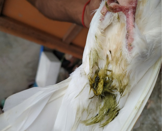
Greenish diarrhea found in the physical examination.
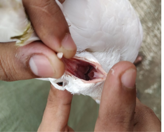
Presence of yellowish-white caseous nodules in the oral cavity.
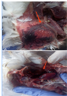
Post-mortem lesions indomestic pigeon– A) Congested proventriculus and nodule in the crop wall. B) Hepatomegaly and Congested heart.
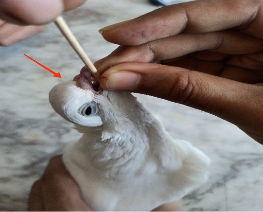
Collection of samplesfrom the oropharynx and crop.
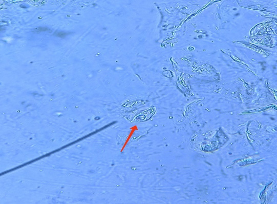
Tropozoid of T. gallinae
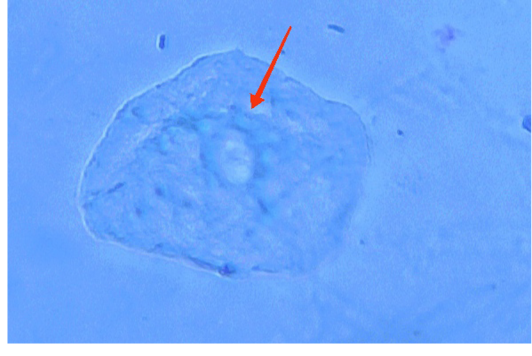
Confirm of Trophozoite by Giemsa staining.
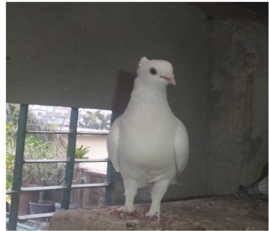
After the treatment, the pigeon was successfully recovered.
References
- Al-Sadi, HI, Hamodi AZ (2011). Prevalence and pathology of trichomoniasis in free-living urban pigeons in the city of Mosul, Iraq. Vet.World., 4(1):12. https://doi.org/10.5455/vetworld.2011.12-14[CrossRef]
- Ali HA, MY Ramadan, AM Ata-Allah (2014). Biochemical effects of trichomonasgallinae on pigeons reared on irrigation channels water and water treated with anti-protozoal agents. Benha. Vet. Med. J., 27: 150-156.
- Anderson NL, Grahn RA, Van Hoosear K, BonDurant RH (2009).Studies of trichomonad protozoa in free ranging songbirds: prevalence of Trichomonasgallinae in house finches (Carpodacusmexicanus) and corvids and a novel trichomonad in mockingbirds (Mimuspolyglottos). Vet. Parasit., 161(3-4): 178-186. https://doi.org/10.1016/j.vetpar.2009.01.023[CrossRef]
- Arenales A, Almeida AC, Prado LF, Garcia S, Luvizotto MC (2014). Cytopathology in the diagnosis of oral trichomoniasis in a rock pigeon (Columba livia). Braz. J. Vet. Pathol., 7(2): 98-99.
- Arfin S, Sayeed MA, Sultana S, Dash AK, Hossen ML (2019). Prevalence of Trichomonasgallinae infection in pigeon of jessore district, Bang. J. Adv. Vet. Anim. Res.,6(4): 549. https://doi.org/10.5455/javar.2019.f381[CrossRef]
- Aydın L, Coșkun Ș, Güleğen E (2000). Efficacy of carnidazole (Spatrix®) and metronidazole (Flagyl®) against Trichomonasgallinae in naturally infected pigeons. Act. Parasit. Turc., 24(1): 65-66.
- Begum N, Mamun MA, Rahman SA, Bari AS (2008).Epidemiology and pathology of Trichomonasgallinae in the common pigeon (Columba livia). J. Bang. Agri. Uni., 6(452-2016-35515).
- Bulbul KH, Shahardar R, Allaie IM, Wani ZA, Tramboo SR, Ashraf A, Maqbool I, Ganaie ZA, Sheikh IU, Shubeena S (2018).Avian trichomonosis with special reference to pigeon. Int. J. Vet .Sci. Anim. Husb., 3: 11-13.
- Bunbury N, Jones CG, Greenwood AG, Bell DJ (2007).Trichomonasgallinae in Mauritian columbids: implications for an endangered endemic. J. of Wild. Dise., 43(3): 399-407. https://doi.org/10.7589/0090-3558-43.3.399[CrossRef]
- Bunbury N, Stidworthy MF, Greenwood AG, Jones CG, SawmyS, Cole RE, Edmunds K, Bell DJ (2008). Causes of mortality in free-living Mauritian pink pigeons Columba mayeri, 2002–2006. End. Spec. res., 9(3): 213-220. https://doi.org/10.3354/esr00088[CrossRef]
- Bunbury N, Jones CG, Greenwood AG, Bell DJ (2008).Epidemiology and conservation implications of Trichomonasgallinae infection in the endangered Mauritian pink pigeon. Bio. Cons., 141(1): 153-161. https://doi.org/10.1016/j.biocon.2007.09.008[CrossRef]
- Dovč A, Zorman-Rojs O, VerglesRataj A, Bole-Hribovšek V, Krapež U, Dobeic M (2004). Health status of free-living pigeons (Columba liviadomestica) in the city of Ljubljana. Act. Vet. Hung., 52(2): 219-226. https://doi.org/10.1556/avet.52.2004.2.10[CrossRef]
- Fadhil LT, Faraj AA, AL-Amery AM (2020).Trichomonasgallinae identification and histopathological study in pigeon (Columba liviadomestica) in Baghdad city, Iraq. The Ira. J. ofVet. Med., 44: 57-63. https://doi.org/10.30539/ijvm.v44i(E0).1022[CrossRef]
- Forzan MJ, Vanderstichel R, Melekhovets YF, McBurney S (2010).Trichomoniasis in finches from the Canadian Maritime provinces an emerging disease. The Cana. Vet. J., 51(4): 391.
- Grunenwald C, Sidor I, Mickley R, Dwyer C, Gerhold R (2018).Tetratrichomonas and trichomonas spp.-associated disease in free-ranging common Eiders (Somateriamollissima) from Wellfleet Bay, MA and description of ITS1 region genotypes. Avi. dise., 62(1): 117-123. https://doi.org/10.1637/11742-080817-Reg.1[CrossRef]
- Ghosh KK, Islam MS, Sikder S, Das S, Chowdhury S, Alim MA (2014). Prevalence of ecto and gastrointestinal parasitic infections of pigeon at Chittagong Metropolitan Area, Bangladesh. TheJ. of Adv. in Parasit.,1(1): 9-11. https://doi.org/10.14737/journal.jap/2014/1.1.9.11[CrossRef]
- Gómez-Muñoz MT, Gómez-Molinero MÁ, González F, Azami-Conesa I, Bailén M, GarcíaPiqueras M, Sansano-Maestre J (2022). Avian OropharyngealTrichomonosis: Treatment, Failures and Alternatives, a Systematic Review. Microorganisms., 10(11): 2297. https://doi.org/10.3390/microorganisms10112297[CrossRef]
- Hamad SS, Hassan HH (2017).Isolation, diagnosis and cultivation of Trichomonasgallinae from domestic pigeon in Kirkuk City, Iraq. Int. J.Curr. Res. Acad. Rev., 5(2):10-18. https://doi.org/10.20546/ijcrar.2017.502.002[CrossRef]
- Höfle U, Gortázar C, Ortiz JA, Knispel B, Kaleta EF (2004). Outbreak of trichomoniasis in a woodpigeon (Columba palumbus) wintering roost. Eur. J. of Wild. Res., 50: 73-77. https://doi.org/10.1007/s10344-004-0043-2[CrossRef]
- Hochleithner M, Hochleithner C (2021). The prevalence of Trichomonasgallinae in budgerigars (Melopsittacusundulatus) in a Veterinary Clinic in Vienna between 2000–2019. Vet. Med., 66: 490–493. https://doi.org/10.17221/110/2020-VETMED[CrossRef]
- Kennedy, M. J. (2001). Trichomoniasis in birds. Alberta Agriculture, Food and Rural Development.
- McDougald LR (2003). Diseases of poultry, 11st Edit. Editorial Broad for American Association of Avian Pathogenesis., 1006-1011.
- Mohamed HM, Saad AS, Khalifa MM, Abdel-Maogood SZ, Awadalla SM, Mousa WM (2023). Detection and molecular characterization of Trichomonasgallinae recovered from domestic pigeons in Egypt. Parasit. Res., 122(1): 257-263. https://doi.org/10.1007/s00436-022-07724-z[CrossRef]
- Paul TK, Amin MR, Alam MA, Rahman MK, Sarker YA, Rizon MK (2015). Occurrence of pigeon diseases at Khulna Sadar, Bangladesh. https://doi.org/10.3329/bjvm.v13i2.26616[CrossRef]
- Pennycott T, Lawson B, Cunningham A, Simpson V, Chantrey J (2005). Necrotic ingluvitis in wild finches. Vet. Rec., 157(12): 360. https://doi.org/10.1136/vr.157.12.360[CrossRef]
- Qiu SB, Yan C, Zhou DH, Hou J, Wang QQ, Lin Y, Fu HC, Zhang J, Weng YB, Song HQ, Lin RQ (2012).High prevalence of Trichomonasgallinae in domestic pigeons (Columba liviadomestica) in subtropical southern China. African. J. Micro. Res., 6(6): 3261-3264. https://doi.org/10.5897/AJMR12.001[CrossRef]
- Qiu SB, Lv MN, He X, Weng YB, Zou SS, Wang XQ, Lin RQ (2017). PCR identification and phylogenetic analysis of Trichomonasgallinae from domestic pigeons in Guangzhou, China. Korean j. of parasi.,55(3): 333. https://doi.org/10.3347/kjp.2017.55.3.333[CrossRef]
- Raza A, Qamar MF, Rubtsova N, Saneela S (2018). Pathogenicity and diagnostic sensitivity of culture media for identification of Trichomonasgallinae in Domestic Pigeons of Lahore, Pakistan. J. of Protoz. Res., 28(1-2): 11-21.
- Seddiek SA, El-Shorbagy MM, Khater HF, Ali AM (2014).The antitrichomonal efficacy of garlic and metronidazole against Trichomonasgallinae infecting domestic pigeons. Parasit. Res., 113: 1319-1329. https://doi.org/10.1007/s00436-014-3771-6[CrossRef]
- Soulsby EJ (1968). Helminths, arthropods and protozoa of domesticated animals. Helminths, arthropods and protozoa of domesticated animals.
- Tietz Marques SA, Marinho de Cuadros RO,Jardim da Silva CI, Baldo M (2007). Parasites of pigeons (Columba livia) in urban areas of lages, Southern Brazil. Parasitologíalatino americana, 62(3-4): 183-187. https://doi.org/10.4067/S0717-77122007000200014[CrossRef]
- Villanúa D, Höfle U, Perez-Rodriguez LO, Gortázar C (2006).Trichomonasgallinae in wintering common wood pigeons Columba palumbus in Spain. Ibis., 148(4): 641-648. https://doi.org/10.1111/j.1474-919X.2006.00561.x[CrossRef]
Services
Article usage statistics
Loading…