
Nanoparticles are gaining popularity due to their increasing biomedical applications as anti-bacterial, anti-fungal and anti-cancer agents. In this work leaf extracts of Melia azedarach (commonly known as dharaik) a species of family Meliaceae and Morus nigra (commonly known as Shahtoot) family Moraceae were used for bio reduction of copper ions to CuO nanoparticles in place of harmful and expensive chemical/physical methods. A cheaper, simple to perform method giving high yield without by products is employed here. The leaf extract act both as reducing and capping agent. The atomic absorption shows a linear curve between concentration and absorbance. The uv-vis spectrum revealed the shift of absorption towards longer wavelength with the increasing concentration of plant extract. XRD indicates CuO Nps particle size is in between 14-20 nm. CuO nanoparticle show very significant antibacterial activity against four bacterial strains three gram negative (Escherichia coli, Proteus mirabilis, Salmonella) and one gram positive (Clostridium tetani). The minimum concentration of leaf extracts needed against these bacterial strains were also calculated. The present study indicated that Melia azedarach and Morus nigra leaf extracts can be used successfully for nanoparticles synthesis and CuO as significantly active antibacterial material. Antibacterial activity of these nanoparticles was observed by agar well diffusion method.
Research Article · Volume 4, Issue 2, 28 March 2019 · Open Access
Synthesis of CuO Nanoparticles by using Leaf Extracts of Melia azedarach and Morus nigra and their Antibacterial Activity
·
Affiliations & Notes ⌄
- 1Department of Chemistry, Lahore College for Women University, Jail Road, Lahore, 54000, Pakistan.
Article Info ⌄
- Journal:
- Journal of Innovative Sciences
- Published:
- 28 March 2019
From the article
- This study concludes that CuO NPS can be prepared from eco-friendly synthesis by using leaf extracts as natural ingredients.
- Confirmation of CuO nanoparticles was done with FTIR, colour change, Atomic Absorption Spectroscopy, UV/Vis spectra and X-ray Diffraction studies.
- Thus, the leaf extracts of Morus Nigra (Shahtoot) and Melia Azedarach (dharaik) successfully worked as natural ingredient.
- Antibacterial studies proved CuO NPs as good inhibiter against three gram negative and one gram positive strain by measuring their Zone of inhibition.
Selected from the article's own text.
Abstract
1. Introduction
Nanotechnology deals with the synthesis of particles and their structures ranging in size from 1-100 nm. Metal nanoparticle are quite significant due to their optical, electrical and electronic properties ; ; Maitinise et al., 2017; . They also have a prominent role as gas sensors ; , electrochromic devices and in solar cells . For increasing their potential, multicomponent oxides are also prepared ; ; . Their biodegradable and biocompatible modifications resulted in reduction of their toxicity ; .
Nanoparticles synthesis involve different methods (physical and chemical) e.g. synthesis of CuO nanoparticles by precipitation method, inverse micro emulsion system, Sol gel method etc. ; ; . Unfortunately, these methods include the use of toxic chemicals and non-eco-friendly by products . Hence, it is the need of the day to opt an alternate environmental friendly synthetic route for obtaining nanoparticles using naturally occurring ingredients as reductants. Thus, Metal oxide nanoparticles by green method are gaining importance . Nanoparticles are mainly classified as inorganic and organic. Inorganic Nps consists of either metal or metal oxides (e.g. NPS of Ag, Fe3O4, TiO2, CuO and ZnO etc). Presently green chemistry involves their biosynthesis through microorganism and plant extracts ;. By using natural extract of Aspalathus Linearis crystalline perovskite ZnSnO3 nanoclusters, NiO, Pd and PdO nanoparticles are biosynthesized ; . Sageretia thea and Moringa Oleifera natural extracts are used as chelating agent to prepare ZnO nanoparticle ; . Ag NPs were synthesized from aqueous Eriobotrya japonica leaf extract , fruit extract of Capsicum frutescence (Sweet pepper) (Shanker et al., 2017) and Mentha asiatica mint extract . In addition to chemical synthesis ; Cu NPS can also be prepared by using natural ingredients (leaf extract) . Organic NPS are mainly polymeric or lipids having 10nm to 1µm diameter . Liposomes, dendrimers, carbon nanomaterials and polymeric micelles are examples of organic nanoparticles.
Inorganic nanoparticles are preferred as antimicrobial agents ; as organic NPs cannot tolerate high temperature (e.g. quaternary ammonium compounds, N-halamine compounds and chitosan etc). The bactericidal effect of inorganic NPS has been attributed to their characteristic micron size less than the pore size of the bacteria and thus, they are capable of easily crossing the cell membranes without any hindrance .
2. Materials and Methods
Due to the local availability and local medical use leaves of Morus nigra (shahtoot) and Melia azedarach (dharaik) were selected (herbarium of botany department LCWU). Powdered 10g/100ml of each aqueous leaf extract was boiled (80°C for 20 min), subjected to shaker (5c/s for 10 minutes), filtered and then refrigerated (4°C) for further experiments. 99.9% pure CuSO4∙5H2O solution (0.01M) of analytical grade was also used.
2.1 Synthesis of CuO Nanoparticles from plants leaf extract
For biosynthesis of nanoparticles, 10 mL of each of leaf extract was mixed with 100 mL of freshly prepared 1×10⁻² M aqueous copper sulphate pentahydrate solution in 250 mL Erlenmeyer flask under continuous magnetic stirring. Formation of CuO nanoparticles was accompanied with colour change (blue to yellow). These were then purified by repeated centrifugation method at 6,000rpm for 25 min. Later the CuO nanoparticles were dried in an oven at 80 ºC for 5 hours.
Table 1 Showing the procedure for the preparation of different concentrations of M. Nigra and M. azedarach.
Table 1: Showing the procedure for the preparation of different concentrations of M. Nigra and M. azedarach.
| Concentration | Procedure |
| 5% | 5mL of the Plant leaf extract was mixed with 95mL of the 0.01M CuSO4 Solution. |
| 15% | 15mL of the Plant leaf extract was mixed with 85mL of the 0.01M CuSO4 Solution. |
| 25% | 25mL of the Plant leaf extract was mixed with 75mL of the 0.01M CuSO4 Solution. |
2.2 Procedure for antibacterial activity
For antibacterial activity agar well diffusion method was employed . Four bacterial strains were selected including Escherichia coli, Proteus mirabilis, Salmonella and Clostridium tetani. Different concentrations of both leaf extracts (5%, 15% and 25%) were checked and their zone of inhibition (ZOI) in cm was measured.
3. Results and Discussion
3.1 Atomic absorption spectrometry
The confirmation of CuO NPS was done by getting straight line between concentration and absorbance. Figure 1and2 shows the increase in absorption with increasing concentration of CuO nanoparticles with the gradual increase of leaf extracts concentration (5 to 25%) for Morus nigra (M.nigra) Figure 1(a, b) and Figure 2(a, b) for Melia azedarach (M.azedarach).
3.2 Visual observation and UV-Vis spectroscopy
On addition of each leaf extract to aqueous CuSO4 solution keeping its concentration same i.e. 0.01M, colour change was observed from pale blue to dark brown due to the conversion of Cu+2 ions to pure CuO nanoparticles Figure 3 (a, b) As the concentration of both leaf extracts (morus nigra and Melia azedarach) increases from (5-25%) the plasmon resonance bands shifts from 250-320nm which is observed by UV-Vis spectroscopy Figure 4(a, b).
3.3 Fourier Transform Infrared Spectroscopy (FTIR)
Three prominent bands of the CuO nanoparticles synthesized from Morus nigra aqueous leaf extract was at 3630 cm-1for OH, 2158 and 2029cm-1 for C≡C and 1024cm-1 is because of C-O stretching Figure 5(a).
For Melia azedarach Figure 5(b) a wide OH band is observed at 3352 cm-1, C≡C peak at 2360 cm-1, C=C group appeared as a weak band at about 1650cm-1.The peak for CuO bond stretching was observed at 507, 543 cm-1 . Carbonyl group and proteins from leaf extract can bind metal, forming a covering on metal nanoparticles. These results shows that some of the bioorganic compounds (leaf extract ) played a double role i.e a strong covering and reducing agent .
3.4 X- Ray diffraction studies
The XRD results of CuO nanoparticles synthesized from Morus nigra exhibits peaks at (32.75°, 35.54°, 38.57° and 48.67° corresponding to planes {110}, {002}, {200}, {202}) and for Melia azedarach exhibits peak at (32.79°, 35.49°, and 37.71° for {110}, {002} and {200}) shows their monoclinic nature Figure 6(a, b).
The diffraction angle at 27.67° may be attributed to Cu nanoparticles instead of CuO nanoparticles. Thus, from these observations it may be concluded that some of the Cu NPs were so stable that they were not oxidized to form CuO nanoparticles.
All the peaks observed are due to Cu NPS and chances of independent crystallization of covering agent is no more because of using centrifugation process done for purification of NPS.
The average nanoparticles size calculated (Debye–Scherrer equation) was 14-20 nm, which may indicate a high surface area-to-volume ratio of nanocrystals.
3.5 Antibacterial activity
Antibacterial activity of CuO nanoparticles was observed against four bacterial strains (gram positive and gram-negative bacteria) Table 2.
Table 2: Showing different bacterial strains.
| Gram Negative | Gram Positive |
| Escherichia coli ATCC 8739 | Clostridium tetani ATCC 10779 |
| Proteus mirabilis ATCC 25933 | |
| Salmonella ATCC 700623 |
3.6 Activity of CuO nanoparticles from Morus nigra and Melia azedarach leaf extract
25% (max conc.) showed maximum activity for both M. nigra and M. azedarach Figure 7(a, b, c, d, e, f). For M. nigra 1.2cm zone of inhibition (ZOI) for Escherichia coli, 0.6cm for Clostridium tetani, 1.3 cm for Proteus mirabilis and 1.2cm ZOI for Salmonella was observed Table 3. For M. Azedarach 1.6cm inhibition for Escherichia coli, 0.7cm for Clostridium tetani, 0.4cm for Proteus mirabilis and 1.1cm inhibition was against Salmonella Table 3.
Table 3: Evaluation of Antibacterial activity of CuO nanoparticles synthesized from Morus nigra and Melia Azedarach.
| Sr. No | Bacterial Isolates | Zone of Inhibition in cm | |||||||
| M. Nigra | M. Azedarach | ||||||||
| 5% | 15% | 25% | 5% | 15% | 25% | ||||
| 1 | Escherichia coli | 0.7 | 1.0 | 1.2 | 0.6 | 0.9 | 1.6 | ||
| 2 | Clostridium tetani | 0.3 | 0.4 | 0.6 | 0.3 | 0.5 | 0.7 | ||
| 3 | Proteus mirabilis | 0.5 | 0.9 | 1.3 | 0.2 | 0.3 | 0.4 | ||
| 4 | Salmonella | 0.6 | 0.9 | 1.2 | 0.3 | 0.7 | 1.1 | ||
Conclusion
This study concludes that CuO NPS can be prepared from eco-friendly synthesis by using leaf extracts as natural ingredients. Confirmation of CuO nanoparticles was done with FTIR, colour change, Atomic Absorption Spectroscopy, UV/Vis spectra and X-ray Diffraction studies. Thus, the leaf extracts of Morus Nigra (Shahtoot) and Melia Azedarach (dharaik) successfully worked as natural ingredient. Antibacterial studies proved CuO NPs as good inhibiter against three gram negative and one gram positive strain by measuring their Zone of inhibition. The Minimum inhibitory concentration of both the extracts were 5%. Thus, the above method is natural, significant, simple, low cost and producing no by-products.
Acknowledgement
All this research is done and supported by central Lab of Lahore College for Women University Jail Road Lahore, Pakistan.
Author’s Contribution
Tahira Moeen Khan: Concept and design of study.
Tahira Moeen Khan, Amat ul Mateen: Data acquisition, analysis and interpretation.
Figures
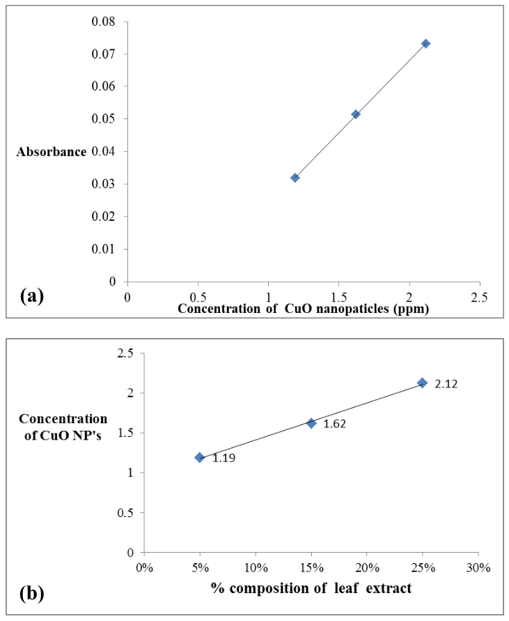
(a) Relation of CuO NPs concentration and Absorbance (Moris Nigra), (b): Effect on the synthesis of CuO NPs due to gradual increase in the M. nigra leaf extract concentration.
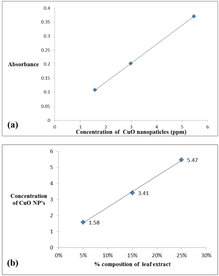
(a) Relation of CuO NPs concentration and Absorbance Melia azedarach (dharaik), (b): Effect on the synthesis of CuO NPs due to the gradual increase in concentration of Melia azedarach (dharaik) leaf extract.
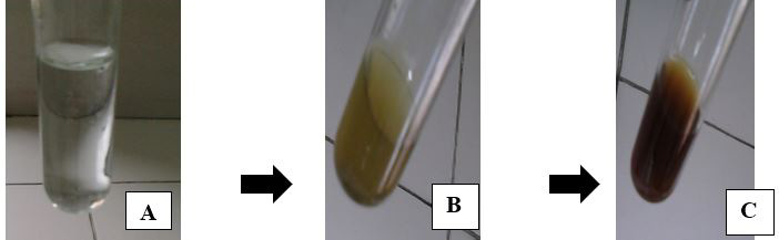
A= pale blue (0.01M CuSO4.5H2O); B= greenish brown (aqueous leaf extract of Morus nigra); C= dark brown (addition of leaf extract into CuSO4.5H2O).
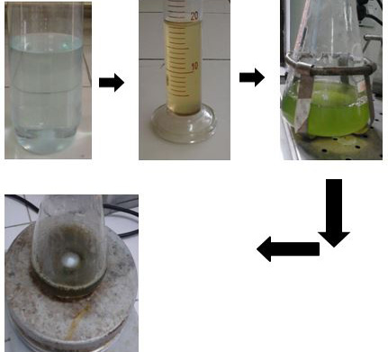
Colour change from pale blue to yellowish green and finally dark brown (left to right) for Melia azedarach.
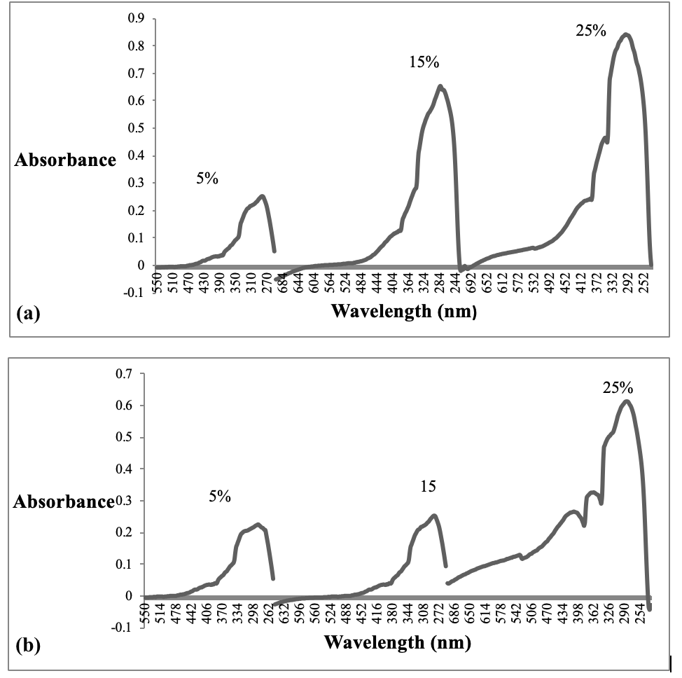
An increase in absorbance is observed with Morus nigra (a) and Melia azedarach (b) increased leaf extract concentration i.e. 5% to 25%.
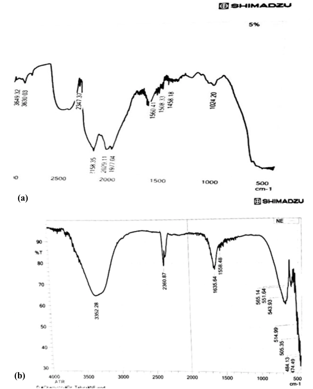
FTIR spectrum of CuO nanoparticles synthesized from M. nigra (a) and Melia azedarach (b) leaves extract.
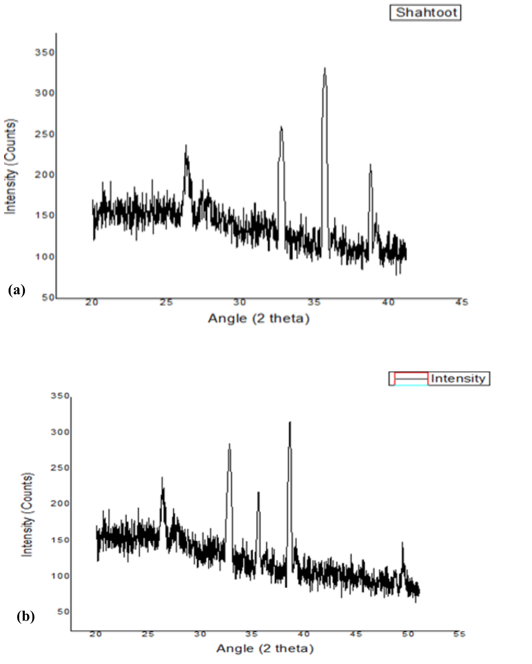
XRD plot of CuO NPs synthesized from M. nigra (a) and Melia azedarach (b) leaf extract.
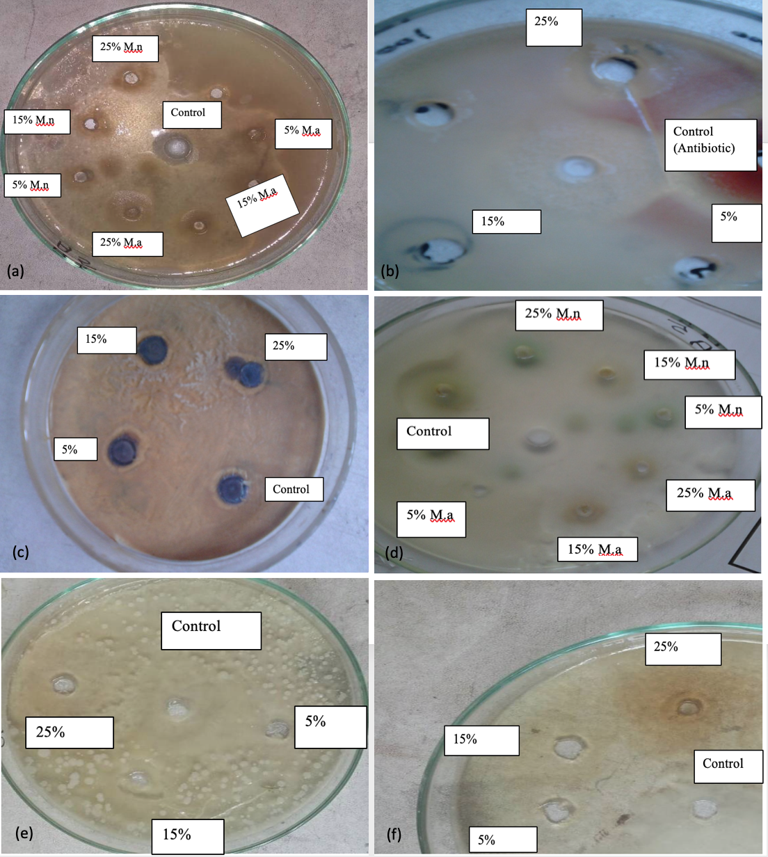
Showing the antibacterial results of CuO NPS synthesized from Morus nigra (M.n) and Melia azedarach (M.a) leaf extracts against a) Eschericia coli b) Clostridium tetani (M.n) c) Clostridium tetani (M.a) d) P. mirabilis e) Salmonella (M.n) f) Salmonella (M.a).
References
- Ahmed, S., Ahmad, M., Babu L.S. and Ikram, S. 2016. Green synthesis of silver nanoparticles using Azadirachta indica aqueous leaf extract. Journal of Radiation Research and Applied Sciences, 9: 1-7. https://doi.org/10.1016/j.jrras.2015.06.006[CrossRef]
- Ahmed, S., Ahmad, M., Swami, B.L. and Ikram, S. 2015. Plants extract mediated synthesis of silver nanoparticles for antimicrobial applications: a green expertise. Journal of Advance Research, 7: 17-28. https://doi.org/10.1016/j.jare.2015.02.007[CrossRef]
- Argazzi, R., Iha, N.Y.M., Zabri, H., Odobel, F. and Bignozzi, C.A. 2004. Design of molecular dyes for application in photoelectrochemical and electrochromic devices based on nanocrystalline metal oxide semiconductors. Coordination Chemistry Reviews, 248: 1299–1316. https://doi.org/10.1016/j.ccr.2004.03.026[CrossRef]
- Couvreur, P. 1988. Polyalkylcyanoacrylates as colloidal drug carriers. Critical Reviews in Therapeutic Drug Carrier System, 5: 1–20.
- Capek, I. 2004. Preparation of metal nanoparticles in water-in-oil (w/o) microemulsions. Advances in Colloid and Interface Science, 110: 49–74. https://doi.org/10.1016/j.cis.2004.02.003[CrossRef]
- Davarpanah, S.J., Karimian, R. and Goodarzi, V. 2015. Synthesis of CuO nanoparticles and its applications as Gas Sensor. Journal of Applied Biotechnology Reports, 2(4): 329-332.
- Deng, Z., Chen, M., Gu, G. and Wu, L. 2008. A facile method to fabricate ZnO hollow spheres and their photocatalytic property. The Journal of Physical Chemistry –B, 112: 16-22. https://doi.org/10.1021/jp077662w[CrossRef]
- Deivasigamani, P., Parthiban, R., Ponnusamy, K.S. and Namasivayam, R.K.S. 2015. Synthesis, Characterization and antibacterial activity of nano zero-valent iron impregnated cashew nut shell. International Journal of Pharmacy and Pharmaceutical Sciences, 7(1): 139-141.
- Hashoosh, S.I., Ayad, M.A., Fadhil and Nabeel, k.Al-Ani. 2014. Production of Ag nanoparticles Using Aloe Vera Extract and its Antimicrobial Activity. Journal of Al-Nahrain University,17 (2): 165-171. https://doi.org/10.22401/JNUS.17.2.22[CrossRef]
- He, Z., Zhong, C., Su, S., Xu, M., Wu, H. and Cao, Y. 2012. Enhanced power conversion efficiency in polymer solar cells using an inverted device structure. Nature Photonics, 6: 591–595. https://doi.org/10.1038/nphoton.2012.190[CrossRef]
- Heiligtag, F.J. and Niederberger, M. 2013.The fascinating world of nanoparticle research. Materials Today, 16 (7–8): 262–271. https://doi.org/10.1016/j.mattod.2013.07.004[CrossRef]
- Hasnidawani, J.N., Azlina, H.N., Norita, H., Bonnia, N.N., Ratim, S. and Ali, E.S. 2016. Synthesis of ZnO nanostructures using Sol Gel Method. Procedia Chemistry, 19: 211-216. https://doi.org/10.1016/j.proche.2016.03.095[CrossRef]
- Jabbar, M.S. 2016. Synthesis of CuO nano structure via sol-gel and precipitation chemical methods. Al-Khwarzami Engineering Journal, 12(4): 126-131.
- Jain, A., Duvvuri, L.S., Farah, S., Beyth, N., Domb, A.J. and Khan, W. 2014. Antimicrobial polymers. Advanced Healthcare Materials, 3: 1969–1985. https://doi.org/10.1002/adhm.201400418[CrossRef]
- Jayaprakash, N., Vijaya, J.J., Kaviyarasu, K., Kombaiaha, K., Kennedy, J., Ramalingam, R.J., Munusamy, M.A. and Al-Lohedan, H.A. 2017. Green synthesis of Ag nanoparticles using Tamarind fruit extract for the antibacterial studies. Journal of Photochemistry and photobiology. B: Biology, 169: 178-185. https://doi.org/10.1016/j.jphotobiol.2017.03.013[CrossRef]
- Khan, A.S., Shahid, S., Sajid, R.M., Noreen, F. and Kanwal, S. 2017. Biogenic synthesis of CuO nanoparticles and their biomedical applications: a current review. International Journal of Advanced Research, 5(6): 925-946. https://doi.org/10.21474/IJAR01/4495[CrossRef]
- Khan, M.Z.H., Tarek, F.K., Nuzat, M., Momin, M.A. and Hasan, M.R. 2017. Rapid biological synthesis of silver nanoparticles from Ocimum sanctum and their characterization. Journal of Nanoscience, 2017: 1-6. https://doi.org/10.1155/2017/1693416[CrossRef]
- Korotcenkov, G. 2007. Metal oxides for solid-state gas sensors: what determines our Choice. Material Science and Engineering, B, 139: 1–23. https://doi.org/10.1016/j.mseb.2007.01.044[CrossRef]
- Li, M., Bala, H., Lv, X., Ma, X., Sun, F., Tang, L. and Wang, Z. 2007. Direct synthesis of monodispersed ZnO nanoparticles in an aqueous solution. Materials Letters, 61: 690-693. https://doi.org/10.1016/j.matlet.2006.05.043[CrossRef]
- Loomba, L. and Scarabelli, T. 2013. Metallic nanoparticles and their medicinal potential, Part II: aluminosilicates, nanobiomagnets, quantum dots and cochleates. Therapeutic Delivery, 4(9): 1179–1196. Matinise, N., Fuku, X.G., Kaviyarasu, K., Mayedwa, N. and Maaza, M. 2017. ZnO nanoparticles via Moringa Oleifera green synthesis: physical properties and mechanism of formation. Applied Surface Science, 406: 339–347. https://doi.org/10.1016/j.apsusc.2017.01.219[CrossRef]
- Matheswari, P.P., Murugan, C., Akila, R., Menisha, S. and Revathi, T. 2018. Punica granatum flower extract mediated synthesis of Copper oxide Nanoparticle and evolution of its antibacterial activity. International Journal of Applied and Pure Science and Agriculture, 4 (1): 7-12.
- Matinise, N., Fuku, X.G., Kaviyarasu, K., Mayedwa, N. and Maaza, M. 2017. ZnO nanoparticles via Moringa oleifera green synthesis: Physical properties and mechanism of formation. Applied Surface Science, 406: 339-347.
- Mayedwa, N., Mongwaketsi, N., Khamlich, S., Kasinathan, K., Matinise, N. and Maaza, M. 2017. Green Synthesis of Zin Tin Oxide (ZnSnO3) Nanoparticles Using Aspalathus Linearis Natural Extracts: Structural, Morphological, Optical and Electrochemistry Study. Applied Surface Science. (Article in press).
- Mayedwa, N., Mongwaketsi, N., Khamlich, S., Kasinathan, K., Matinise, N. and Maaza, M. 2018. Green synthesis of nickel oxide, palladium and palladium oxide synthesized via Aspalathus linearis natural extracts: physical properties and mechanism of formation. Applied Surface Science, 446: 266-272. https://doi.org/10.1016/j.apsusc.2017.12.116[CrossRef]
- Mayedwa, N., Khalil, A.T., Mongwaketsi, N., Matinise, N., Shinwari, Z.K. and Maaza, M. 2017. The Study of Structural, Physical and Electrochemical Activity of ZnO Nanoparticles Synthesized by Green Natural Extracts of Sageretia thea. Nano Research and Applications, 3: 2. https://doi.org/10.21767/2471-9838.100026[CrossRef]
- Perez, J., Bax, L. and Escolano, C. 2005. Roadmap Report on Nanoparticles. Willems and van den Wildenberg (WandW) Espana sl: Barcelona-Spain.
- Prabu, D., Parthiban, R., Kumar, P.S. and Namasivayam, K.R. 2015. Synthesis, Characterization and antibacterial activity of nano zero-valent iron impregnated cashew nut shell. International Journal of Pharmacy and Pharmaceutical Sciences, 7: 139-141.
- Prabu, J.H. and Johnson, I., 2015. Plant mediated biosynthesis and characterization of silver nanoparticle by leaf extracts of Tragia involucrata, Cymbopogon citronella, Solanum verbascifolium and Tylophora ovata. Karbala International Journal of Modern science,1 (4): 237-246.
- Rao, B. and Tang, R.C. 2017. Green synthesis of silver nanoparticles with antibacterial activities using aqueous Eriobotrya japonica leaf extract. Advances in Natural Sciences: Nanoscience and Nanotechnology, 8: 1-8. https://doi.org/10.1088/2043-6254/aa5983[CrossRef]
- Sadri, F., Ramazani, A., Massoudi, A., Khoobi, M., Tarasi, R., Shafiee, A., Azizkhani, V. and Dolatyari, L. 2014. Green oxidation of alcohols by using hydrogen peroxide in water in the presence of magnetic Fe3O4 nanoparticles as recoverable catalyst. Green Chemistry Letters and Reviews, 7(3): 257-264. https://doi.org/10.1080/17518253.2014.939721[CrossRef]
- Sarkar, D. and Paul, G. 2017. Green synthesis of silver nanoparticles using Mentha asiatica (Mint) extract and evaluation of their antimicrobial potential. International Journal of Current Research in Biosciences and Plant Biology, 4(1): 77-82. https://doi.org/10.20546/ijcrbp.2017.401.009[CrossRef]
- Shankar, T., Karthiga, P., Swarnalatha, K. and Rajkumar, K. 2017. Green synthesis of silver nanoparticles using Capsicum frutescence and its intensified activity against E. coli. Resource-Efficient Technologies, 3(3): 303-308. https://doi.org/10.1016/j.reffit.2017.01.004[CrossRef]
- Sone, B.T., Manikandan, E., Gurib-Fakim, A. and Maaza, M. 2015. Sm2O3 nanoparticles green synthesis via Callistemon viminalis’ extract. Journal of Alloys and Compounds, 650: 357-362. https://doi.org/10.1016/j.jallcom.2015.07.272[CrossRef]
- Wadkar, P., Bauskar, D. and Patil, P. 2013. High performance H2 sensor based on ZnSnO3 cubic crystallites synthesized by a hydrothermal method. Talanta, 105: 327-332. https://doi.org/10.1016/j.talanta.2012.10.051[CrossRef]
- Xu, J.Q., Jia, X., Lou, X. and Shen, J. 2006. One-step hydrothermal synthesis and gas sensing property of ZnSnO3 microparticles. Solid-State Electronics, 50(3): 504-507. https://doi.org/10.1016/j.sse.2006.02.001[CrossRef]
- Zeng, Y., Zhang, T., Fan, H., Lu, G., Kang, M. 2009. Synthesis and gas-sensing properties of ZnSnO3 cubic nanocages and nanoskeletons. Sensors and Actuators B: Chemical, 143(1): 449-453. https://doi.org/10.1016/j.snb.2009.07.021[CrossRef]
- Zhang, X., Luo, J., Tang, P., Ye, X., Peng, X., Tang, H., Sun, S.G. and Fransaer, J. 2017. A universal strategy for metal oxide anchored and binder-free carbon matrix electrode: a supercapacitor case with superior rate performance and high mass loading. Nano Energy, 31: 311–321. https://doi.org/10.1016/j.nanoen.2016.11.024[CrossRef]
- Zhou, T., Zhang, T., Deng, J., Zhang, R., Lou, Z. and Wang, L. 2017. P-type Co3O4 nanomaterials- based gas sensor: preparation and acetone sensing performance. Sensors and Actuators B: Chemical, 242: 369–377. https://doi.org/10.1016/j.snb.2016.11.067[CrossRef]
Services
Article usage statistics
Loading…