
Three cases, including a buffalo and two cows were presented with complaint of persistent unilateral hind limb extension. The animals were unable to flex the limb during walk. Diagnosis was based on case history, clinical sign and clinical examination. Upward fixation of patella was diagnosed in all three animals. After careful palpation of the ligament, medial patellar desmotomy was performed in lateral recumbence, under local anesthesia achieved by 2% lignocaine hydrochloride. The skin was apposed with simple interrupted sutures using silk. Post-operative care was continued for five days. Clinical signs abated in all operated animals, proving that the technique employed was effective and of simple execution for the treatment of dorsal patellar fixation in bovine.
Short Communication · Volume 49, Issue 2, 5 April 2017 · Open Access
Surgical Correction of Intermittent Unilateral Hind Limb Extension in Buffalo and Cattle
· · · ·
Affiliations & Notes ⌄
- 1University of Agriculture, Faisalabad, Pakistan 2Livestock and Dairy Development Department, Punjab, Pakistan 3Department of Pathology, University of Veterinary and Animal Sciences, Lahore, Pakistan 4Department of Pathobiology, Faculty of Veterinary and Animal Sciences, PMAS Arid Agriculture University, Rawalpindi, Pakistan
Article Info ⌄
- Journal:
- Pakistan Journal of Zoology
- Published:
- 5 April 2017

From the article
- Following medial patellar desmotomy, all the animals showed immediate recovery signs with no evidence of jerky movement.
- We suggest medial patellar desmotomy as an efficient surgical technique for the treatment of persistent unilateral hind limb extension in dairy animals.
Selected from the article's own text.
Abstract
Materials and methods
On 2nd June 2015 two Sahiwal cows from the same herd were presented with the complaint of difficulty in walking. Both had previous history of treatment one week ago with inj. Loxicom® (meloxicam, Norbrook, UK) at the dose rate of 0.5mg/kg for 3 days, but no apparent recovery was observed. One of the cow was having 7 months pregnancy and the other one had recently perpetrated. On 10th July 2015, another case of a 5 year old Nili-Ravi buffalo was presented in the field with 4 months prior history of showing the signs of lameness of right hind leg.
All the three animals were in excellent body condition and appeared alert. Their appetite and bowel motility was normal, temperature, pulse and respiration (TPR) were in normal range, auscultation of heart and lung sounds were also normal. When the animals were made to move, all of them showed stiffness of hind leg with no or very little flexion of stifle joint along with dragging of toe. However, palpation revealed no pain over the stifle region. The buffalo (Fig. 1) and a cow had right unilateral hind limb affected while other cow had left hind limb affected.
The history, owner’s description and clinical features of upward fixation of patella are almost pathognomonic ; . A diagnosis of intermittent upward fixation of patella was made based on the signs of hesitation of the stride, dragging of the toe along the ground, and subsequent hyperflexion of the stifle. MPD were suggested to alleviate the condition. MPD is a surgical technique that aims at disrupting the locking mechanism, and is well described in the literature . This procedure is usually performed under local anaesthetic, so that the medial patellar ligament is clearly defined.
In all three animals surgery was performed in lateral recumbency as previously described by Singh et al. (2015). The tibia-femoral-patellar articulation area was appropriately prepared, and antisepsis was done by a disinfectant solution containing iodophor, diluted in water as recommended. Local anesthesia was achieved in all animals by infiltrating approximately 10 ml of inj. lignocaine (xylocaine® Barrett Hodgson, Pakistan) along with xylazine hydrochloride (Alfazyne®, Egevet, Turkey @ 0.3mg per kg of body weight) into the gap located between tibia crest and the medial and intermediate patellar ligaments. All cases were operated by using the open method of medial patellar desmotomy ; . By placing the thumb and the medium finger, respectively at the tibial tuberosity and at the upper spot of the femoral medial trochleal crest, the medium point between these two anatomic references was established using the index finger. This procedure facilitated the identification of the medial patellar ligament. A small linear incision of 3cm was made 0.5cm lateral to medial patella ligament. The fascia was bluntly dissected to expose the medial patellar ligament. The ligament was exteriorized with the help of a curved scissor near the ligament’s insertion point at the tibial tuberosity. The ligament was then sectioned using a scalpel. The operation site was further explored and undivided fibers were sectioned by using scissors. The skin edges were apposed with#2 monofilament nylon (Ethilon,Ethicon, Inc., Somerville, New Jersey 00876). The cow was released and walked with a normal stride.
The wound was dressed daily with a povidone iodine solution till complete healing. Post-operative care include administration of Procaine penicillin (Penbiotic®, PDH Pakistan, @ 20,000 IU/ Kg live weight, every 48 hours) and inj. meloxicam, (Loxicom® Norbrook, UK) (@ 0.5mg/ body weight) injected intramuscularly for 5 days.
Results
Following medial patellar desmotomy, all the animals showed immediate recovery signs with no evidence of jerky movement. Skin sutures were removed 15 days post operation. All animals completely recovered within 15 to 30 days (Fig. 2).
Conclusion
We suggest medial patellar desmotomy as an efficient surgical technique for the treatment of persistent unilateral hind limb extension in dairy animals.
Conflict of interest statement
We declare that we have no conflict of interest.
Figures
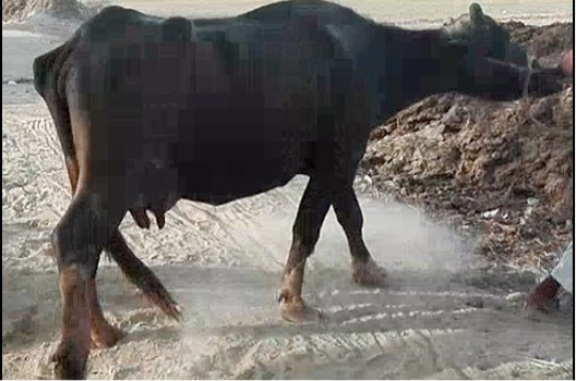
Prior to surgery. Animal showing sign of extended hind limbs and lameness.
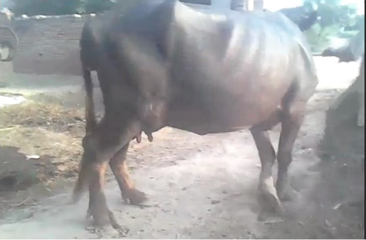
Post-surgery. Photograph demonstrating normal stride immediately after medial patellar desmotomy.
References
- Baird, A., Angel, K., Moll, H., Wolfe, D., Morris, D., Welch, R., Hooper, R. and Wenzel, J., 1993. J. Am. Vet. med. Assoc., 202: 434-436.
- Belge, A., Yaygingul, R., Sarierler, M. and Tatli, Z.B., 2016. Kafkas Univ. Vet. Fakul. Derg. J., 22: 853-857.
- Ducharme, N.G., 2004. In: Farm animal surgery (eds. S. Fubini and N. Ducharme), Elsevier, Missouri, pp. 497-501.
- Kim, N.S., Alam, M.R., Lee, J.I., Park, Y.J., Choi, I.H. 2005. J. Vet. Med. Sci., 67: 723-725. https://doi.org/10.1292/jvms.67.281[CrossRef]
- Kilic, E., Ozaydin, I., Aksoy, O. and Ozturk, S., 2008. Kafkas Univ. Vet. Fakul. Derg. J., 14: 185-190.
- Kofler, J., 1999. Vet. J., 158: 21-32. https://doi.org/10.1053/tvjl.1998.0329[CrossRef]
- Kramer, J., 2006. In: Manual of equine field surgery (eds. D.A. Wilson, J. Kramer and G.M. Constantinescu), Saunders-Elsevier, St. Louis, Missouri, pp. 76-79.
- Meungthong, N., Jenajaroen, K., Sukjaroen, R. and Cherdchutham, W., 2007. Dorsal patellar luxation management in the beef cow. Proc. Chulalongkorn Univ. Vet. Sci. Annu. Conf., 92: 2007.
- Mondal, S., Karnam, S., Baranwal, A. and Das, P., 2013. Explor. Anim. med. Res., 3: 184-185.
- Naveen, M., Kumar, D.D., Shivaprakash, B., Usturge, S., Pawar, A. and Patil, N., 2013. Indian J. Vet. Surg., 34: 94-96.
- Read, R.A., 1999. Waltham Focus, 9: 28-31.
- Singh, A.K., Gangwar, A., Devi, K.S. and Singh, H., 2015. Vet. World, 8: 221-224. https://doi.org/10.14202/vetworld.2015.1331-1339[CrossRef]
- Shivaprakash, B.V. and Usturge, S.M., 2004. Buffalo Bull., 23: 58-63.
Services
Article usage statistics
Loading…