There is a great similarity between the digestive system of rabbits and humans, so rabbits can be considered a good model for the study of gastrointestinal diseases (Kararli, 1995). After 10 days the control rabbits group (Group III) which was given a (physiological saline solution) for the period of the experiment was characterized by normal weight and normal appearance of the histological sections in small intestine and large intestine Figures (1a, 2a, 3a & 4a). The features of intestine were normal microvilli as well as the natural appearance of the mucosa goblet cells compared with animal in positive control which refuse food after 10 days and show decrease in weight. The sections of the tissue that obtained from intestine animals infected with the E.histolytica group (Group IIa) appeared many changes in the small intestine and large intestine Figures (1b, 2b, 3b & 4b), Cecal lesion and infiltration of inflammatory cells, mucosal ulceration, sever destruction in intestinal microvilli, hyperplasia in goblet cells and intestinal mucosal degeneration in colon section. These results were in agreement with (AL-Nafouli., 2004). The section also showed sloughing in epithelial cells and presence of trophozoit of E.histolytica and this is in agreement with the results by (Chadee and Meerovitch, 1985).
The histological sections of K-antigen group show hyperplasia and increases in length of intestinal microvilli in the duodenum (Fig. 1c). In ileum there is apoptosis in epithelial cell lining microvilli and increase in length of intestinal microvilli (Fig. 3c) .The section in jejunum showed hyperplasia in mucosa and increased thickness of jejunum’s walls (Fig. 2c). These changes are due to the immune response to the (capsular antigen) given to the animals. (AL-Kabi., 2006) was pointed to these results. The rabbits can be immunized by used the protein of the outer membrane and the capsule of K. pneumonia.
The results of a study achieved by (Cryz et al., 1985) showed increases in IgG and IgM titers after immunization with capsulated polysaccharide. The IgG antibodies obtained from immune serum was given a significant effect in preventing deadly burn wound sepsis in experimental mice due to K. pneumoniae K1.
The histological sections of the Group Ia treated with (K-Antigen) showed simple histological changes Figures ( 1c, 2c, 3c & 4c). The appearance of the colon is close to normal, and also having normal of mucosa except the hyperplasia (Fig. 4c). The changes in jejunum show increases in goblet cells and increase in length of mucosa region lining jejunum (Fig. 2c).
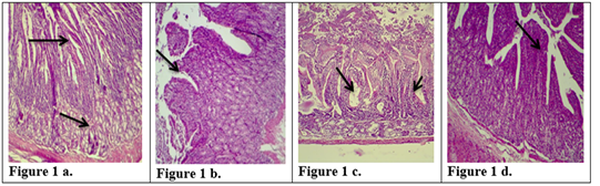
Figure 1: Description of histological changes in duodenum sections. a): Group III, negative control, animal feeding with saline solution. Normal appearance of microvilli, Brunner’s glands in duodenum. (200 X),(H & E)). b): Group II, positive control, animal feeding with E.histolytica cyst, hyperplasia and increases in microvilli and mucosal glands of duodenum. (200 X),(H & E). c): Group I a, section in the animal duodenum immunized by K-antigen, showing hyperplasia increases in length of intestinal microvilli (200 X),(H & E) d): Group I b, section in the duodenum shows the presence of hypoplasia of the intestinal microvilli with an increase in the mucous glands after infected immunized animal with E.histolytica (200 X)(H & E).
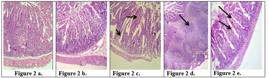
Figure 2: Description of histological changes in the jejunum sections. a): Group III, negative control, (200 X) (H & E). b): Group II, positive control, hyperplasia, (200 X) (H & E). c): Group I a, the section in jejunum shows increases in goblet cells and increases in length of mucosa region lining jejunum after an immunized animal with K-antigen (200 X) (H & E). d): Group I b, section in jejunum shows hyperplasia in the lymphatic glands (peyer’s patch) L.N. of submucosa with germinal center this refers to induce immunity (100 X) (H & E). e): Group I b, section in jejunum after infection with E.histolytica shows simple degenerations in the cells surrounding microvilli with filtration of inflammatory cells inside the cavity of intestinal microvilli and decrease in goblet cells (200 X)(H & E).
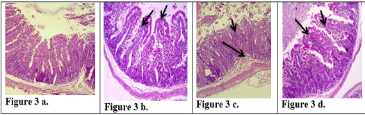
Figure 3: Description of histological changes in ileum sections. a): Group III, negative control animal feeding with saline solution, normal appearance of ileum (200 X)(H & E). b): Group II, positive control, animal feeding with E.histolytica cyst, (200 X)(H & E). c): Group I a, after injection with k-antigen the section in the ileum shows increases in length of intestinal microvilli and apoptosis in epithelial cell lining microvilli (200 X)(H & E). d): Group I b, section in jejunum after infection with E.histolytica shows simple degenerations in the cells surrounding microvilli with filtration of inflammatory cells inside the cavity of intestinal microvilli and decrease in goblet cells (200 X) (H & E)
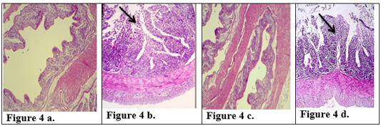
Figure 4: Description of histological changes in colon sections. a): Group III, negative control, normal appearance of colon (200 X) (H & E). b): Group II, positive control.(200 X) (H & E) submucosal ulceration. c): Group Ia ,section in the colon looks like normal and there is no hyperplasia in lining of colon mucosa after immunized animal with K-antigen (200 X) (H & E) d): Group I b, colon section shows superficial mucosal ulceration with heavy filtration of inflammation cells in and between folded intestinal microvilli after infected immunized animal with E.histolytica (200 X) (H & E).
The Group Ib infected with E.histolytica after immunization with K- antigen did not show severe histological changes (Fig 1d, 2d, 2e, 3d, 4d) compared with positive control. The Section in jejunum showed simple degenerations in the cells surrounding microvilli with filtration of inflammatory cells inside the cavity of intestinal microvilli (Fig. 2e) shows hyperplasia in lymphatic glands (peyer’s patch) L.N. of submucosa with germinal center (Fig 2d) and this result points to induced immunity. Colon section showed superficial mucosal ulceration with heavy filtration of inflammation cells in and between folded intestinal microvilli and increase in goblet cells (Fig 4d). Compared with positive control which showed submucosal ulceration (Fig 4b), this increase in goblet cells leads to produce mucus, which is a protective layer against pathogen invasiveness. Resistance to damage to the laboratory animals intestine that infected with parasite may be due to the fact that the polysaccharide injected into the rabbits has produced an immune response that prevented the damage caused by E. histolytica.These results are agree with the study carried out by (Al-Kabi, 2006). Which was pointed in his study to increase the phagocytic cells after immunization with lipopolysaccharide, which is consistent with the results of (AL-Taei, 1996) who realized increasing the phagocytic process rate in (rats) immunized with (polysaccharides) isolated from (Rhizobium leguminosarum) before and after infected by cysts of (Echinococcus granulosus). A study by (Houpt et al., 2004) indicated that immunization with the Gal/GalNAc lectin can protect mouse intestine from amebiasis. (Gilchrist et al., 2015) found that fecal IgA was correlated with prevent recurrent infection with E. histolytica. Study by (Burgess et al., 2014) suggested that colonization of the gut with the bacteria (commensal Clostridia) was protective during infection with E. histolytica. Another study was shown that the polysaccharide processed and presented on MHC II) like the processing of proteins ,to activate CD4+ (helper T-cells) and stimulation of( immune responses) dependent to (T-cell) (Cobb et al., 2004). Our study suggested that immunization with capsulated polysaccharide k-antigen isolated from k.pneumoniae may be used as immunizer against infection with E.histolytica. Research has proved its safe use as a vaccine in humans against infection with k.pneumoniae (Opoku-Temeng et al., 2019).

