
Although the autophagic effect and mechanism of spermidine have been reported, the role of spermidine-induced autophay in cancer cell survival has not been fully elucidated yet. In the present study, we aim to investigate the effect and toxic mechanism of spermidine on cell survival using Hela cells, a cervical cancer cell line as a model. Cell viability was assessed by MTT assay. LDH leakage was determined using a LDH assay kit. Mitochondria membrane potential (MMP) was measured by flow cytometry. LC3 conversion and p62 expression were evaluated by western blot analysis. MTT assay revealed that spermidine decreased the viability of Hela cells in a dose-dependent manner in conjunction with disrupted morphology. Moreover, spermidine caused cell damage as evidenced by elevated LDH leakage and compromised MMP as demonstrated by flow cytometry. Concomitantly, western blot analysis showed that spermidine induced autophagy by activating the LC3 conversion and p62 degradation at different concentrations and durations. However, inhibition of autophagy by 3-MA rescued the survival of Hela cells. This study demonstrated that the potential effect of spermidine on decreasing the survival of Hela cells is attributable to autophagy induction.
Research Article · Volume 53, Issue 1, 26 December 2020 · Open Access
Spermidine-Induced Autophagy Regulates the Survival of HeLa Cells
· · · ·
Affiliations & Notes ⌄
- 1Gansu Key Laboratory of Biomonitoring and Bioremediation for Environmental Pollution, School of Life Sciences, Lanzhou University, 222 South Tianshui R.D., Lanzhou 730000, China , * Corresponding authors: qiym@lzu.edu.cn
- 2ymzhang@lzu.edu.cn
Article Info ⌄
- Journal:
- Pakistan Journal of Zoology
- Published:
- 26 December 2020

From the article
- In this study, we found that exogenous supply of spermidine reduced the survival, caused cell damage and compromised MMP of Hela cells.
- In this study, spermidine led to autophagy activation as well as cell survival inhibition.
- As expected, induction of autophagy by spermidine is accompanied by decreased level of p62 in Hela cells.
- This study clearly shows that autophagy fulfills the critical functions in the death of Hela cells.
Selected from the article's own text.
Abstract
Introduction
Spermidine, a type of natural polyamines, participates in various biological processes . Many studies indicate that cellular polyamines can modulate normal and cancer cell survival ; . Most of the studies acknowledge the role of spermidine as a facilitator of cell survival. However, it is reported that spermidine also has a role in promoting cell death ; . The controversial effect of spermidine on survival of cancer cells is not fully clarified yet. Thus, the effect of spermidine on viability of cervical cancer HeLa cells is investigated in this study.
The basis of these diverse cellular responses to spermidine is currently not known, but the induction of autophagy may serve as a great contributor. Autophagy is known to be a cellular degradation pathway that is responsible for the breakdown of damaged or redundant macromolecules at critical circumstances such as cellular stress . LC3 (microtubule-associated protein light chain 3), typically characterized as an autophagosome marker, is one of the most widely monitored autophagy-related proteins in mammalian cells . Besides, the p62 protein can be incorporated into the autophagosome and be degraded in autolysosomes, thus decreased p62 levels are associated with autophagy activation .
Although autophagy mostly serves a cytoprotective function against certain stresses such as mitochondrial membrane potential (MMP) collapse, it has also been linked to cell death ; . In certain conditions, autophagy may activate cell death, and depending on context, autophagy may promote survival of a given cell species ; . Despite the mounting knowledge about autophagy, the mechanisms through which the autophagic machinery regulates cancer cell survival are not entirely understood. It has been reported that spermidine induces autophagy in cultured yeast, immune cells, nematodes and flies ; . Thus, the effects of external spermidine on autophagy and cell viability are investigated, using HeLa cell line as a model.
The present study explores the effects of spermidine on autophagy and viability and their relationships in HeLa cells. It reveals that spermidine induces autophagy and decreases viability of HeLa cells. Moreover, 3-MA pretreatment affects the autophagy process dramatically and restores the viability of HeLa cells, indicating autophagy is a cause of decreased cell survival. The purpose of this study is to illuminate the complexity of the role of spermidine in cancer cell survival and delineate the function of autophagy in this process.
Materials and Methods
Chemicals
Spermidine, 3-methyladenine (3-MA), carbonyl cyanide 3-chlorophenylhydrazone (CCCP) were obtained from Sigma-Aldrich Inc.
Cell culture and treatments
HeLa cells, obtained from the Cell Bank of Type Culture Collection of Chinese Academy of Sciences (Shanghai, China), were cultured in DMEM (Gibco) supplemented with 10% FBS (Royabio), 100 IU/ml of penicillin & streptomycin at 37°C in a 5% CO2 incubator.
Cell viability and morphology
Cell viability was carried out using MTT assay as our previous study . Briefly, HeLa cells were seeded into 96-well plates (5000 cells/well) and treated with spermidine (0.1, 0.5, 1 mM) for 8 h. Then, MTT was added to each well at a final concentration of 0.5 mg/mL and further incubated for 4 h. Media were removed and 150 mL DMSO was added to dissolve the formazan crystals. The absorbance was detected at 570 nm using a spectrophotometer (Bio-Rad, UltramarkTM Microplate System).
Meanwhile, the morphology of HeLa cells was visualized with an inverted fluorescence microscope (Axio Observer Z1, Carl Zeiss) after the same treatment as above.
LDH assay
LDH, released into the culture media by damaged cells, was measured using LDH assay kits (Beyotime Biotechnology) according to the manufacturer’s protocol. Briefly, after exposure to spermidine, 100 μL LDH reaction solutions were added to the supernatants of HeLa cells (10 μL/well) and incubated for 30 min. The absorbance was quantified at 450 nm using a spectrophotometer (Bio-Rad, UltramarkTM Microplate System).
Transmission electron microscopy
HeLa cells were treated with 0.5 mM spermidine for 8 h and then fixed with 1 ml of 2.5% glutaraldehyde. The samples were sent to the electron microscope center of Lanzhou University for evaluation using a transmission electron microscope (JEOL JEM-1230). Micrographs were taken using a Gatan Erlangshen ES500W camera.
Western blotting
The western blotting was performed as our previous study . Briefly, proteins were separated by SDS-PAGE and transferred to a PVDF membrane, and then incubated with the following primary antibodies: rabbit anti-LC3 and anti-p62 (1:1000, L7543 and P0067, Sigma-Aldrich Inc.). The samples were incubated with HRP conjugated secondary antibodies and developed with the Immobilon Western Chemiluminescent reagents (Millipore).
MMP measurement
HeLa cells were incubated with 1 μg/ml JC-1 (excitation, 507 nm; emission, 530 nm) at 37°C for 30 min. Cells with healthy mitochondria were detected by measuring the increase in the red fluorescence due to JC-1aggregates. While cells that have depolarized Δψm emitting JC-1 monomers were detected by increase in the green fluorescence. The fluorescence intensity was monitored by flow cytometer (BD Biosciences).
Statistical analysis
Statistical analyses were performed using SPSS 19.0 (IBM, USA) and graphed with Origin 8.0 (Origin Lab, USA). Multiple comparisons were performed by ANOVA with LSD post hoc test. Differences were considered significant at p < 0.05.
Results
Spermidine decreased the viability of HeLa cells
The effect of spermidine on the survival of HeLa cells was investigated by MTT assay. As shown in Figure 1A, 0.1, 0.5, 1.0 mM spermidine significantly reduced the cell viability to 83.92%, 65.45% and 67.80%, respectively when compared to control (p<0.05).
Next, we defined whether spermidine led to a change in morphology of HeLa cells. According to Figure 1B, spermidine caused deterioration of HeLa cells as indicated by more cytoplasmic vacuolation compared to control.
Spermidine induced damage of HeLa cells
To confirm the adverse effect of spermidine on HeLa cells, we measured the LDH levels in the supernatant. After 8 h of treatment, LDH levels were significantly increased in supernatants of cells at 0.5 and 1.0 mM spermidine (Fig. 2).
Spermidine induced mitochondrial depolarization in HeLa cells
As demonstrated in Figure 3, 0.5 and 1.0 mM spermidine significantly decreased the MMP relative to control (p<0.05). The mitochondrial uncoupler CCCP as a positive control showed more potent effect.
Spermidine induced autophagy in HeLa cells
Autophagy induction was evaluated by observing the formation of autophagic vacuole. As shown in Figure 4A, 0.5 mM spermidine encapsulated the cytosolic contents within autophagosome.
To confirm the effect of spermidine on autophagy, we examined the conversion of LC3I to LC3II as the amount of LC3II is clearly correlated with the number of autophagosomes. HeLa cells were exposed to 0.5 mM spermidine for different time-points (0, 2, 4, 6, 8, 10 h) and starvation for 5 h as a positive control. Spermidine significantly increased the ratio of LC3II / LC3I at 4, 6 and 8 h (p<0.05) with the most dramatic effect at 8 h (Fig. 4B).
In addition to LC3, receptor protein p62 was used as an autophagy marker. p62 expression was reduced by 10% and 38% when HeLa cells were treated with 0.5 and 1.0 mM spermidine for 8 h (p<0.05) (Fig. 4C).
Spermidine-induced autophagy caused the decrease of cell viability
Autophagy inhibitor 3-MA (5 mM) cotreated with 0.5 mM spermidine significantly restored cell viability when compared to spermidine alone (p<0.05). 5.0 mM 3-MA alone slightly decreased cell viability (Fig. 5).
Discussion
Excessive accumulation or depletion of spermidine may disrupt many cellular functions, including mitochondrial integrity, leading to oxidative stress, autophagy or apoptosis ; ; . In this study, we found that exogenous supply of spermidine reduced the survival, caused cell damage and compromised MMP of Hela cells. Meanwhile, spermidine elicited autophagy and inhibition of autophagy by 3-MA restored the viability, indicating that induction of autophagy is essential for the decreased survival of Hela cells.
It is well accepted that spermidine is a strong inducer of autophagy ; ; . The consequence of promoting autophagy depends on multiple factors, including extent of induction, duration, and cellular context. Although the multiple roles of autophagy in cancer require further clarification, it is obvious that autophagy is directly involved in many important physiological processes such as response to stress, and cell death pathways in cancer cells ; . Autophagy is generally a stress-responsive, survival mechanism. However, it may also be a cell death mechanism (autophagic cell death) ; . The present study showed that direct induction of autophagy through spermidine is sufficient to inhibit cell survival and collapse MMP of Hela cells. Although it is found that addition of spermidine could increase the survival of human peripheral blood mononuclear cells (PBMC), this effect was obtained at relatively low concentrations (around 20 nM) . When applied at millimolar concentrations as in this study, spermidine efficiently elicited autophagy and decreased the survival of Hela cells. These differences may be either due to cell-type-specific differences in the metabolic pathway of spermdine or to differences in the sensitivity to cell death.
Accumulating evidence demonstrates interesting links between autophagy and cancer cell survival ; . In this study, spermidine led to autophagy activation as well as cell survival inhibition. Remarkably, inhibition of autophagy by 3-MA restored the viability of Hela cells. Therefore, autophagy seems to be an important player in the life and death of Hela cells. It is interesting to note that a recent study confirmed this by demonstrating that spermidine induced growth inhibition and apoptosis by autophagic activation in Hela cells .
Unraveling the complex molecular regulation of autophagy is pivotal in understanding the mechanism of cancer cell survival. Recent progress in autophagy research has been led by the identification of a number of autophagy-related proteins. Among these proteins, LC3 has been extensively studied . We found that the conversion of LC3-I to LC3-II was obviously increased when Hela cells were exposed to spermidine. In addition to LC3, p62 is also served as a protein marker of autophagy, which links LC3 and ubiquitinated substrates ; . As expected, induction of autophagy by spermidine is accompanied by decreased level of p62 in Hela cells.
Conclusion
This study clearly shows that autophagy fulfills the critical functions in the death of Hela cells. However, a complete understanding of the autophagic process and its crosstalk with cell death pathways represents an evolving story that will only be clarified with additional research.
Acknowledgements
This work was supported by National Natural Science Foundation of China (31300941) and Fundamental Research Funds for the Central Universities (lzujbky-2018-kb18).
Statement of conflict of interest
The authors declare that there is no conflict of interests regarding the publication of this article.
Figures
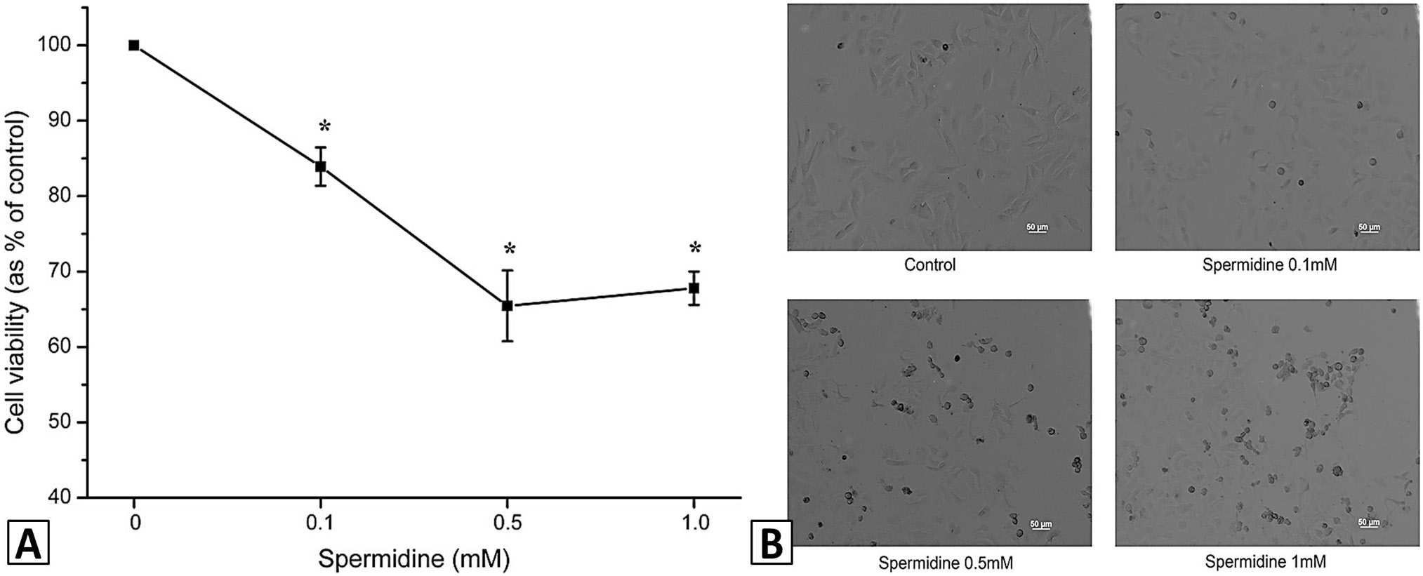
Spermdine reduced the viability of HeLa cells. Cells treated with spermidine (0, 0.1, 0.5, 1.0 mM) for 8 h. Cell viability was measured using the MTT assay. Results are expressed as the percentage of control and represent the mean ± S.E.M. of three independent experiments performed with triplicate cultures. *p < 0.05, compared with control (A). Images of cells were taken after exposure to spermidine as described above (B).
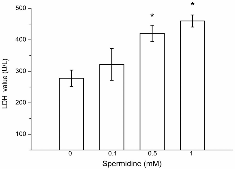
Spermdine increased the LDH leakage of HeLa cells. LDH levels in the supernatant was assessed using the LDH assay kit. Results are expressed as the percentage of control and represent the mean ± S.E.M. of three independent experiments. *p < 0.05, compared with control.
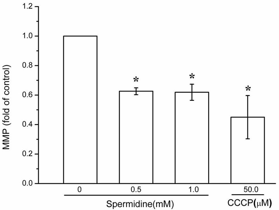
Spermdine collapsed MMP in HeLa cells. Cells were treated with spermidine (0, 0.5, 1.0 mM) for 8 h or 50 μM CCCP for 3 h. MMP was measured by flow cytometry. Results are expressed as the percentage of control and represent the mean ± S.E.M. of three independent experiments. *p < 0.05, compared with control.
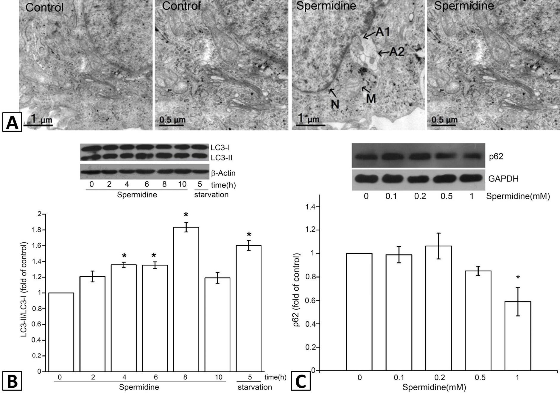
Spermidine induced autophagy in HeLa cells. HeLa cells were treated with 0.5 mM spermidine for 8 h. The formation of autophagic vacuole was observed under TEM (A). The nucleus is marked with letter N, letter M indicates intact mitochondria. autophagic vacuole is labeled as A1, A2. Cells were treated with 0.5 mM spermidine for 2, 4, 6, 8, 10 h or starved for 5 h. The expression of LC3 protein was assessed by western blotting and followed by grey scale analysis (B). Alternatively, the cells were treated with indicated concentrations of spermidine for 8 h and followed by western blotting of p62 (C). *p < 0.05, compared with control.
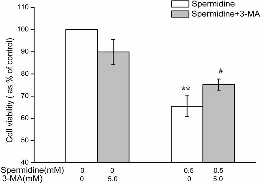
Spermidine-induced autophagy reduced viability of HeLa cells. Cells were treated with spermidine and 3-MA alone or simultaneously for 8 h. Cell viability was measured as in Figure 1. **p < 0.01, compared with control. #p < 0.05, compared with spermidine treatment alone.
References
- Alva, A.S., Gultekin. S.H. and Baehrecke, E.H., 2004. Autophagy in human tumors: Cell survival or death? Cell Death Different., 11: 1046-1048. https://doi.org/10.1038/sj.cdd.4401445[CrossRef]
- Chen, Y., Henson, E.S., Xiao, W., Huang, D., McMillan-Ward, E.M., Israels, S.J. and Gibson, S.B., 2016. Tyrosine kinase receptor EGFR regulates the switch in cancer cells between cell survival and cell death induced by autophagy in hypoxia. Autophagy, 12: 1029-1046. https://doi.org/10.1080/15548627.2016.1164357[CrossRef]
- Chen, Y., Zhuang, H.F., Chen, X.R., Shi, Z.Q. and Wang, X., 2018. Spermidine-induced growth inhibition and apoptosis via autophagic activation in cervical cancer. Oncol. Rep., 39: 2845-2854. https://doi.org/10.3892/or.2018.6377[CrossRef]
- Classen, F., Kranz, P., Riffkin, H., Pompsch, M., Wolf, A., Gopelt, K., Baumann, M., Baumann, J., Brockmeier, U. and Metzen, E., 2018. Autophagy induced by ionizing radiation promotes cell death over survival in human colorectal cancer cells. Exp. Cell Res., 374: 29-37. https://doi.org/10.1016/j.yexcr.2018.11.004[CrossRef]
- Das, G., Shravage, B.V. and Baehrecke, E.H., 2012. Regulation and function of autophagy during cell survival and cell death. Cold Spring Harbor Perspect. Biol., 4: pii: a008813. https://doi.org/10.1101/cshperspect.a008813[CrossRef]
- Eisenberg, T., Knauer, H., Schauer, A., Buttner, S., Ruckenstuhl, C., Carmona-Gutierrez, D., Ring, J., Schroeder, S., Magnes, C. and Antonacci, L., 2009. Induction of autophagy by spermidine promotes longevity. Nat. Cell Biol., 11: 1305-1314. https://doi.org/10.1038/ncb1975[CrossRef]
- Gomes, A.P., Schild, T. and Blenis, J., 2017. Adding polyamine metabolism to the mtorc1 toolkit in cell growth and cancer. Develop. Cell, 42: 112-114. https://doi.org/10.1016/j.devcel.2017.07.004[CrossRef]
- Gu, X., Qi, Y., Feng, Z., Ma, L., Gao, K. and Zhang, Y., 2018. Lead (Pb) induced ATM-dependent mitophagy via PINK1/Parkin pathway. Toxicol. Lett., 291: 92-100. https://doi.org/10.1016/j.toxlet.2018.04.012[CrossRef]
- Henson, E., Chen, Y. and Gibson, S., 2017. EGFR family members’ regulation of autophagy is at a crossroads of cell survival and death in cancer. Cancers, 9: 27. https://doi.org/10.3390/cancers9040027[CrossRef]
- Igarashi, K. and Kashiwagi, K., 2000. Polyamines: Mysterious modulators of cellular functions. Biochem. biophys. Res. Commun., 271: 559-564. https://doi.org/10.1006/bbrc.2000.2601[CrossRef]
- Jiang, P. and Mizushima, N., 2015. LC3- and p62-based biochemical methods for the analysis of autophagy progression in mammalian cells. Methods, 75: 13-18. https://doi.org/10.1016/j.ymeth.2014.11.021[CrossRef]
- Kabeya, Y., Mizushima, N., Ueno, T., Yamamoto, A., Kirisako, T., Noda, T., Kominami, E., Ohsumi, Y. and Yoshimori, T., 2000. LC3, a mammalian homologue of yeast Apg8p, is localized in autophagosome membranes after processing. EMBO J., 19: 5720-5728. https://doi.org/10.1093/emboj/19.21.5720[CrossRef]
- Kovacs, J.R., Li, C., Yang, Q., Li, G., Garcia, I.G., Ju, S., Roodman, D.G., Windle, J.J., Zhang, X. and Lu, B., 2012. Autophagy promotes T-cell survival through degradation of proteins of the cell death machinery. Cell Death Different., 19: 144-152. https://doi.org/10.1038/cdd.2011.78[CrossRef]
- Levine, B. and Yuan, J., 2005. Autophagy in cell death: an innocent convict? J. clin. Investig., 115: 2679-2688. https://doi.org/10.1172/JCI26390[CrossRef]
- Madeo, F., Eisenberg, T., Buttner, S., Ruckenstuhl, C. and Kroemer, G., 2010. Spermidine: a novel autophagy inducer and longevity elixir. Autophagy, 6: 160-162. https://doi.org/10.4161/auto.6.1.10600[CrossRef]
- Morselli, E., Marino, G., Bennetzen, M.V., Eisenberg, T., Megalou, E., Schroeder, S., Cabrera, S., Benit, P., Rustin, P. and Criollo, A., 2011. Spermidine and resveratrol induce autophagy by distinct pathways converging on the acetylproteome. J. Cell Biol., 192: 615-629. https://doi.org/10.1083/jcb.201008167[CrossRef]
- Navarro-Yepes, J., Burns, M., Anandhan, A., Khalimonchuk, O., del Razo, L.M., Quintanilla-Vega, B., Pappa, A., Panayiotidis, M.I. and Franco, R., 2014. Oxidative stress, redox signaling, and autophagy: Cell death versus survival. Antioxid. Redox Signal., 21: 66-85. https://doi.org/10.1089/ars.2014.5837[CrossRef]
- Pankiv, S., Clausen, T.H., Lamark, T., Brech, A., Bruun, J.A., Outzen, H., Overvatn, A., Bjorkoy, G. and Johansen, T., 2007. p62/SQSTM1 binds directly to Atg8/LC3 to facilitate degradation of ubiquitinated protein aggregates by autophagy. J. biol. Chem., 282: 24131-24145. https://doi.org/10.1074/jbc.M702824200[CrossRef]
- Rajeeve, V., Pearce, W., Cascante, M., Vanhaesebroeck, B. and Cutillas, P.R., 2013. Polyamine production is downstream and upstream of oncogenic PI3K signalling and contributes to tumour cell growth. Biochem. J., 450: 619-628. https://doi.org/10.1042/BJ20121525[CrossRef]
- Ryter, S.W., Bhatia, D. and Choi, M.E., 2018. Autophagy: A lysosome-dependent process with implications in cellular redox homeostasis and human disease. Antioxid. Redox Signal., 30: 7518. https://doi.org/10.1089/ars.2018.7518[CrossRef]
- Schultz, C.R., Geerts, D., Mooney, M., El-Khawaja, R., Koster, J. and Bachmann, A.S., 2018. Synergistic drug combination GC7/DFMO suppresses hypusine/spermidine-dependent eIF5A activation and induces apoptotic cell death in neuroblastoma. Biochem. J., 475: 531-545. https://doi.org/10.1042/BCJ20170597[CrossRef]
- Shen, H.M. and Codogno, P., 2012. Autophagy is a survival force via suppression of necrotic cell death. Exp. Cell Res., 318: 1304-1308. https://doi.org/10.1016/j.yexcr.2012.02.006[CrossRef]
- Stefanelli, C., Stanic, I., Zini, M., Bonavita, F., Flamigni, F., Zambonin, L., Landi, L., Pignatti, C., Guarnieri, C. and Caldarera, C.M., 2000. Polyamines directly induce release of cytochrome c from heart mitochondria. Biochem. J., 347: 875-880. https://doi.org/10.1042/bj3470875[CrossRef]
- Thomas, T. and Thomas, T.J., 2001. Polyamines in cell growth and cell death: molecular mechanisms and therapeutic applications. Cell. mol. Life Sci., 58: 244-258. https://doi.org/10.1007/PL00000852[CrossRef]
- Yu, L., Wan, F., Dutta, S., Welsh, S., Liu, Z., Freundt, E., Baehrecke, E.H. and Lenardo, M., 2006. Autophagic programmed cell death by selective catalase degradation. Proc. natl. Acad. Sci. U.S.A., 103: 4952-4957. https://doi.org/10.1073/pnas.0511288103[CrossRef]
Services
Article usage statistics
Loading…